


APA 7: TWs Editor & ChatGPT. (2023, October 7). Researchers Advance Brain Injury Treatment with Innovative 3D Printing Technique. PerEXP Teamworks. [Article Link]
A pioneering method, devised by researchers from the University of Oxford, holds the promise of offering personalized remedies for individuals coping with brain injuries. In a groundbreaking achievement, the researchers have shown that it’s possible to 3D print neural cells to replicate the intricate structure of the cerebral cortex. These findings have been officially published in the journal Nature Communications, marking a significant milestone in the field.
Injuries to the brain, whether stemming from trauma, stroke, or surgical procedures to treat brain tumors, commonly result in substantial harm to the cerebral cortex, the outer layer of the human brain. This damage often leads to challenges in cognitive function, mobility, and communication. For instance, annually, approximately 70 million individuals worldwide experience traumatic brain injuries (TBI), with 5 million of these cases categorized as severe or fatal. Presently, there are no efficacious therapies available for severe brain injuries, resulting in profound implications for the affected individuals’ quality of life.
The future of treating brain injuries might lie in regenerative therapies focused on tissue repair, particularly approaches involving the use of patients’ own stem cells. Yet, until recently, a crucial challenge has remained unresolved: the absence of a method to guarantee that transplanted stem cells accurately replicate the intricate structure of the brain.
In their latest research, scientists from the University of Oxford employed 3D printing techniques to craft a bilayered brain tissue using human neural stem cells. Once introduced into sections of mouse brain tissue, these cells seamlessly integrated both structurally and functionally with the existing host tissue, yielding compelling results.
This advance marks a significant step towards the fabrication of materials with the full structure and function of natural brain tissues. The work will provide a unique opportunity to explore the workings of the human cortex and, in the long term, it will offer hope to individuals who sustain brain injuries.
Yongcheng Jin
Lead author
Department of Chemistry, University of Oxford
The cortical structure was engineered from human induced pluripotent stem cells (hiPSCs), which possess the capacity to generate various cell types found throughout the human body. An essential benefit of harnessing hiPSCs for tissue regeneration is their ability to be readily derived from a patient’s own cells, eliminating the risk of provoking an immune response.
Human-induced pluripotent stem cells (hiPSCs) underwent differentiation into neural progenitor cells, each destined for distinct layers of the cerebral cortex. This transformation was achieved through precise combinations of growth factors and chemical stimuli. Subsequently, these specialized cells were suspended in a solution, generating two unique ‘bioinks,’ which were then meticulously 3D printed to create a bilayered structure. Remarkably, these printed tissues retained their stratified cellular organization for an extended period in culture, a phenomenon confirmed by the continued expression of layer-specific biomarkers.
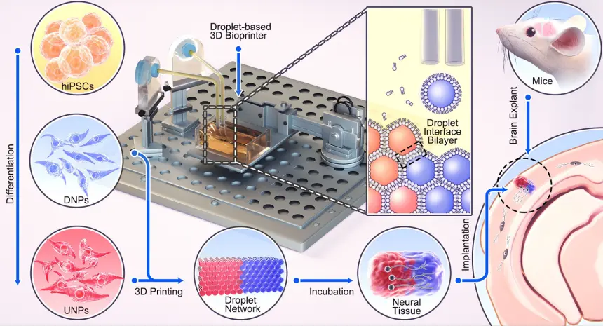
Upon implantation into mouse brain slices, the printed tissues exhibited robust integration. This was evidenced by the extension of neural processes and the migration of neurons across the boundary between the implant and host tissue. Furthermore, the implanted cells displayed signaling activity that corresponded with that of the host cells. This interaction indicated effective communication between human and mouse cells, highlighting both structural and functional integration.

The researchers are now committed to advancing the droplet printing method to develop intricate, multi-layered cerebral cortex tissues that closely emulate the architectural complexity of the human brain. Beyond their potential for treating brain injuries, these engineered tissues hold promise for applications in drug evaluation, investigations into brain development, and enhancing our comprehension of the foundations of cognition.
This latest breakthrough extends the team’s impressive legacy of over a decade in pioneering and patenting 3D printing techniques for the creation of synthetic tissues and the cultivation of cells.
Our droplet printing technique provides a means to engineer living 3D tissues with desired architectures, which brings us closer to the creation of personalized implantation treatments for brain injury.
Linna Zhou
Senior author
Department of Chemistry, University of Oxford
The use of living brain slices creates a powerful platform for interrogating the utility of 3D printing in brain repair. It is a natural bridge between studying 3D printed cortical column development in vitro and their integration into brains in animal models of injury.
Francis Szele
Senior author
Department of Physiology, Anatomy and Genetics, University of Oxford
Human brain development is a delicate and elaborate process with a complex choreography. It would be naïve to think that we can recreate the entire cellular progression in the laboratory. Nonetheless, our 3D printing project demonstrates substantial progress in controlling the fates and arrangements of human iPSCs to form the basic functional units of the cerebral cortex.
Zoltán Molnár
Senior author
Department of Physiology, Anatomy and Genetics, University of Oxford
This futuristic endeavour could only have been achieved by the highly multidisciplinary interactions encouraged by Oxford’s Martin School, involving both Oxford’s Department of Chemistry and the Department of Physiology, Anatomy and Genetics.
Hagan Bayley
Senior author
Department of Chemistry, University of Oxford
Resources
- NEWSPAPER Oxford News & Events. (2023, October 4). Oxford researchers develop 3D printing method that shows promise for. Oxford News & Events. [Oxford News & Events]
- JOURNAL Jin, Y., Mikhailova, E., Lei, M., Cowley, S. A., Sun, T., Yang, X., Zhang, Y., Liu, K., Da Silva, D. C., Soares, L. C., Bandiera, S., Szele, F. G., Molnár, Z., Zhou, L., & Bayley, H. (2023). Integration of 3D-printed cerebral cortical tissue into an ex vivo lesioned brain slice. Nature Communications, 14(1). [Nature Communications]


