



Cancer detection often depends on finding cellular warning signs before malignant cells spread into distant body parts. In Canada, cancer stands as the leading cause of death, where two in five people develop tumors during their lifetimes and nearly 700 individuals receive a new diagnosis each day. According to the Canadian Cancer Society, common malignancies include lung, breast, prostate, and colorectal cancers, but survival rates drop when tumors are found late. Doctors rely on tissue biopsies, but teams are now developing liquid biopsy methods that detect circulating tumor traces in blood samples [3]. Liquid biopsy blood tests aim to reveal tumor biology without hospital surgery [2].
How Liquid Biopsy Detects Circulating DNA
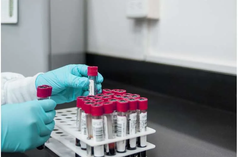
Standard diagnostic biopsies require surgeons to cut tissue fragments from suspected tumors in hospital operating suites. The surgical procedure carries bleeding risks and causes pain that limits repeat patient testing. Outpatient blood tests spare people from surgical trauma. By relying on a simple blood collection, liquid biopsy allows medical staff in local clinics to evaluate disease without hospital admissions [3].
Engineering professor Alexandre Pellan Cheng leads cancer detection studies at ÉTS Montréal and the CHUM Research Centre, focusing on how dying tumor cells release genetic material into the bloodstream. For decades, oncologists evaluated protein biomarkers like prostate-specific antigen (PSA) to track disease recurrence. PSA tests lack specificity because protein levels often rise during benign inflammation. Circulating tumor DNA provides a precise biological signal because it carries genetic alterations present in the tumor [3].
Dying tumor cells shed fragments of genetic code into plasma streams as cellular membranes rupture. Spotting these shed molecules lets laboratory teams evaluate cancer genetics without cutting into fragile organs. Blood travels through every human organ. A single blood tube captures shed fragments from multiple metastatic sites at once, giving liquid biopsy a diagnostic reach that localized needle biopsies cannot match when sampling solid tumors [3].
Limits of Standard Tissue Sampling
Small cell lung cancer accounts for roughly 15% of all lung cancer cases, according to data from the Centers for Disease Control and Prevention. The illness spreads early through lymphatic channels and grows fast compared with other pulmonary malignancies. Patients often arrive with metastases at their first consultation. Tumors recur within months following chemotherapy, so doctors need prompt biological data to guide treatment choices [2].
Repeat surgical biopsies present severe hazards for individuals whose pulmonary lesions return after first-line chemotherapy, carrying serious risks of pneumothorax, arterial bleeding, and respiratory failure. Sparing sick patients from invasive procedures remains an urgent medical goal. Scarce biopsy tissue often leaves hospital laboratories without enough cells for genomic subtyping. Without adequate cell counts, doctors cannot identify secondary drug vulnerabilities [2].
Senior author Anish Thomas, M.D., the Roswell Park Professor of Hematology and Oncology at Northwestern University, pointed out that tumor biology varies between patients. “Although SCLC has historically been treated largely as a single disease, we now recognize that there are important biological differences between tumors,” said Thomas, who also conducts oncology research at the Robert H. Lurie Comprehensive Cancer Center of Northwestern University. Tumor diversity demands personalized treatment strategies [2]. Because small cell lung cancer (SCLC) progresses fast and spreads early, doctors need blood-based profiling tools that track biological evolution before recurring tumors resist treatment [1], [2].
Gene Expression in Small Cell Tumors
Northwestern Medicine investigators partnered with a Jerusalem-based laboratory headed by Dr. Nir Friedman at Hebrew University to overcome biopsy barriers [1], [2]. The collaborative team used cell-free chromatin immunoprecipitation sequencing (cfChIP-seq) to read functional genetic states from a single milliliter of patient blood [1],. “SCLC presents a unique challenge,” Thomas said. “Most existing liquid biopsy technologies are very good at detecting DNA mutations, but in SCLC, much of the important biology is driven by differences in gene expression rather than mutations.” [2] Functional chromatin analysis reveals which cellular pathways remain active within living tumor cells [1].
Standard liquid biopsies look for point mutations in circulating DNA, but small cell lung tumors depend on transcriptional reprogramming. By isolating circulating nucleosomes—coils of genomic DNA wrapped around histone proteins—the cfChIP-seq assay maps chemical modifications on gene promoters. Lead author Gavriel Fialkoff and colleagues observed strong promoter signals at key transcription factor genes, including ASCL1, NEUROD1, POU2F3, and ATOH1. Promoter marks matched the transcriptional subtypes discovered in patient tumor RNA [1].

Mapping lineage-specific transcription factors gives doctors a reliable blueprint of small cell tumor biology. Each molecular subtype relies on separate regulatory networks that create unique treatment vulnerabilities. Epigenetic profiling reveals tumor adaptation over time. Tracking active nucleosomes bridges a major gap in cancer medicine, allowing teams to evaluate disease biology without needle punctures into chest cavities [1].


Clinical Trials and Data Processing
Detecting tiny amounts of tumor DNA among billions of healthy host fragments remains a major obstacle for pathology laboratories. Alexandre Pellan Cheng noted that early tumors shed minute quantities of genetic material into circulation, pushing sequencing instruments to their detection limits. “The smaller the tumour, the less DNA it releases into the blood,” Cheng explained in his report. Shed DNA fragments degrade quickly in blood and need careful purification before sequencing instruments can read them [3].
Each blood sample yields terabytes of data with billions of genetic sequences. Cheng’s engineering team at ÉTS Montréal designs artificial intelligence algorithms to separate genuine cancer mutations from background machine noise. Pathology laboratories must test these automated computational models in clinical trials before adopting them for regular patient screening [3].

Bioinformatics pipelines must process immense genomic datasets while maintaining high diagnostic accuracy under real-world clinical conditions, correcting for sequencing errors and eliminating background noise caused by normal cellular turnover in the bloodstream. Specialized software compares shed fragments against validated cancer databases, pinpointing malignant alterations with high statistical certainty. Computational filtering makes blood testing reliable across diverse patient groups [3].
Advances in Liquid Biopsy Profiling
In the study published in the Journal of Clinical Investigation, investigators analyzed 441 plasma samples from patients with small cell lung cancer, other neuroendocrine carcinomas, non-SCLC cancers, and healthy control donors. The research team created a quantitative SCLC score that tracked tumor volume across serial computed tomography (CT) scans [1]. “A particular strength of our study was the availability of matched blood and tumor samples from several patients,” Thomas said. “This allowed us to validate the blood-based measurements against gene expression in the tumors themselves.” [2]
The blood assay classified molecular subtypes across 73 matched patient samples, including 41 time-matched pairs collected during disease evolution. Testing needs only one milliliter of plasma, so hospital staff can collect samples during routine clinical visits [1], [2]. The cfChIP-seq platform provided an accurate reflection of tumor burden, confirming that circulating nucleosomes carry reliable diagnostic information [1].
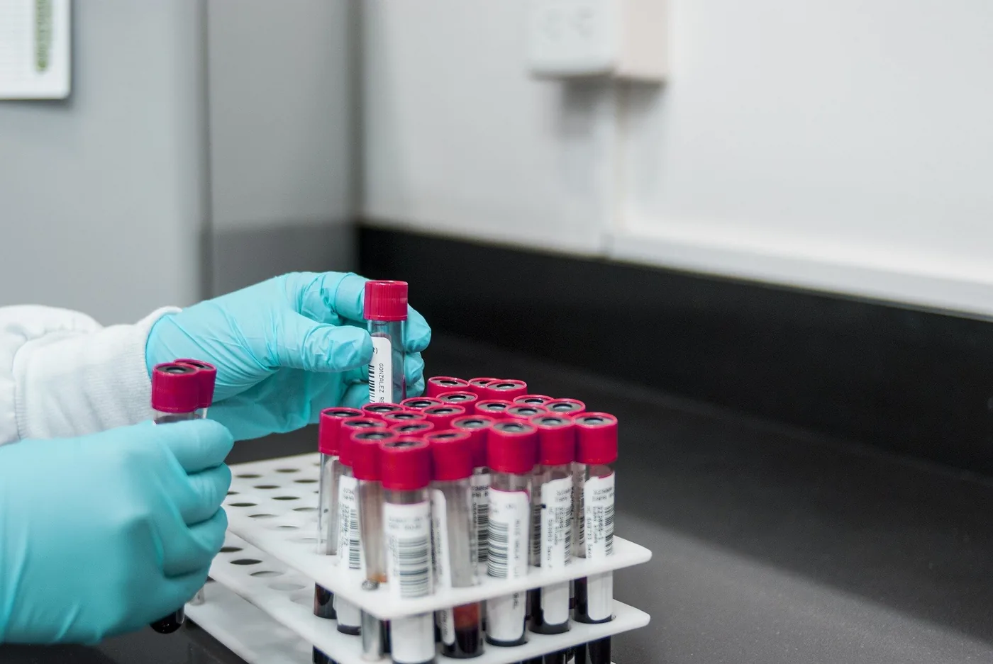
Comparing blood plasma profiles with matched solid tissue transcriptomes proved that circulating nucleosomes preserve cellular identity, allowing the assay to distinguish small cell lung cancer from other neuroendocrine malignancies without diagnostic confusion. Capturing both transcriptional subtypes and total tumor burden gives oncologists a dual-purpose diagnostic tool. The method advances liquid biopsy capabilities [1].
Monitoring Treatment Response Over Time
Serial blood tests allow oncologists to observe how tumor biology shifts during active treatment regimens [2],. When patients receive cytotoxic drugs, measuring whether circulating tumor DNA levels rise or fall helps doctors evaluate therapeutic efficacy [3]. Quick drops in tumor markers indicate that cancer cells are dying, while rising concentrations warn doctors of emerging drug resistance weeks before new tumors appear on radiology scans [2],. Real-time blood data helps care teams adjust medications and protect patients from failing therapies [3].
In recurring small cell lung cancer, malignant cells develop resistance to platinum chemotherapy regimens. Investigating resistance mechanisms has been difficult because obtaining repeat lung biopsies from sick patients carries severe medical risks [2]. Tracking shifts in circulating nucleosomes over time lets doctors spot resistance pathways as they appear [1], [2]. Early molecular detection could allow oncologists to switch patients to alternative therapies before substantial tumor progression occurs [1], [2].
Northwestern Medicine researchers are now planning larger prospective trials to track patients across multiple treatment cycles. “Our hope is that approaches like this will help bring precision medicine to SCLC, where it has been particularly challenging,” Thomas said. Broad hospital implementation will require multicenter trial validation, but blood-based molecular tests offer a practical path toward personalized cancer care [2], [3]. Simple blood draws could soon become routine tools for managing complex cancer cases worldwide [2], [3].
- ACADEMIC JOURNAL Fialkoff, G., Takahashi, N., Sharkia, I., Gutin, J., Hermoni, N., Nirula, M., Kumar, R., Pongor, L., Nichols, S., Sciuto, L., Parmar, K., Desai, P., Suresh, P., Abel, M., El Meskini, R., Maoz, M., Rottenberg, Y., Nevo, S., Nechushtan, H., & Temper, M. (2026). Plasma nucleosome profiling reports on tumor burden and molecular subtypes in small cell lung cancer. Journal of Clinical Investigation, 136(19). [Article Link]
- ONLINE NEWS Dimmer, O. (2026). Blood test offers new way to track lung cancer. Medical Xpress. [Article Link]
- ONLINE NEWS Cheng, A. P. (2026). A simple blood test could detect cancer. The Conversation. [Article Link]
APA 7: PerEXP Teamworks. (2026, October 30). Small Cell Lung Cancer Burden Tracked Through Liquid Biopsy. PerEXP Teamworks. https://perexpteamworks.com/en/liquid-biopsy-cancer-blood-test/


