






Can an everyday camera detect nerve damage in babies before permanent disability takes hold? Clinicians diagnosing spinal muscular atrophy face an urgent challenge because motor neurons die quickly without therapy [4]. In 2019, Zolgensma made news as the world’s most expensive medicine at nearly 2 million euros per injection [3]. While the drug stops motor neurons from dying, it can’t bring lost cells back to life [4]. A French study shows that an ordinary video camera paired with machine learning can spot early movement changes within sixty seconds [2].
What Is Spinal Muscular Atrophy?
Spinal muscular atrophy is a rare genetic disorder in which motor nerve cells that govern infant muscle activity gradually break down [4]. Because those spinal neurons cannot send signals to skeletal tissue, affected infants experience progressive muscle wasting and quickly lose fundamental motor milestones. In its most acute presentation, termed type 1 spinal muscular atrophy, newborn babies quickly lose the ability to lift their limbs, sit upright, and eventually breathe on their own, meaning that life expectancy rarely exceeds two years without treatment [3]. Although therapeutic drugs can halt this neurodegenerative process, early diagnosis is essential because medical therapies cannot regenerate nerve cells that have already died [4].
In France, clinicians diagnose approximately one infant among 6,000 to 10,000 births [4]. France added the condition to its routine neonatal screening program in 2025 [3]. However, many hospital systems lack universal screening, leaving doctors dependent on bedside clinical evaluations to spot infants who need confirmatory genetic tests [4].
The resulting weakness shares clinical similarities with wider forms of muscle loss and neuromuscular deterioration observed in other clinical domains [4]. If medical teams wait until an infant displays overt paralysis, vulnerable neuronal pathways have already suffered irreversible damage that no current therapy can reverse [3]. Early detection is the single most reliable strategy to preserve motor function in vulnerable infants [4]. Every lost week reduces what medical therapy can save [3].
Clinical Challenges in Detecting Newborn Hypotonia
Before genetic laboratories confirm a suspected diagnosis, pediatricians rely on bedside clinical examinations of neonatal reflexes and muscle tone [4]. Doctors evaluate babies for signs of hypotonia, a clinical condition marked by reduced muscle tone and a noticeable deficit in spontaneous movements [3]. When held by an examiner, an affected newborn appears limp, and its arms and legs droop downward without resistance against gravity [4]. Pediatric specialists often describe this appearance as a “hypotonic infant,” but subjective visual scoring varies substantially between clinicians [3]. Because early motor deficits can appear subtle during brief examinations, objective quantification is necessary to prevent missed diagnostic opportunities [4].
Dozens of illnesses cause infant hypotonia [4]. Mild motor lag often escapes notice when a baby is tired or fussy [3]. By the time motor delays become undeniable to family members, the underlying disease has already progressed significantly [4]. Pediatric wards need reliable tools to quantify motor function objectively [3].
Researchers Imen Trabelsi, François Jouen, and Jean Bergounioux shared their preliminary findings in a preprint paper that has not yet undergone peer review, demonstrating how video cameras can assist clinical evaluations [2]. The investigators said that because the therapeutic window in spinal muscular atrophy is narrow, subjective clinical scoring too often delays essential genetic testing [4]. An automated computer vision tool provides a quick objective indicator that guides clinicians within minutes, enabling prompt medical intervention before permanent neuron loss takes hold [3].
How Video Cameras and AlphaPose Track Infant Motion
To eliminate subjectivity from bedside examinations, the French research team developed an automated motion capture system powered by a standard consumer video camera [4]. The approach intentionally avoids wearable sensors, attached markers, or complex clinical hardware that might distress fragile newborns or distort their natural motor behaviors [3]. During assessment, an infant lies comfortably on a plain contrasting background while an ordinary camera records spontaneous movements for exactly sixty seconds [4]. Machine learning algorithms then examine the resulting video footage frame by frame to extract precise motion trajectories [3]. This non-invasive setup allows hospital staff to conduct fast motor evaluations directly in pediatric intensive care units [4].
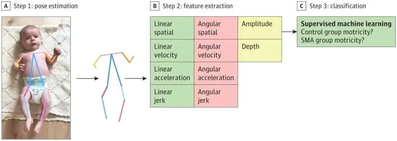
The automated pipeline operates through three steps [4]. First, the system reconstructs a digital skeleton using a real-time pose estimation framework called AlphaPose [3]. The model maps 12 anatomical joints, eight limb segments, and four motion angles in every recorded video frame [4]. This digital skeleton tracks infant movement without skin contact [3].

From this dynamic skeleton, the software extracts 108 features that capture gesture amplitude, movement frequency, bilateral symmetry, and motion depth in three-dimensional space [4]. The computational framework detects minute motor variations that human clinicians cannot reliably quantify during routine clinical exams [3]. As observed in broader diagnostic machine learning, medical vision systems must avoid pitfalls like healthcare AI bias in clinical datasets to ensure dependable diagnostic support in varied patient populations [4]. Here, computational vision transforms subjective clinical impressions into reproducible quantitative curves that reveal motor deficits with mathematical rigor [3].


How Depth Movement Signals Spinal Muscular Atrophy
When the researchers evaluated the 108 extracted movement features, one specific parameter distinguished infants with neurological illness from healthy controls with remarkable clarity [4]. That critical metric was the depth of movement, which measures how effectively an infant lifts its arms and legs into the three-dimensional space above the mattress [3]. Neurologically healthy babies frequently propel their limbs upward against gravity during spontaneous motor activity [4]. In contrast, infants affected by spinal muscular atrophy show altered motor skills and struggle to generate upward vertical motion along the depth axis [3]. The camera system measures this movement limitation with sub-millimeter precision, converting a clinician’s intuitive observation of weakness into verified numerical data [4].
Infants with the genetic condition showed a depth-axis deficit with sensitivity greater than 97% [4]. While affected babies can still slide their hands across a flat sheet, raising limbs against gravity requires muscle strength they lack [3]. Gravity exposes motor neuron loss almost immediately [4]. The algorithm quantifies this deficit objectively [3].

To make algorithm outputs interpretable for medical practitioners, the research team implemented Shapley Additive Explanations (SHAP) [3]. This mathematical framework illustrates which specific movement parameters exerted the greatest influence on each algorithmic classification [4]. Rather than presenting a black-box conclusion, SHAP allows doctors to examine visual plots detailing depth limitations and bilateral asymmetries for each patient, helping pediatric specialists explain automated findings to families before genetic tests confirm the result [3].
Testing the XGBoost Algorithm on Clinical Cases
To validate the video analysis pipeline, investigators conducted a clinical trial involving 25 infants hospitalized in pediatric intensive care units [4]. The study cohort comprised five patients with genetically confirmed spinal muscular atrophy alongside 20 infants who presented normal neurological evaluations [3]. Researchers used this dataset to train and validate a supervised machine learning model using XGBoost, a scalable tree boosting system introduced by Chen and Guestrin [1]. The algorithm learned to differentiate normal spontaneous infant motor patterns from the “altered” movement profiles caused by early motor neuron degeneration, accurately separating the two infant groups in each tested trial [3].
The model classified both patient cohorts with 97% accuracy. The trial evaluated 25 infants [4]. Five patients had confirmed SMA [3]. The sixty-second video capture caused no stress to fragile newborns [4]. Computational analysis completed in just a few minutes, delivering prompt results [3].

The authors noted that their study took place prior to 2025, when France had not yet established systematic genetic screening for newborn infants [4]. Scientific editors Sadie Harley and Robert Egan reported that this retrospective analysis demonstrates why early bedside screening mattered during that pre-screening era [3]. Even where universal neonatal genetic testing exists, an optical motion tracking tool provides valuable real-time clinical confirmation at birth, offering guidance long before blood test results return from central laboratories [4].
Expanding Early Screening Beyond SMA in Pediatric Care
The study authors said that this computer vision tool is not intended to replace pediatricians or medical specialists [4]. Instead, the system functions as an objective, non-invasive screening aid that equips bedside doctors with actionable motor metrics within minutes, requiring no complex hospital machinery [3]. Beyond spinal muscular atrophy, dozens of genetic and metabolic conditions cause infant hypotonia during early developmental stages [4]. The research group is now expanding the video analysis pipeline to screen for other rare neuromuscular disorders that reduce infant muscle tone [3]. Early automated detection could accelerate diagnoses in pediatric movement disorders [4].
Research funding was provided through the Axa Foundation for Human Progress, which unites commitments from Axa Group and Mutuelles d’Assurances in science, nature, solidarity, and culture. Previously, global philanthropy was managed by the Axa Research Fund, which supported over 750 projects worldwide since 2007. This funding helped researchers build accessible diagnostic tools [4].
Because early diagnosis allows newborns to receive gene therapies before permanent motor neuron death takes place, integrating computer vision into neonatal screening programs offers profound clinical benefits. As journal information published in JAMA Pediatrics and republished under Creative Commons indicates, video tracking makes subtle motor deficits visible to clinical teams within minutes of birth [3]. When every passing week dictates whether an infant maintains the ability to move and breathe, artificial intelligence provides pediatricians with an objective ally at the bedside [4].
- CONFERENCE PAPER Chen, T., & Guestrin, C. (2016). XGBoost. Proceedings of the 22nd ACM SIGKDD International Conference on Knowledge Discovery and Data Mining, 785-794. [Article Link]
- PREPRINT Trabelsi, I., Jouen, F., & Bergounioux, J. (2026). A camera combined with AI could help further diagnosis of movement disorders like spinal muscular atrophy in newborns. [Article Link]
- ONLINE NEWS Trabelsi, I., Jouen, F., & Bergounioux, J. (2026, October 6). A camera combined with AI could help further diagnosis of movement disorders in newborns. Medical Xpress. [Article Link]
- ONLINE NEWS Jouen, F., Trabelsi, I., & Bergounioux, J. (2026, October 6). A camera combined with AI could help further diagnosis of movement disorders like spinal muscular atrophy in newborns. The Conversation. [Article Link]
APA 7: TWs Editor. (2026, October 7). How Everyday Video Cameras Spot Spinal Muscular Atrophy. PerEXP Teamworks. https://perexpteamworks.com/en/spinal-muscular-atrophy-video-ai/


