




Can bioengineered cellular scaffolds permanently eradicate residual cancer cells following surgical resection? Precision oncology faces a persistent challenge when micro-tumors survive operative interventions, prompting researchers to develop spatiotemporal drug delivery systems that coordinate biochemical suppression with cellular cytotoxicity. Natural killer cells recognize malignant tissues through an immunological synapse (a specialized junction coordinating receptor interactions and toxic granule release). However, postoperative inflammation and dense biochemical barriers frequently dismantle these critical immune contacts before destruction occurs. By synchronizing local anti-inflammatory release with progressive cell deployment, biomedical researchers demonstrated that bioengineered scaffolds suppress local tumor recurrence across multiple preclinical animal surgical models. The platform transforms surgical oncology by targeting residual disease directly at the operative margin [1].
- How Does Spatiotemporal Drug Delivery Work?
- Engineering Mechanical Synapses for Armored Killer Cells
- How Localized Drug Delivery Calms Resection Cavities
- Why Five Percent of Tumor Cells Resist Attack
- Can Spatiotemporal Delivery Dismantle Protective Tumor Niches?
- Translating Bioengineered Cellular Platforms into Oncology Practice
How Does Spatiotemporal Drug Delivery Work?
Spatiotemporal drug delivery functions by releasing therapeutic agents in a strictly controlled physical location and sequence. In systemic chemotherapy, active pharmacological agents disperse randomly, causing severe toxicity while failing to accumulate at therapeutic concentrations within targeted surgical margins. Lead investigator Junzhu Shi and senior author Lian Li at Sichuan University addressed this issue with a semi-spike patch (a semi-spike scaffold engineered for sequential release) inserted directly into postoperative cavities [1].
Surgical intervention causes localized tissue trauma and acute wound-healing cascades that recruit immunosuppressive myeloid cells to shield surviving tumor fragments from immune destruction. Fast-acting therapeutic intervention disrupts this harmful pathway. By loading the rapidly dissolving surface of the semi-spike patch with dexamethasone, investigators established a permissive wound environment that calms initial inflammation. Co-authors Ping Zhang, Shenao Yan, and Mengyang Liu at Sichuan University confirmed that this local biochemical suppression prevents cytokine surges and ensures that subsequent cytotoxic effectors remain viable and active within host tissue beds [1].
Sustained cellular release follows this preliminary anti-inflammatory phase over several days. Rather than releasing cells immediately into inflamed tissue, the scaffold matrix gradually delivers armed natural killer cells against residual margins. Similar principles guide adoptive cellular reprogramming in CAR-T therapy, where controlled cellular activation prevents premature lymphocyte exhaustion while directing sustained cytotoxic activity toward persistent malignant targets. Co-investigators Junlin Li and Guangsheng Du at Sichuan University confirmed that orderly timing ensures effective clearance [1].
Engineering Mechanical Synapses for Armored Killer Cells
Normal lymphocytes bind tumor targets through delicate surface receptor pairings to defend against malignant transformation by forming an immunological synapse (an organized cell-to-cell interface coordinating cytotoxic granule secretion). Malignant cells downregulate surface adhesion molecules, causing effector cells to detach prematurely without delivering their toxic cargo. These abortive contacts, termed transient ‘kiss-and-run’ events, allow cancer cells to survive and seed metastases [1].
Bioengineers equipped natural killer cells with multivalent galectin-3 binding polymers that function like prehensile mechanical arms to overcome premature detachment. These fortified cells, designated a-NKs, anchor firmly to abundant galectin-3 molecules on tumor cell membranes, extending contact duration and strengthening receptor contacts across the immunological synapse. Contacts become a ‘kiss of death’. The enforced proximity stimulates extensive synapse remodeling and triggers massive cytotoxic degranulation, converting ineffective glancing contacts into decisive cellular lysis. Armed killer cells secrete key chemokines that attract host CD8 T cells, bridging innate cytotoxicity with adaptive immune activation to clear residual postoperative tumor cells [1].
Preclinical testing in female mouse models of postoperative breast cancer demonstrated the therapeutic efficacy of this mechanical enhancement. Delivered straight into resection cavities, armored killer cells adhered tightly to tumor margins, suppressing local recurrence significantly better than unmodified cells. Senior investigator Lian Li and researcher Guangsheng Du at Sichuan University secured funding from the National Natural Science Foundation of China (grants 82373818 and 82574346) to support these comprehensive in vivo preclinical validations published in Nature Communications [1].
How Localized Drug Delivery Calms Resection Cavities
The physical architecture of resection cavities presents severe biological obstacles to cellular immunotherapies. Surgical excision damages blood vessels, triggers sudden cytokine surges, and creates elevated interstitial pressure that washes away non-adherent immune cells. Applying localized drug delivery mechanics through a semi-spike patch architecture anchors the therapeutic platform securely into cavity margins. Specialist Yuanhao Zhao at Sichuan University fabricated and characterized the specialized (Dex→a-NK)@ssPatch device to ensure structural resilience in moving tissue beds [1].
Tumor cells could not detach. Uncontrolled surgical inflammation accelerates micro-tumor seeding while exhausting newly arrived killer lymphocytes in preclinical animal models [1].
Dexamethasone released during the initial phase suppresses acute inflammatory cascades without permanently harming arriving lymphocytes. This temporary chemical modulation quells tissue inflammation and neutralizes hostile reactive oxygen species that would otherwise inactivate therapeutic cells. As acute swelling subsides, the biodegradable patch initiates sustained release of a-NK cells directly onto residual tumor beds, ensuring that therapeutic effectors deploy into a receptive microenvironment to maximize cytotoxic clearance of residual breast cancer cells across the surgical margin [1].


Why Five Percent of Tumor Cells Resist Attack
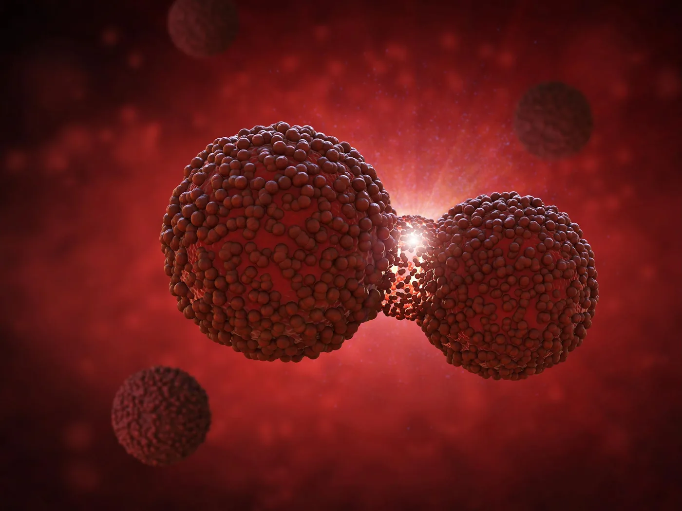
Genetic heterogeneity within solid tumors creates specialized microenvironments that protect malignant clones from immune destruction. In pancreatic ductal adenocarcinoma, where the five-year survival rate lingers around 13 percent, standard immunotherapies routinely fail to penetrate dense tumor tissue. Researchers at the Icahn School of Medicine at Mount Sinai investigated why these neoplasms exclude immune cells so efficiently. Utilizing Perturb-map, an innovative spatial genomics platform combining multiplexed genetic editing, molecular barcodes, and high-resolution imaging, scientists examined how distinct cellular subpopulations organize their immediate microenvironments. Science writer Juan Martinez at SciTechDaily highlighted how these localized shields prevent cytotoxic lymphocytes from reaching their targets. Mount Sinai teams mapped these niches [2].
The investigation revealed that resistance does not require genetic changes across every malignant cell. Five percent proved sufficient. When merely 5 percent of pancreatic cancer cells produced the protein PAI1, that minor fraction organized a localized biochemical shield protecting neighboring malignant cells from immune surveillance. First author Chiara Falcomatà, postdoctoral fellow at Mount Sinai who published the study in Nature, stated: “small groups of cancer cells create localized neighborhoods that protect themselves and nearby cancer cells from immune attack” [2].
Two serpin family proteins, PAI1 and PAI2, build this barrier by blocking enzymes that dissolve fibrin (a mesh-forming protein involved in clotting and tissue repair). Fibrin shields the local niche. This dense protein scaffold captures macrophages and instructs them to suppress killer T cells. Similar obstacles appear when tracking malignant cell signaling in solid glioma tumors, where local matrix remodeling hinders immune infiltration [2].
Can Spatiotemporal Delivery Dismantle Protective Tumor Niches?
Spatiotemporal delivery dismantles protective tumor niches by coordinating localized molecular inhibition with direct cytotoxic cell deployment. In Mount Sinai preclinical models, genetic deletion of PAI1 or PAI2 decreased tumor burden by more than half while doubling CD8 T cell infiltration. Senior investigator Brian Brown at Mount Sinai emphasized that “small groups of cancer cells create localized neighborhoods” that shelter malignant tissue from surveillance. Co-authors Maximilian Schaefer, Bhavya Singh, Divya Chhamalwan, and Alexander Tepper at Mount Sinai documented how these distinct mechanisms operate across diverse solid tumor models [2].
Pharmacological targeting reinforces these genetic findings in living animals. Pairing PAI-039, an experimental inhibitor of PAI1, with anti-PD-1 immunotherapy extended survival in two pancreatic cancer models where checkpoint blockade alone showed minimal efficacy. Median survival reached 47 days. Genetic deletion of PAI1 combined with anti-PD-1 therapy increased median survival from 24 to 47 days in mice, proving that opening tumor shields restores immune eradication. Mount Sinai investigators Sebastian Nielsen, Hunter Potak, Maxime Dhainaut, and Gurkan Mollaoglu confirmed these survival dynamics [2].
Integrating molecular disruption with engineered cellular scaffolds represents an attractive clinical paradigm. While PAI1 inhibition dismantles fibrin barriers, armored natural killer cells provide the direct cytotoxicity required to eliminate surviving tumor cells [1, 2]. Through bioengineered cellular release, a patch device can deliver targeted inhibitors to break down fibrin shields before releasing armed lymphocytes [1]. Mount Sinai teams confirmed this [2].
Translating Bioengineered Cellular Platforms into Oncology Practice
Moving bioengineered cellular delivery platforms into human oncology requires extensive preclinical testing and validation. Mount Sinai researchers Matthew Park, Miriam Merad, and Alessia Baccarini demonstrated how cellular heterogeneity governs clinical responses in patient tissues. While mouse breast and pancreatic cancer models demonstrate robust anti-tumor responses, human surgical cavities present greater tissue volume and biomechanical diversity [1, 2]. Clinical translation depends on ensuring that semi-spike matrices adhere firmly to irregular resection beds without generating adverse foreign-body reactions [1].
Targeting serpin activity offers therapeutic possibilities beyond oncology. High PAI1 levels correlate strongly with deep vein thrombosis, cardiovascular dysfunction, and biological aging. Modulating the fibrin-macrophage axis could aid neurodegenerative treatments, including therapies for Alzheimer’s disease where fibrous scarring restricts neural tissue repair. Through coordinated postoperative therapy, bioengineered spatiotemporal platforms achieve targeted therapeutic effects in in vivo tissues while preventing systemic toxicity across non-target vital organ systems [1, 2].
Clinical oncology continues to shift toward synchronized multi-agent platforms placed directly into resection sites, where deploying spatiotemporal drug delivery patches enables surgical teams to eliminate residual micro-metastases before recurrent tumors take root. Supported by the Science & Technology Department of Sichuan Province and the 1.3.5 Project for Disciplines of Excellence (grant numbers 2026NSFSC0564 and ZYYC25001), investigators Yining Xu and Yuan Huang at Sichuan University continue expanding patch design. Fellow researchers at Sichuan University and the Fundamental Research Funds for the Central Universities (grant YJ202412) continue refining these surgical applications. These engineering innovations offer a practical roadmap for preventing postoperative relapse in clinical oncology [1].
- ACADEMIC JOURNAL Shi, J., Li, J., Zhang, P., Li, B., Yan, S., Liu, M., Du, G., Xu, Y., Huang, Y., & Li, L. (2026). Spatiotemporal delivery of immune-synapse-stabilizing NK cells for postoperative tumor therapy. Nature Communications. [Article Link]
- ACADEMIC JOURNAL Falcomatà, C., Schaefer, M. M., Singh, B., Chhamalwan, D., Tepper, A., Nielsen, S. R., Potak, H. T., Dhainaut, M., Mollaoglu, G., Park, M. D., Merad, M., Baccarini, A., & Brown, B. D. (2026). A serpin–myeloid axis in pancreatic cancer heterogeneity and immune evasion. Nature. [Article Link]
APA 7: PerEXP Teamworks. (2026). Armed NK Cells Attack Tumors via Spatiotemporal Drug Delivery.


