




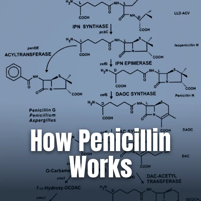
Penicillin stops bacterial illness by preventing multiplying microbes from building their outer peptidoglycan walls. A clear view of how penicillin works reveals that beta-lactam compounds block the transpeptidase enzymes that stitch wall layers together during cell division [6]. When drug units bind to these proteins, dividing cells cannot withstand the steep fluid pressure generated by their own cytoplasm. Water rushes inward across the weakened envelope until the swollen bacterium bursts [5].
Peptidoglycan Cell Wall Mesh
In 1895, Vincenzo Tiberio at the University of Naples reported that water well molds in Arzano killed bacteria, while Costa Rican scientist Clodomiro Picado Twight recorded Penicillium antibiotic actions at the Pasteur Institute in 1923. In 1924, André Gratia and Sara Dath at the Free University of Brussels found that a mold extract called mycolysate destroyed Staphylococcus aureus [11]. Bacteria rely on a tough outer casing to preserve their shape and survive harsh osmotic conditions. Under this casing lies the fragile plasma membrane [5]. The main polymer providing strength to the bacterial envelope is peptidoglycan, a giant mesh built from repeating amino sugars that form glycan strands linked by short peptide chains [6].
As a bacterium multiplies, it must constantly expand this outer casing through tightly controlled enzymatic reactions that make room for new cell mass while preserving structural stability. Bacterial autolysins cut tiny openings into the existing peptidoglycan mesh so that newly made sugar-peptide building blocks can slip into place [5]. If this remodeling pauses while growth proceeds, the envelope quickly weakens, making the microbe helpless against osmotic forces [6]. Newly produced peptidoglycan strands must quickly fill every gap created during cell division, keeping the outer barrier unbroken as the single rod-shaped organism elongates [5].
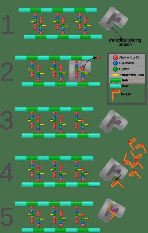
Stitching newly inserted peptidoglycan strands together needs transpeptidase enzymes embedded in the plasma membrane. These penicillin-binding proteins act as molecular bridges connecting adjacent peptide side chains into an interlocking three-dimensional lattice that completely encircles the bacterial cytoplasm. The resulting covalent bonds convert loose glycan strands into a tough framework capable of resisting high mechanical stress. Without functional PBPs, new wall sections remain completely unbonded and porous, meaning the microbe produces raw building units but cannot weave them into a protective shield against external fluid pressures [6]. Fleming first noted this lysis on St Mary culture plates [12].
DD-Transpeptidase as the Target Enzyme
In 1927, Irish physician Joseph Warwick Bigger and his students C.R. Boland and R.A.Q. O’Meara at Trinity College Dublin studied variant colonies of Staphylococcus aureus isolated from an axillary abscess, inspiring later work on chemical inhibitors. Bigger published these findings in The Journal of Pathology [12]. All beta-lactams share a strained four-membered ring that mimics the terminal D-alanyl-D-alanine peptide sequence of natural cell wall precursors, causing bacterial DD-transpeptidase to bind the drug rather than its authentic biological substrate [6].
Once positioned inside the catalytic pocket of the target enzyme, the strained four-membered beta-lactam ring undergoes a chemical reaction that permanently inactivates the protein [6]. The catalytic serine residue within the transpeptidase attacks the carbonyl carbon of the beta-lactam ring, forming an irreversible covalent penicilloyl-enzyme complex that the cell cannot break apart [7]. Because the drug stays locked in the active site, the transpeptidase cannot release itself to cross-link adjacent glycan chains or take part in further wall construction. Deprived of cross-linking enzymes, dividing cells continue to synthesize linear glycan strands without the peptide bridges needed to secure them into a cohesive lattice. Each blocked enzyme permanently loses its catalytic role during cell division [6].
Targeting transpeptidases explains why penicillin works with high safety in clinical medicine because human cells lack peptidoglycan walls entirely [8]. Mammalian tissues possess flexible lipid membranes, whereas pathogens like Staphylococcus aureus rely on transpeptidases for basic physical survival. Human cells contain no cell walls or transpeptidase targets [6]. Medical providers at Cleveland Clinic rely on this clear biological contrast when prescribing beta-lactam drugs for bloodstream infections, pneumonia, and severe systemic illness [1].
Osmotic Turgor Pressure and Lysis
In 1910, Paul Ehrlich established modern chemotherapy by discovering arsphenamine (Salvarsan) to treat Treponema pallidum, proving that targeted synthetic chemicals could attack microbes without harming the human host. Ehrlich later introduced neoarsphenamine (Neosalvarsan) to improve clinical tolerability while maintaining antibacterial potency [4]. Bacterial cells keep high internal solute levels compared to human tissue fluid, creating an osmotic gradient that drives water inward across the fragile cytoplasmic membrane. This inward flow creates substantial turgor pressure that healthy, cross-linked peptidoglycan walls easily withstand [5].
As multiplying bacteria grow in the presence of penicillin, the imbalance between wall cleavage and blocked cross-linking creates gaping structural tears across the envelope. Autolytic enzymes continue cleaving old peptidoglycan strands to prepare space for cell elongation, but no cross-linked polymers arrive to seal the expanding gaps [5]. This mechanism reveals why dormant bacteria survive exposure: the drug needs active cell division and ongoing wall synthesis to exert its lethal bactericidal action [6]. Surrounding fluids flood across the exposed plasma membrane along the steep osmotic gradient, stretching the lipid bilayer beyond its elastic breaking point. Internal turgor forces the membrane to herniate through newly opened gaps [5].

Eventually, the swelling microbe reaches a critical physical threshold where the stretched plasma membrane tears apart under internal turgor pressure, producing explosive osmotic lysis that empties the cell [5]. In 1940, Oxford researchers confirmed this rapid lysis in animal trials [4]. Penicillin is classified as a bactericidal drug because it directly destroys living pathogens rather than merely suppressing their ongoing reproduction [1].
Gram-Negative Outer Membrane Defenses
In 1935, French scientists showed that Prontosil, discovered by Nobel laureate Gerhard Domagk, metabolized into active sulfanilamide to fight virulent bacterial infections [4]. Bacterial envelope anatomy dictates whether classic penicillin molecules can reach their target enzymes. Gram-positive pathogens, including Streptococcus pneumoniae, Listeria, and diverse staphylococcal strains, possess a thick peptidoglycan wall measuring several layers deep that directly interfaces with the surrounding fluid environment. Because these microbes lack an outer lipid membrane, penicillin diffuses freely through the porous sugar mesh to bind PBPs on the cell surface. This direct access makes Gram-positive cocci highly sensitive to early penicillins [6].
Gram-negative bacteria display a more intricate architecture that obstructs classic penicillin molecules. Although they retain a peptidoglycan layer, it is thin and shielded beneath an outer membrane rich in lipopolysaccharide (LPS) complexes. This outer lipid bilayer acts as a dense barrier that prevents hydrophobic compounds and large polar antibiotics from diffusing toward the underlying periplasmic space [6]. Because natural penicillins like penicillin G cannot penetrate this lipid barrier in high amounts, early mold broths failed to inhibit Gram-negative pathogens like Salmonella typhimurium and Shigella [9]. Fleming confirmed this Gram-negative resistance in his 1929 report [12].
To solve this physical barrier, chemists developed aminopenicillins like ampicillin that pass through outer membrane protein channels called porins into the periplasmic space. Through these water-filled channels, ampicillin reaches PBPs in organisms like Escherichia coli, broadening clinical coverage for urinary, ear, and respiratory tract infections [7]. Beecham researchers isolated the 6-APA core in 1959 to create these broad-spectrum drugs. Overcoming outer membrane defenses allowed beta-lactam therapy to expand far beyond its original Gram-positive targets [3].



Scaling Production in Peoria
Alexander Fleming began the antibiotic era in August 1928 at St Mary’s Hospital in London, returning from a Suffolk vacation at The Dhoon to find staphylococci lysed on a Petri dish contaminated with Penicillium notatum [8]. Fleming published his findings in 1929 in the British Journal of Experimental Pathology, officially naming the substance penicillin [11]. In 1930, his student Cecil George Paine successfully used mold juice to cure neonatal conjunctivitis in human infants [10].
At Oxford, Howard Florey, Ernst Chain, and Norman Heatley purified penicillin and tested it in eight laboratory mice on May 25, 1939, confirming that treated animals survived virulent streptococcal infections. Chain called the treated animals’ survival “a miracle”. The team published their work in The Lancet in August 1940 and treated an Oxford policeman with severe abscesses in February 1941 [4]. In March 1942, Anne Miller became the first American civilian successfully cured of streptococcal sepsis [5]. Meanwhile, Dutch scientists at the Netherlands Yeast and Spirit Factory in Delft produced penicillin under the code name Bacinol, marketing it in 1946 to expand global supplies [4].
Florey and Heatley traveled to Peoria, Illinois, in June 1941 to collaborate with the USDA Northern Regional Research Laboratory, where Charles Thom, Andrew Moyer, Kenneth Raper, and Robert Coghill scaled production using corn-steep liquor in submerged deep tanks. In 1943, laboratory technician Mary Kay Hunt isolated Penicillium chrysogenum strain NRRL 1951 from a moldy Peoria market cantaloupe [2]. This melon strain produced six times more antibiotic than Fleming’s mold [4]. While Hunt detailed finding the melon in 1962, Raper noted in 1976 that an unnamed woman delivered it, and a 1948 Abbott Laboratories painting by Douglas Gorsline credited Mary Lou Finley Roberson. Governor J.B. Pritzker designated NRRL 1951 the State Microbe of Illinois in August 2021 [2].

Core Penam Ring and Side Chains
All penicillins share a bicyclic core termed 4-thia-1-azabicyclo (3.2.0) heptane, known simply as penam, which consists of a four-membered beta-lactam ring fused to a five-membered sulfur-containing thiazolidine ring. An intact beta-lactam ring remains essential for antibacterial activity because chemical cleavage of this strained cyclic amide bond completely destroys its ability to bind transpeptidases [8]. Chemical removal of the natural acyl side chain from penicillin G leaves 6-aminopenicillanic acid (6-APA), an adaptable nucleus that enabled chemists to build hundreds of semisynthetic antibiotic derivatives. British biochemist Dorothy Hodgkin solved this three-dimensional structure using X-ray crystallography, earning the 1964 Nobel Prize in Chemistry [10].
Natural penicillins divide clinically into benzylpenicillin (penicillin G) and phenoxymethylpenicillin (penicillin V) [10]. Penicillin G is rapidly destroyed by gastric acid, making it ineffective when swallowed, whereas penicillin V resists acid degradation [8]. Culturing the mold with phenoxyacetic acid yields penicillin V, an oxygen-bearing compound that provides approximately 60% oral bioavailability in ambulatory patients. Pharmacologists advise taking oral penicillin V thirty minutes before meals or two hours after food because meals reduce peak blood levels [7]. Penicillin G is administered intravenously as Pfizerpen for neurosyphilis and endocarditis, or intramuscularly as long-acting benzathine benzylpenicillin to prevent recurrent infections [9]. One unit of penicillin G equals 0.600 micrograms, while 400,000 units of penicillin V standardizes at 250 milligrams [10].
Adding bulky acyl side chains created penicillinase-resistant drugs like methicillin, nafcillin, and oxacillin, which protect the beta-lactam ring from enzymatic attack through steric hindrance [8]. Adding polar amino groups yielded ampicillin and amoxicillin, while ureidopenicillins like piperacillin were developed to kill resilient Pseudomonas aeruginosa pathogens [7]. Ernst Chain discussed these synthetic modifications with Nelson Kardos at Imperial College London in 1973 [3].
How Penicillin Works Against Sensitive Strains
Bacteria have developed several sophisticated defensive strategies to evade beta-lactam therapy [5]. The most widespread bacterial defense is the secretion of beta-lactamase enzymes, such as staphylococcal penicillinase, which hydrolyze the amide bond of the four-membered beta-lactam ring before the drug can reach PBPs [6]. Once cleaved into inactive penicilloic acid, the altered molecule cannot attach to bacterial transpeptidase enzymes [7]. Bacterial populations spread beta-lactamase genes through plasmid conjugation, enabling rapid horizontal transfer of resistance traits between diverse pathogen strains [5]. Most clinical Staphylococcus aureus isolates now produce beta-lactamase [6].
A second major resistance pathway alters the molecular shape of the target transpeptidase enzyme [5]. Methicillin-resistant Staphylococcus aureus (MRSA) produces an alternative transpeptidase called PBP2a with an extremely low binding affinity for beta-lactam molecules. This acquired enzyme continues cross-linking peptidoglycan chains with normal efficiency even when therapeutic levels of methicillin, nafcillin, or flucloxacillin saturate the surrounding host tissues. Because beta-lactams cannot bind PBP2a, the cell wall remains fully intact throughout active bacterial multiplication. MRSA demonstrates blanket clinical resistance across all standard beta-lactams [6].

Gram-negative bacteria also deploy multi-drug efflux pumps and selective porin mutations to prevent beta-lactams from accumulating in the periplasm. To overcome enzymatic destruction, pharmacologists combine vulnerable penicillins with beta-lactamase inhibitors like clavulanic acid, tazobactam, or sulbactam, creating combination drugs like Augmentin, Unasyn, and Zosyn. These sacrificial inhibitor compounds bind and neutralize bacterial enzymes, allowing the companion penicillin to cross-link targets and destroy dividing microbes [7]. The World Health Organization warned in 2014 that antibiotic resistance poses a global health crisis [6].
Penicillin Mechanism in Clinical Care
Penicillins distribute well into lung tissue, pleural fluid, and inflamed joints, but undergo rapid renal elimination where active tubular secretion clears most of the dose within hours [7]. The antigout medication probenecid (Benemid) competitively blocks this renal tubular secretion, maintaining high therapeutic blood levels during intensive intravenous therapy [9]. Because renal clearance is slower in neonates and elderly patients, physicians modify dosing schedules to prevent drug accumulation and neurotoxicity [8]. Probenecid raises circulating penicillin levels by blocking renal transporters [9].
Approximately 10% of hospital patients report a penicillin allergy, yet true hypersensitivity remains remarkably rare in medical practice. Formal skin testing and oral challenges show that fewer than 5% of labeled individuals suffer genuine IgE-mediated allergic reactions. Studies confirm that true penicillin allergy wanes over time, with approximately 80% of allergic patients becoming completely tolerant after a decade of avoidance [10]. Unverified allergy labels routinely force physicians to prescribe expensive broad-spectrum reserve antibiotics that raise patient toxicity and increase the incidence of dangerous Clostridioides difficile colitis [1]. Cross-reactivity between penicillins and newer cephalosporins occurs in only about 2% of patients, enabling safe alternative prescribing [10].
A comprehensive assessment of where penicillin works best confirms that it remains the first-line treatment for syphilis, streptococcal pharyngitis, and cellulitis, a skin infection affecting 14.5 million Americans annually. In a randomized controlled trial published in the New England Journal of Medicine, low-dose penicillin prophylaxis delayed recurrent cellulitis from 532 days to 626 days [9]. Patients must complete their full prescribed course to ensure complete pathogen eradication and prevent surviving bacteria from acquiring resistance mutations [1]. Sir Almroth Wright urged proper medical recognition for Fleming in The Times in 1942 [4].
- WEBSITE Cleveland Clinic. (2024). What Is Penicillin? [Article Link]
- WEBSITE Suszkiw, J. (2021). The Enduring Mystery of “Moldy Mary”. USDA Agricultural Research Service. [Article Link]
- ACADEMIC JOURNAL Kardos, N., & Demain, A. L. (2011). Penicillin: the medicine with the greatest impact on therapeutic outcomes. Applied Microbiology and Biotechnology, 92(4), 677–687. [Article Link]
- ACADEMIC JOURNAL Gaynes, R. (2017). The Discovery of Penicillin—New Insights After More Than 75 Years of Clinical Use. Emerging Infectious Diseases, 23(5), 849–853. [Article Link]
- ONLINE NEWS Newman, T. (2018). Penicillin: Function, history, and resistance. Medical News Today. [Article Link]
- WEBSITE Chow, S. (2010). Penicillin Mechanism. News-Medical. [Article Link]
- WEBSITE Boucher, M. (2026). Penicillin Antibiotics: Mechanism, Spectrum & Pharmacokinetics for Pharmacy and Medical Students. TeachMePharmacy. [Article Link]
- WEBSITE Meštrović, T. (2010). What is Penicillin? News-Medical. [Article Link]
- WEBSITE Ogbru, O. (2022). What Is Penicillin Used For? Antibiotic Types, Side Effects, Drug Names. MedicineNet. [Article Link]
- WEBSITE Wikipedia contributors. (2026). Penicillin. Wikipedia, The Free Encyclopedia. [Article Link]
- WEBSITE Wikipedia contributors. (2026). History of penicillin. Wikipedia, The Free Encyclopedia. [Article Link]
- WEBSITE Wikipedia contributors. (2026). Discovery of penicillin. Wikipedia, The Free Encyclopedia. [Article Link]


