






When people ask what are alveoli, the direct anatomical answer points to millions of tiny, cup-shaped air sacs (singular: alveolus) packed at the terminal ends of the human airway tree. Mammalian lungs rely on these delicate structures to move oxygen into circulating blood while expelling carbon dioxide produced by ongoing cellular metabolism during ordinary rest or intense physical exertion [8]. A healthy adult possesses roughly 480 million of these units, creating a combined surface area of 70 to 80 square meters [12]. Without such an expansive and thin architecture, human respiration would quickly fail to meet everyday metabolic demands across vital organs [11].
- Structural Organization of the Respiratory Zone
- What Are Alveoli Made of?
- Why Alveoli Do Not Collapse Under Surface Tension
- Mechanisms of Gas Diffusion Across the Blood-Air Barrier
- Capillary Perfusion and Fluid Balance in Alveolar Walls
- Alveolar Maturation From Gestation to Old Age
- Clinical Conditions Disrupting Pulmonary Alveoli
- Recent Research on Epithelial Repair and Injury
Structural Organization of the Respiratory Zone
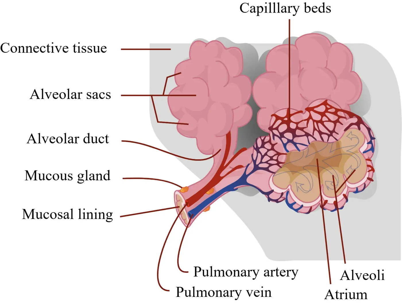
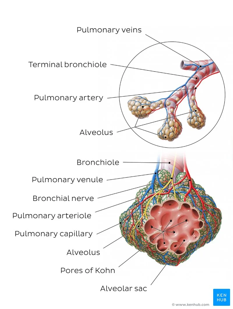
Inhaled air travels through branching conduits until it arrives at the terminal respiratory unit known as an acinus, where tiny air pockets first emerge along the walls of respiratory bronchioles before opening into alveolar ducts. Each bronchiole divides into 2 to 11 alveolar ducts, and each duct terminates in 5 to 6 alveolar sacs [9].
Alveolar sacs form polyhedral clusters of 20 to 30 individual air spaces that open into a shared central space called an atrium. In a typical adult, human lungs contain roughly 480 million alveoli, although anatomical surveys by Kenhub report a broad range between 274 million and 790 million with a coefficient of variation of 37% [8]. Kenhub notes that these tiny hollow cavities make up about 90% of total lung volume, while individual adult diameters average 0.2 to 0.3 millimeters [12].
Thin alveolar septa separate nearby air sacs and hold a dense capillary network that envelopes three-quarters of each sac’s outer surface area. Connective tissue inside septal walls holds fibroblasts, pericytes, and elastic fibers made of elastin and collagen that permit steady expansion and passive recoil during ordinary breathing cycles [12]. Pores of Kohn, measuring 10 to 15 micrometers in diameter, form small openings within these septa to connect adjacent air spaces. By equalizing ventilation pressure across nearby lobules, these collateral channels allow air to bypass small obstructions if mucus blocks a feeding bronchiole [7].
What Are Alveoli Made of?
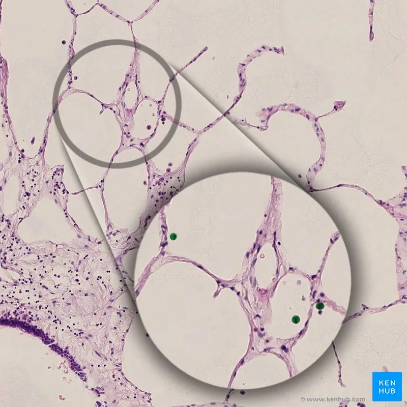
The alveolar wall features an epithelial layer supported by an extracellular matrix and surrounded by blood vessels, where squamous Type I pneumocytes span roughly 95 percent of the internal airspace lining to provide an ultra-thin barrier for gas diffusion. Cytoplasmic extensions of these cells become so thin that their thickness drops to roughly 25 nanometers in some peripheral regions [12]. Because visible light wavelengths measure hundreds of nanometers, researchers must rely on transmission electron microscopy to view these delicate cell borders clearly, where organelles cluster around the central nucleus and leave broad sheets of bare cytoplasm. Type I cells cannot divide.
Cuboidal cells called Type II pneumocytes make up roughly 60% of all epithelial cells in the alveolus, yet they cover only 5% of its total surface [10]. Storage organelles known as lamellar bodies allow these compact cells to synthesize and secrete pulmonary surfactant directly into the air space [12]. When toxic chemicals or viral infections destroy fragile Type I cells, surviving Type II cells proliferate and differentiate to rebuild the epithelial barrier [10].
Mobile scavenger cells known as alveolar macrophages, often called dust cells, wander through the air spaces and connective tissue to provide immune protection by engulfing foreign debris, soot particles, and airborne bacteria [12]. Roughly 100 million expired macrophages ride up the mucociliary escalator each day before being swallowed at the esophagus and digested [8].
Why Alveoli Do Not Collapse Under Surface Tension
Because alveoli are small and curved, the thin water film coating their inner lining generates inward surface tension that pulls the walls together. How do fragile microscopic air spaces stay inflated against inward surface tension? Under the physical law of Laplace, smaller spheres generate higher collapsing pressure than larger spheres when wall tension remains constant, prompting progressive lung collapse known as atelectasis [11].
To prevent collapse, Type II pneumocytes continuously release pulmonary surfactant through exocytosis into the liquid layer, producing a lipid-protein blend consisting mostly of dipalmitoylphosphatidylcholine with proteins SP-A through SP-D [10]. Lipid particles align at the air-water boundary, where their hydrophilic heads sit in liquid and hydrophobic tails project into air. When alveoli contract during exhalation, surfactant lipids pack tightly together, lowering surface tension toward zero and keeping small air sacs stable [11].

Recent biomechanical simulations have shed fresh light on how surfactant films behave in whole lungs during regular breathing. A 2025 study led by R. Li and co-authors integrated alveolar surfactant dynamics into a multiscale computational model connecting acinar mechanics with the full bronchial tree. Computer simulations showed that surfactant generates pronounced hysteresis between inhalation and exhalation, keeping lungs compliant and easy to inflate under varying tidal volumes while preventing severe breathing difficulties [5]. Without this dynamic regulation, the mechanical work of breathing would quickly overwhelm respiratory muscles and cause widespread airspace collapse.
Mechanisms of Gas Diffusion Across the Blood-Air Barrier
Gas movement between inspired air and capillary blood relies on passive diffusion governed by Fick’s law of diffusion, where gas transfer is proportional to total surface area and partial pressure difference, but inversely proportional to membrane thickness. The alveolar blood-air barrier is exceedingly thin, averaging 0.2 to 0.6 micrometers between alveolar air and red blood cells across a three-layer boundary that consists of the Type I pneumocyte, a shared basement membrane, and the capillary endothelium. Red blood cells measure about 7.5 micrometers in diameter, meaning they must deform slightly and travel in single file through tiny capillaries to let oxygen cross the barrier in a fraction of a second [8].
Partial pressure differences supply the driving force that pushes gases in opposite directions through the respiratory membrane. In fresh alveolar air, oxygen partial pressure sits near 100 mmHg, compared to roughly 40 mmHg in deoxygenated blood arriving from the right ventricle. The pressure drop drives oxygen through the membrane and into erythrocytes, where it binds with hemoglobin. Carbon dioxide moves in reverse down its own gradient, diffusing from venous blood at 45 mmHg into alveolar air at 40 mmHg so it can be exhaled [10].
Although oxygen molecules are smaller, carbon dioxide dissolves in blood plasma far more readily. Because of its high solubility, carbon dioxide has a diffusion coefficient roughly 20 times higher than oxygen, allowing it to cross the alveolar membrane just as quickly despite having a much smaller partial pressure difference [10].

Capillary Perfusion and Fluid Balance in Alveolar Walls
Because gases diffuse too slowly through liquid to oxygenate blood effectively, alveoli must stay mostly free of fluid. Mean capillary blood pressure averages only 10 mmHg, whereas plasma oncotic pressure measures about 28 mmHg, creating a strong inward osmotic pull that draws excess fluid back into blood vessels (a balance described by Starling principles) [8].
Capillary blood flow inside alveolar walls fluctuates dynamically during every breath. In a 2022 research paper, X. A. Si and J. Xi created an advanced 3D computational model of an expanding and contracting alveolus-capillary unit. Simulated blood flow showed natural non-uniformity across capillary meshes, with rhythmic breathing movements increasing this spatial variability. A static geometry model underestimated blood perfusion by 11% during normal respiration, showing that dynamic lung motion directly alters how oxygen enters passing erythrocytes [4].
Optimal respiration requires close alignment between air ventilation and capillary blood flow, producing a ventilation-perfusion ratio that averages about 0.8 in healthy lungs [10]. When foreign fluids disturb this balance, tissue damage follows. In a 2012 study, K. Westphalen and co-authors studied what happens when gastric acid enters rodent alveoli during aspiration. Microinjecting acid at pH 1.5 produced transient nanoscale pores in the alveolar membrane, allowing cytosolic dye to escape before the cell layer rapidly resealed. Calcium entry through these pores triggered reactive oxygen species generation in nearby blood vessels without causing permanent cellular necrosis [3].
Alveolar Maturation From Gestation to Old Age
Human lung development begins around day 22 after fertilization as a tiny bud branching from the foregut endoderm, with respiratory growth advancing through embryonic, pseudoglandular, canalicular, saccular, and alveolar stages [12]. By week 20 in the canalicular phase, cuboidal epithelial cells begin differentiating into Type I and Type II pneumocytes [10].
Type II cells step up surfactant production around week 32 of pregnancy, preparing the fetus for air breathing at delivery. At birth, human lungs contain between 50 million and 70 million immature air saccules. Secondary septation subsequently subdivides these saccules into mature alveoli across early childhood. Alveoli multiply rapidly during the first two years of life, tripling the gas exchange surface from roughly 3 to 5 square meters at birth to 10 to 15 square meters by age two, with new alveoli continuing to form until about eight years of age [10].
As alveolar surface area expands during childhood, resting respiratory rates drop from 40 to 60 breaths per minute in newborns down to 12 to 20 breaths per minute in adults. Once people reach adulthood, alveolar numbers peak and eventually begin a slow decline. After age 50, alveolar septa gradually thin and lose elastic recoil, reducing total functional surface area by 10% to 20% in late adulthood [10]. This natural loss of surface area lowers diffusing capacity in elderly individuals [1].
Clinical Conditions Disrupting Pulmonary Alveoli
When babies are born prematurely before 35 weeks of gestation, their immature lungs cannot produce adequate surfactant, triggering infant respiratory distress syndrome that leads to alveolar collapse and severe breathing failure. Premature infants require supplemental surfactant and mechanical ventilation to prevent harmful atelectasis [11].
Long-term exposure to airborne irritants produces chronic damage in adult alveoli. In emphysema, cigarette smoke triggers an imbalance between neutrophil elastase and alpha-1-antitrypsin (an enzyme inhibitor that protects lung tissue). Without adequate defense, elastase destroys the elastic fibers that support alveolar septa, causing air spaces to enlarge permanently and lose their elastic recoil while trapping stale air during exhalation [11]. In chronic bronchitis, airways fill with thick mucus, blocking airflow into alveolar ducts. In rare conditions like pulmonary alveolar microlithiasis, tiny calcium phosphate stones accumulate inside air sacs, stiffening lung tissue and slowing gas transfer [9].
Infections and acute trauma can flood the delicate air spaces. In pneumonia, bacteria or viruses trigger inflammation that fills alveolar cavities with fluid and pus, drastically cutting the surface area available for gas diffusion [12]. Deep tissue destruction can also cause giant bullae to form in lung tissue. In a 2019 clinical case report, S. K. Valasareddy and co-authors described the anesthetic management of a patient with giant pulmonary bullae undergoing surgery for carcinoma upper alveolus, explaining how careful pressure control avoids rupture of fragile air pockets [6].
Recent Research on Epithelial Repair and Injury
For decades, medical textbooks maintained that damaged adult alveoli could never regenerate. However, an influential review by D. Massaro and G. D. Massaro overturned this assumption by showing that alveolar turnover programs are conserved from rodents to humans. Animal models reveal that targeted signaling molecules can stimulate surviving Type II pneumocytes to proliferate and form new alveolar septa. Investigators hope that activating these dormant regenerative pathways will one day help restore lost surface area in patients suffering from chronic lung diseases [1].
Laboratory methods for studying alveolar injury have evolved beyond simple cell cultures. In a 2020 study, F. Zhang and co-authors created a 3D coculture microfluidic pulmonary alveolus system that mimics the human alveolar-capillary barrier. By culturing alveolar epithelial cells opposite microvascular endothelial cells under continuous microscale fluid flow, the team examined pollutant-induced inflammation with realistic tissue junctions. Testing on this in vitro platform showed how particulate pollution damages the blood-air interface, offering a versatile tool for screening protective compounds without using live animal lungs [2].
Pharmacological discoveries are revealing new ways to protect alveolar cells from infectious stress, where the MUC1 gene serves as a key biomarker for Type II pneumocytes in clinical studies. During the COVID-19 pandemic, clinical reviews detailed evidence that selective serotonin reuptake inhibitors such as fluvoxamine and fluoxetine can protect Type II pneumocytes from viral inflammation [12].
- ACADEMIC JOURNAL Massaro, D., & Massaro, G. D. (2006). Toward Therapeutic Pulmonary Alveolar Regeneration in Humans. Proceedings of the American Thoracic Society, 3(8), 709-712. [Article Link]
- ACADEMIC JOURNAL Zhang, F., Liu, W., Zhou, S., Jiang, L., Wang, K., Wei, Y., Liu, A., Wei, W., & Liu, S. (2020). Investigation of Environmental Pollutant-Induced Lung Inflammation and Injury in a 3D Coculture-Based Microfluidic Pulmonary Alveolus System. Analytical Chemistry, 92(10), 7200-7208. [Article Link]
- ACADEMIC JOURNAL Westphalen, K., Monma, E., Islam, M. N., & Bhattacharya, J. (2012). Acid contact in the rodent pulmonary alveolus causes proinflammatory signaling by membrane pore formation. American Journal of Physiology-Lung Cellular and Molecular Physiology, 303(2), L107-L116. [Article Link]
- ACADEMIC JOURNAL Si, X. A., & Xi, J. (2022). Pulmonary Oxygen Exchange in a Rhythmically Expanding–Contracting Alveolus–Capillary Model. Journal of Respiration, 2(4), 159-173. [Article Link]
- ACADEMIC JOURNAL Li, R., Clark, A. R., Tawhai, M. H., & Burrowes, K. (2025). In Silico Modeling of Pulmonary Surfactant Dynamics From Alveolus to Whole Lung. Journal of Biomechanical Engineering, 147(10). [Article Link]
- ACADEMIC JOURNAL Valasareddy, S. K., Rajasree, O., Vijayan, J., Koshy, R. C., & Pillai, A. R. (2019). Anaesthetic Management of a Patient with Giant Pulmonary Bullae for Surgical Resection of Carcinoma Upper Alveolus: A Case Report. JOURNAL OF CLINICAL AND DIAGNOSTIC RESEARCH. [Article Link]
- WEBSITE Anatomy Note. (2026). Pulmonary Alveolus Anatomy and Microvascular Gas Exchange. [Article Link]
- WEBSITE Kenhub. (2014). Alveoli. [Article Link]
- WEBSITE VEDANTU. (2021). Pulmonary Alveolus Structure and Function Guide. [Article Link]
- WEBSITE Grokipedia. (2026). Pulmonary alveolus. [Article Link]
- WEBSITE New World Encyclopedia. (2026). Pulmonary alveolus. [Article Link]
- WEBSITE Wikipedia. (2026). Pulmonary alveolus. [Article Link]
- WEBSITE Wikipedia. (2026). Alveolus. [Article Link]
Cover image: https://www.scientificanimations.com / Wikimedia Commons, CC BY-SA 4.0


