



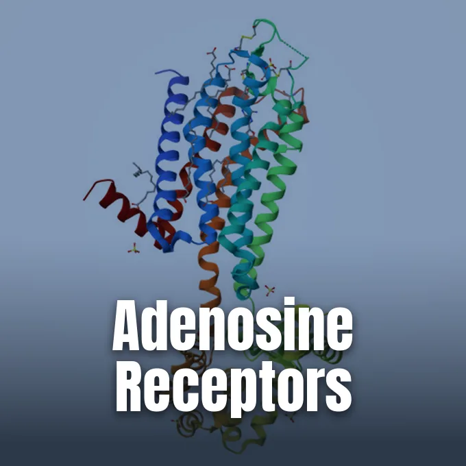
Adenosine receptors are cell-surface proteins that detect free purines to regulate everyday body functions, ranging from neural sleep drive to vascular muscle tone and immune responses. Belonging to the family of purinergic G protein-coupled receptors, these sensors translate changing cell energy states into specific biochemical signals. When working organs burn fuel and release purine molecules, these receptors alter inner signals to slow tissue work and guard stressed cells [5].
- What Are Adenosine Receptors?
- Four Separate Subtypes in Human Tissue
- Signaling Cascades and G-Protein Coupling
- How Adenosine Receptors Control Sleep Pressure
- Cardiovascular Regulation and Blood Flow
- Caffeine Competition at Adenosine Receptors
- Immune Suppression in Tumor Environments
- Therapeutic Targets in Clinical Drug Trials
What Are Adenosine Receptors?
How do human cells sense the gradual breakdown of their fuel supply during waking hours? They rely on a family of surface proteins known as P1 receptors, which share a common design consisting of seven transmembrane alpha helices threading back and forth through the lipid bilayer. Each receptor protein presents an N-terminus exposed to outer fluid and a C-terminus anchored inside the cytoplasm. Helix 8 guides G-protein signals. Four separate subtypes handle differing energy demands across human physiology. These surface sensors help organs and tissues adjust their work rhythm based on nearby purine levels in the bloodstream. Each subtype belongs to the P1 class of purinergic receptors, distinguishing them from P2 receptors that respond directly to ATP or ADP [5].
Adenosine serves as the natural activator for these adenosine receptors, forming when cell metabolism breaks down ATP during ongoing physical work. As fuel use climbs, outer purine levels rise and bind to these surface pockets to initiate defense loops. Kenneth A. Jacobson and Zhan-Guo Gao reported that synthetic targeted agonists now allow researchers to stimulate each receptor type without causing broad off-target complications [1].
Four Separate Subtypes in Human Tissue
Human cells express four separate varieties of adenosine receptors—A1, A2A, A2B, and A3—with the A1 subtype appearing widely across human organs and reaching its highest density in the brain, but it also maintains functional pools in the heart, pancreas, and testis. In contrast, the A2A receptor, encoded by the human ADORA2A gene, concentrates in the striatum and basal ganglia, as well as vascular smooth muscle cells, blood platelets, and immune T lymphocytes. Both A1 and A2A bind purines at nanomolar levels. Individual genes reside on separate chromosomes [5].
The remaining two varieties occupy unique control roles in peripheral and neural tissues. In the brain, the A2B receptor responds to high purine levels during heavy neuronal firing by stimulating glial glucose metabolism in astrocytes to nourish neighboring neurons. Research shows that A2B also binds with netrin-1 to guide developing axon paths. Meanwhile, the A3 receptor exhibits more restricted expression and suppresses specific growth pathways in human melanoma cell cultures [5].
Skeletal biology illustrates how these subtypes balance bone remodeling. The A1 receptor stimulates osteoclast differentiation. Chemical blockade of A1 suppresses bone breakdown and increases mineral density. In contrast, the A2A receptor halts osteoclast maturation and activates bone-forming osteoblasts. Research shows that A2B directs mesenchymal stem cells to become osteoblasts rather than chondrocytes via Runx2 gene expression. This pathway establishes a molecular framework for bone repair [5].
Signaling Cascades and G-Protein Coupling
Downstream cellular outcomes depend on the specific G protein partners that associate with adenosine receptors. When an agonist binds to the A1 subtype, it prompts coupling to inhibitory Gi1, Gi2, Gi3, or Go proteins, which promptly halt adenylate cyclase activity and cause a decrease in cyclic AMP levels. The pathway also activates phospholipase C to raise inner levels of inositol triphosphate and diacylglycerol, after which DAG lipase cleaves diacylglycerol into free arachidonic acid to initiate downstream lipid signals. On the cell surface, A1 binding opens inward potassium channels and closes voltage-sensitive N-type, P-type, and Q-type calcium channels. Membrane shifts dampen synaptic vesicle release at nerve endings and hyperpolarize membranes to quiet overactive neural circuits. Through coordinated molecular actions, A1 signals function as a steady calming brake that protects vulnerable cells from metabolic overwork [7].
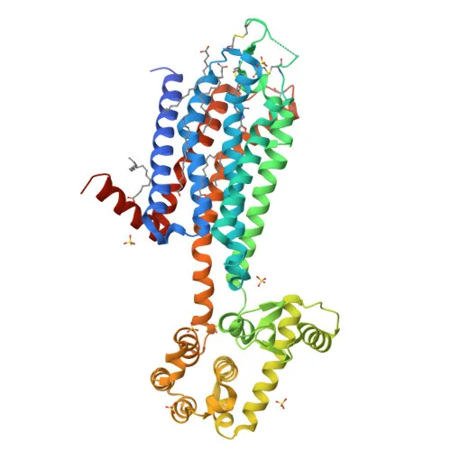
In contrast, the A2A receptor binds heterotrimeric Gs proteins containing alpha, beta, and gamma subunits to stimulate adenylyl cyclase and elevate inner cyclic AMP. Jaakola and Stevens mapped this architecture at The Scripps Research Institute in La Jolla by solving the 2.6 angstrom crystal structure of human A2A complexed with the blocker ZM241385 in Science under PDB code 3EML. Lebon and Tate at the MRC Laboratory of Molecular Biology in Cambridge later solved agonist-bound structures in Nature under PDB code 2YDO, showing that adenosine makes polar contacts with Ser 277 and His 278 [2].

How Adenosine Receptors Control Sleep Pressure
The progressive sensation of fatigue that develops during waking hours illustrates purine signaling in everyday life, as active brain cells consume ATP to sustain attention and mental processing as outer adenosine steadily accumulates in sleep-regulating forebrain networks. This buildup meter triggers sleep pressure. A1 receptors in the basal forebrain play a leading role by binding excess adenosine to quiet wake-promoting cholinergic neurons, which gradually dials down cortical activation across the cerebral hemispheres. Prolonged wakefulness forces purine levels even higher. Animal studies confirm that local adenosine levels drop during restful slumber, which lets waking circuits fully reset before the next daily cycle begins [7].
A1 dampens cholinergic arousal centers, but A2A receptors contribute a complementary sedative effect within the striatum and basal ganglia. A2A proteins form physical heterodimers with dopamine D2 receptors. These hybrid signaling complexes lower the affinity of D2 receptors for dopamine. Blunting of dopaminergic signals reduces voluntary locomotion and fosters the behavioral stillness required for deep sleep. Striatal circuits thereby coordinate physical relaxation together with mental drowsiness [6].
Higher-order receptor mosaics add further nuance to this control mechanism. Neurobiologists have observed heteromers combining A2A with cannabinoid CB1 receptors, glutamate mGluR5 receptors, and CB1/A2A/D2 heterotrimers in synaptic membranes. Once restful sleep arrives, enzymatic clearance removes outer adenosine, releasing the purine brake. Cholinergic and dopaminergic pathways resume normal signaling. This clearance helps a rested person awaken with clear focus and renewed energy [6].

Cardiovascular Regulation and Blood Flow
Purinergic signals govern human circulatory pacing. In the heart, cardiac adenosine receptors suppress electrical conduction across the sinoatrial node, the atrioventricular node, and the His-Purkinje system by opening potassium channels and blocking both the inward calcium current (ICa) and the hyperpolarization-activated funny current (If). A1 activation also prevents local sympathetic nerves from releasing norepinephrine, which blunts adrenergic stimulation, slows the heartbeat, and reduces heart oxygen demand to shield cardiac muscle during ischemic stress. Under healthy baseline conditions, this pathway shields heart muscle against excessive workload by slowing electrical conduction through the bundle of His [7].
Emergency physicians utilize this rapid electrical blockade to manage acute supraventricular tachycardias in clinical settings. A rapid intravenous push of adenosine causes momentary cardiac standstill at the atrioventricular node, interrupting abnormal electrical loops and restoring regular sinus rhythm. However, during severe hypotension or circulatory shock, excess endogenous adenosine can harm patients by preventing the compensatory tachycardia needed to maintain cerebral blood flow [7].
A1 moderates cardiac rhythm, but vascular A2A receptors regulate coronary arterial tone to ensure adequate myocardial perfusion. Binding of adenosine relaxes smooth muscle within coronary vessels, boosting blood supply to working cardiac myocytes during exercise. Fuel balance shifts under purine control. A1 lowers circulating insulin and slows glycolysis, but A2A stimulation raises plasma glucose and lactate. This metabolic shift supplies cardiac myocytes with ready fuel during physical strain [7].
Caffeine Competition at Adenosine Receptors
Every morning, billions of people alter their purine signaling by consuming natural methylxanthines, such as caffeine in coffee, theophylline in tea, and theobromine in chocolate. Because the xanthine core shares structural similarities with endogenous adenosine, caffeine fits into the orthosteric binding pocket of both A1 and A2A receptors without triggering the helix shifts needed for G-protein activation, which physically blocks adenosine from delivering its fatigue signal to cholinergic and striatal neurons. In addition to caffeine, theobromine in cocoa and theophylline in brewed teas act as non-selective antagonists across both heart and brain tissue. This blockade keeps brain circuits alert. The stimulant effect persists until hepatic enzymes metabolize caffeine and clear it from the bloodstream [5].
In hospital neonatal units, this antagonistic effect provides life-saving respiratory support for vulnerable newborns. Physicians administer caffeine and theophylline to premature infants to stimulate immature respiratory control centers in the brainstem. Pharmacological studies suggest that suppressing A1 activity helps protect developing white matter against hypoxia-induced ventriculomegaly. Breathing patterns stabilize significantly as prolonged infant respiratory pauses decline under treatment [5].
Because caffeine and theophylline also inhibit phosphodiesterase enzymes, they produce bronchodilator actions useful in treating asthma. To achieve greater experimental precision, chemists designed subtype-selective blockers like 8-cyclopentyl-1,3-dipropylxanthine (DPCPX), cyclopentyltheophylline (CPT), bamifylline, and rolofylline (KW-3902) for A1, along with SCH-58261 and istradefylline (KW-6002) for A2A. These selective compounds help scientists isolate individual receptor pathways without phosphodiesterase interference. These tools give scientists clearer insights into purinergic pharmacology [5].
Immune Suppression in Tumor Environments
Purinergic signaling supplies a necessary regulatory brake that prevents runaway tissue injury during systemic inflammation. In a 2002 study in Zeitschrift für Gastroenterologie, Bilzer, Akio Ohta, Michail Sitkovsky, and A. L. Gerbes demonstrated that sub-threshold inflammatory stimuli causing minimal damage in normal mice triggered extensive tissue necrosis, sustained cytokine storms, and lethal shock in male mice lacking A2A receptors, proving that A2A receptors play a non-redundant role in limiting inflammation in vivo. High levels of pro-inflammatory cytokines accompanied this tissue destruction, confirming that A2A receptors terminate systemic immune reactions. Without this checkpoint, white blood cells destroy healthy tissues [3].
This feedback shields healthy tissues during infection, but solid tumors hijack the pathway to escape immune destruction. Hypoxic stress within tumors upregulates the transcription factor HIF-1α, prompting cell-surface CD39 and CD73 enzymes to convert outer ATP into abundant adenosine. When adenosine binds to A2A receptors on tumor-infiltrating T lymphocytes, elevated cyclic AMP works through protein kinase A to upregulate inhibitory PD-1 receptors and TGF-β. Stimulating FOXP3 further converts CD4+ T-cells into regulatory Treg cells, shutting down antitumor immunity [6].
To dismantle this protective shield, oncologists are combining A2A receptor blockers with modern immune checkpoint therapies. The selective antagonist CPI-444 cleared tumors in nearly 90% of experimental mice when paired with anti-PD-L1 or anti-CTLA-4 antibodies. Furthermore, when cured mice were rechallenged with cancer cells, tumor growth remained completely blocked. This complete block demonstrated long-term immunological memory that halted cancer recurrence [6].
Therapeutic Targets in Clinical Drug Trials
The biological footprint of adenosine receptors has turned all four subtypes into candidates for clinical drug development. In a review in Expert Opinion on Emerging Drugs, Kenneth A. Jacobson and Zhan-Guo Gao reported that over a dozen targeted agonists had entered clinical trials for human diseases. Adenosine itself stays the sole agent with regulatory approval for treating acute arrhythmias, but synthetic compounds offer far greater selectivity. Clinical teams evaluated selective A1 agonists such as tecadenoson and selodenoson for controlling arrhythmias and reducing neuropathic pain, and selective A2A agonists like CGS-21680 and ATL-146e entered trials for myocardial perfusion imaging. Programs also tested capadenoson (BAY68-4986), BnOCPA, and CCPA to explore cardiovascular pathways. Selective A1 agonists reduce pain signaling without depressing respiration, and selective A2A agonists permit clear visualization of coronary artery flow during stress testing [1].
Parallel research programs focus on selective blockers for neurodegenerative and inflammatory disorders. In neurology, A2A antagonists such as istradefylline (KW-6002) and KW-6356 advanced through clinical trials to relieve motor symptoms in Parkinson’s disease by balancing striatal dopamine activity. Preclinical studies evaluate A2B blockers for cardiac ischemia and tumor control, and selective A3 agonists such as Cl-IB-MECA and MRS3558 undergo trials for rheumatoid arthritis and colorectal cancer. Selective A3 blockers such as MRS1191, MRS1523, and MRE3008F20 provide additional chemical tools to investigate cancer immunology [1].
- ACADEMIC JOURNAL Gao, Z., & Jacobson, K. A. (2007). Emerging adenosine receptor agonists. Expert Opinion on Emerging Drugs, 12(3), 479-492. [Article Link]
- ACADEMIC JOURNAL Jaakola, V. P., Griffith, M. T., Hanson, M. A., Cherezov, V., Chien, E. Y., Lane, J. R., IJzerman, A. P., & Stevens, R. C. (2008). The 2.6 Angstrom Crystal Structure of a Human A2A Adenosine Receptor Bound to an Antagonist. Science, 322(5905), 1211-1217. [Article Link]
- ACADEMIC JOURNAL Bilzer, M., Ohta, A., Sitkovsky, M., & Gerbes, A. L. (2002). Role of G-protein-coupled Adenosine Receptors in Downregulation of Inflammation and Protection from Tissue Damage. Zeitschrift für Gastroenterologie, 40(7), 543-544. [Article Link]
- ACADEMIC JOURNAL Lebon, G., Warne, T., Edwards, P. C., Bennett, K., Langmead, C. J., Leslie, A. G. W., & Tate, C. G. (2011). Agonist-bound adenosine A2A receptor structures reveal common features of GPCR activation. Nature, 474(7352), 521-525. [Article Link]
- WEBSITE Wikipedia. (n.d.). Adenosine receptor. [Article Link]
- WEBSITE Wikipedia. (n.d.). Adenosine A2A receptor. [Article Link]
- WEBSITE Wikipedia. (n.d.). Adenosine A1 receptor. [Article Link]


