





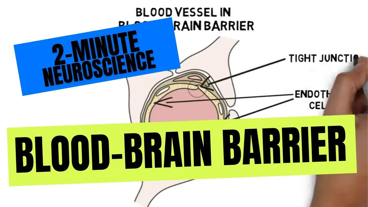
The blood-brain barrier, commonly called the BBB, is a selective cellular border that seals central nervous system (CNS) blood vessels from erratic chemical swings in the bloodstream. Body capillaries let fluid and small solutes leak freely through loose cellular gaps, but vessels in the brain knit together into a tight continuous wall to keep neural circuits stable [1]. Capillary endothelial cells (ECs) regulate the passage of essential fuels like glucose while turning away blood toxins, white blood cells, and pathogens. Because of this tight defense, the barrier also turns away more than ninety-eight percent of small-molecule drugs and virtually all large biopharmaceuticals, creating a profound delivery bottleneck for modern medicine [2].
- Anatomy of the Neurovascular Unit
- Blood-Brain Barrier Molecular Seals
- Transport Routes Across Brain Capillaries
- What Can Cross the Capillary Wall?
- Circumventricular Organs and Permeable Windows
- How Does the Blood-Brain Barrier Break Down?
- Spinal Cord and Developmental Differences
- Bypassing the Blood-Brain Barrier for Medical Therapy
Anatomy of the Neurovascular Unit
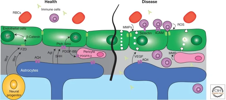
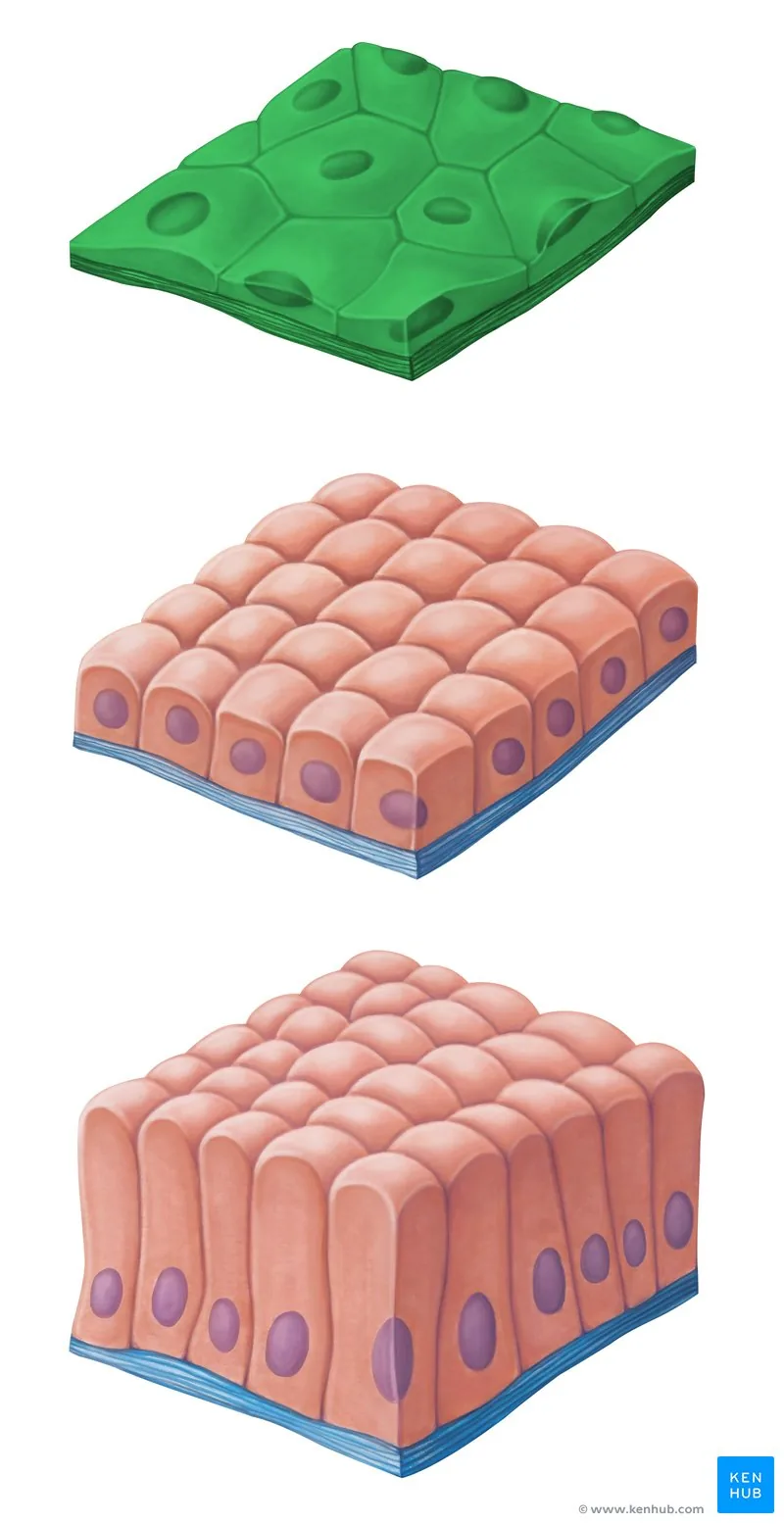
Brain vessels rely on the neurovascular unit. In 2007, William C. Aird showed that continuous nonfenestrated CNS vessels create the most restrictive vascular bed in the body. In 2008, Berislav Zlokovic detailed how endothelial cells (ECs), pericytes, and astrocytes maintain CNS homeostasis. Unlike vessels in skin or muscle, brain ECs lack fenestra pores completely. In 1985, Ann Coomber and Patrick Stewart measured these brain vessels, showing they are thirty-nine percent thinner than muscle endothelium, leaving under a quarter of a micron between blood and brain parenchyma [1].
Pericytes wrap around the ablumenal surface of these vessels, embedded in a shared basement membrane. Brain capillaries have the highest pericyte coverage of any vascular bed, with an EC-to-pericyte ratio between 1:1 and 3:1. Muscle vessels, by contrast, have a ratio of roughly 100:1. In 2006, Claire Peppiatt showed that brain pericytes contract to control capillary blood flow. In 2011, Annika Armulik confirmed that pericytes regulate vessel stability through peg-and-socket junctions held by N-cadherin [1].
Astrocytes build the outer cellular sheath of this vessel defense system. Star-shaped astrocyte end-feet encircle more than ninety percent of the capillary wall. These glial cells express aquaporin-4 water channels to anchor their membranes to the basal matrix, helping clear fluid and buffer excess potassium ions during intense electrical firing in nearby neurons. Glial cues instruct ECs to tighten their seals. In simpler invertebrate brains without blood vessels, glial cells carry out this entire tissue isolation alone [2].
Blood-Brain Barrier Molecular Seals
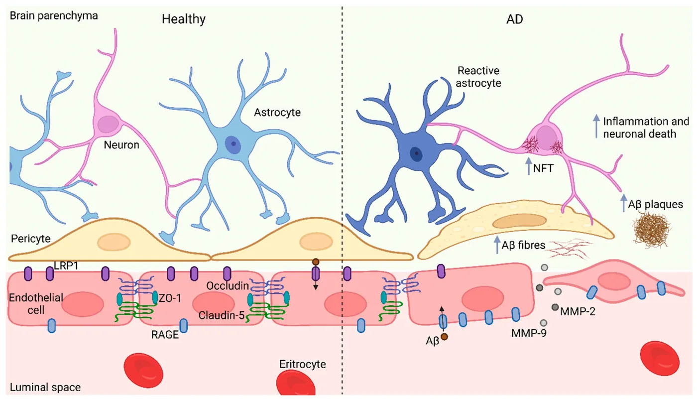
Tight junction strands between adjacent ECs create the primary seal of the barrier. In landmark electron microscopy studies, Thomas Reese and Morris Karnovsky showed that these intercellular joints block the passive diffusion of solutes between blood and brain parenchyma [1]. The junctional meshwork, historically known as the zonula occludens, gives cerebral capillaries an exceptionally high electrical resistance. Transmembrane strands consist of claudin-5, occludin, and junctional adhesion molecules like JAM-A, which bind tightly to identical proteins on neighboring cells to close the paracellular cleft [9].
Cytoplasmic scaffolding proteins anchor these transmembrane strands to the cellular skeleton beneath the membrane. Scaffolding proteins including ZO-1 and ZO-2 connect claudins and occludins to actin support filaments. If these scaffolding anchors uncouple during trauma, the tight junction strands pull apart, letting fluid leak freely into nervous tissue [9].
Vesicle transport rarely happens across healthy brain vessels. Endothelial cells suppress pinocytotic vesicle formation almost completely, shutting down the bulk transfer of fluid and dissolved proteins through the cell interior. To power active transport gradients, cerebral ECs pack far more mitochondria than ordinary body vessels. High mitochondrial volume provides the ATP needed to hold ions away from neural circuits [1].
Transport Routes Across Brain Capillaries
Nutrients cannot slip between sealed cells. They rely on carrier proteins embedded within the endothelial lining to reach brain tissue [4]. The organ runs mainly on glucose, burning about 120 grams of glucose each day, which accounts for roughly sixty percent of all sugar consumed by the human body [7]. To satisfy this high energetic appetite, capillary walls use glucose transporter 1 (GLUT1) to move sugar down its concentration gradient. Other carriers, like MCT1 and MCT2, ferry lactate and ketones when metabolism shifts during fasting or intense exercise [4].
Larger peptides cross through receptor-mediated transcytosis. Blood-borne molecules attach to target surface receptors along the vessel interior, prompting the membrane to form an internal vesicle that carries cargo to the brain side. Iron enters the brain through the transferrin receptor, which shuttles the metal across the cytoplasm before releasing it into interstitial fluid [7]. Efflux pumps act in reverse. P-glycoprotein, an ATP-dependent pump, intercepts lipophilic foreign chemicals inside the cell membrane and ejects them back into blood flow [2].
Macromolecules can also cross via adsorptive-mediated transcytosis based on electrostatic attraction between cationic molecules and negatively charged membrane domains. While nutrient carriers import essential amino acids, these proteins can mistakenly carry heavy metals like lead or mercury into brain tissue. When metals accumulate, severe neurotoxicity follows [4].
What Can Cross the Capillary Wall?
Molecules that cross the capillary wall through passive diffusion must meet narrow physical rules. Fat-soluble chemicals with a molecular weight under 400 to 600 daltons dissolve through endothelial membranes without needing transport proteins [2]. Small nonpolar gases like oxygen and carbon dioxide diffuse freely down concentration gradients. When a compound carries high polarity or forms more than eight hydrogen bonds, its chance of crossing the lipid bilayer drops sharply [8].
Why do psychoactive compounds (such as caffeine, nicotine, and alcohol) act on the brain within seconds? These chemicals are tiny and lipid-soluble, slipping through endothelial cells before efflux pumps can clear them [7]. Larger circulating hormones, body antibodies, and neurotransmitters like dopamine cannot enter the brain from the bloodstream. When treating Parkinson’s disease, doctors cannot give dopamine directly; instead, they prescribe the precursor levodopa, which crosses via amino acid transporters before brain enzymes convert it into active dopamine [3].
Pharmacologists track drug delivery using the brain-to-blood ratio. Propranolol, a fat-soluble beta-blocker, easily crosses into neural tissue with a human brain-to-blood ratio between 15:1 and 26:1. Atenolol stays largely in body blood, showing a low ratio of roughly one to five [11].


Circumventricular Organs and Permeable Windows
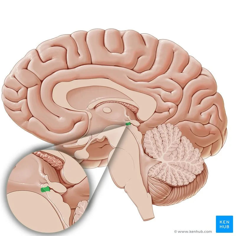
Although most cerebral capillaries keep tight seals, small midline structures bordering the third and fourth ventricles deliberately leave their vessels fenestrated. These specialized regions, known as circumventricular organs (CVOs), allow the brain to sample blood chemistry and release neuroendocrine hormones into general circulation. These sensory gateways consist of three specialized structures: the subfornical site near the foramen of Monro, the vascular organ of the lamina terminalis (OVLT), and the paired area postrema. Situated on the floor of the fourth ventricle, the area postrema serves as a trigger zone for vomiting, detecting emetic toxins that cannot cross standard brain capillaries [4].
Secretory circumventricular organs release neural products directly into blood or spinal fluid. Melatonin from the pineal gland enters blood vessels supplied by the posterior choroidal artery, while the posterior pituitary releases hypothalamic oxytocin and vasopressin. The nearby subcommissural organ secretes SCO-spondin, a glycoprotein that aggregates into Reissner’s fibers within the third ventricle to maintain cerebral aqueduct patency during early development [4].
Capillaries at the margins of CVOs transition gradually into standard barrier vessels. Scientists identified specialized hybrid capillary zones, such as the area postrema border, where vessels show intermediate permeability. These hybrid zones allow local neural circuits to sample blood-borne peptide signals while shielding deeper brain nuclei [9].
How Does the Blood-Brain Barrier Break Down?
Trauma and ischemic stroke cause immediate vessel collapse. In traumatic brain injury, torn vessel walls let albumin and red blood cells flood into brain tissue within minutes, sparking acute swelling and reactive glial inflammation [10]. When stroke blocks blood flow, oxygen deprivation starves endothelial cells of energy, causing ion pumps to fail and tight junction strands to uncouple. Vascular collapse unleashes secondary inflammatory cascades that expand brain tissue damage over the days following the initial injury [7].
Chronic neuroinflammatory diseases weaken barrier integrity through enzymatic destruction of vessel scaffolding. In multiple sclerosis, autoreactive leukocytes cross capillary walls by releasing matrix metalloproteinases (specifically MMP-2 and MMP-9), which cleave dystroglycan receptors anchoring astrocyte end-feet to the basement matrix. Immune infiltration drives demyelination and progressive lesion formation [12]. These inflammatory breaches closely involve microglia and their defensive roles in the human brain, showing how vascular disruption and innate immune signaling intersect during central nervous pathology.
Vessel walls also deteriorate during aging and progressive neurodegeneration. In Alzheimer’s disease, impaired vascular clearance of amyloid-beta damages capillary cells and accelerates microvascular leaks [7]. These vascular deficits provide key context when studying the biological mechanisms of dementia, where early barrier breakdown can precede classic amyloid accumulation and hasten cognitive decline.
Spinal Cord and Developmental Differences
The blood-spinal cord barrier (BSCB) protects the spinal cord. Compared with brain capillaries, spinal vessels express fewer ZO-1 and occludin junctional strands, rendering the spinal endothelium noticeably more permeable to circulating solutes and inflammatory cytokines. Spinal vessels also maintain less P-glycoprotein, slowing the elimination of harmful chemicals. Osmotic mannitol crosses spinal capillaries far more easily than it crosses cerebral capillaries [10].
When mechanical impact damages the spinal column, this vulnerability triggers rapid barrier failure that can last for weeks. Animal models indicate that capillary permeability remains elevated for fourteen to fifty-six days after spinal injury. Persistent protein leakage and oxidative stress can lead to post-traumatic syringomyelia, where expanding fluid cysts inside the spinal cord compress neural tracts and worsen paralysis [10].
An old medical myth claimed that human newborns have an immature, leaky blood-brain barrier, but modern developmental biology shows that cerebral tight junctions function well before birth. Neonatal vulnerability to neurotoxins stems instead from immature metabolic clearance and differences in cerebrospinal fluid transport. During neonatal jaundice, fetal hemoglobin breaks down into insoluble bilirubin, which binds to serum albumin. If bilirubin production exceeds albumin binding capacity, excess free bilirubin crosses into the brain and causes kernicterus, an avoidable bilirubin neuropathy [4].
Bypassing the Blood-Brain Barrier for Medical Therapy
The protective efficiency of cerebral capillaries presents a major bottleneck for drug development. Leading neurovascular expert William Pardridge calculated that the barrier excludes more than ninety-eight percent of small-molecule drugs and nearly all large biopharmaceuticals [7]. Consequently, experimental drugs that easily kill glioblastoma cells or dissolve amyloid plaques in laboratory petri dishes fail in human clinical trials because therapeutic doses never reach their targets in brain parenchyma [6].
Clinicians at UT MD Anderson Cancer Center test several methods to open capillary gates safely. Infusing mannitol draws water out of endothelial cells to separate tight junctions, though researcher Joseph McCarty said this aggressive approach is “almost like a bomb” that risks neuroinflammation. A localized technique uses focused ultrasound with microbubbles. Acoustic waves cause microbubbles to oscillate and briefly open tight junctions, letting chemotherapy enter tumor tissue before the barrier reseals [5].
Other teams use Trojan horse strategies or direct delivery to bypass capillary walls. At UT MD Anderson, endovascular surgeon Christopher Young inserts microcatheters through arm vessels into brain arteries, while researchers Frederick Lang and Juan Fueyo test direct delivery of oncolytic viruses. Teams led by Betty Kim and Wen Jiang package messenger RNA into engineered extracellular vesicles that bind glioblastoma markers [5]. These therapies build on a century of discovery. In 1900, Max Lewandowsky hypothesized a vascular barrier, and Paul Ehrlich observed that aniline dyes spared brain tissue. In 1913, Edwin Goldmann proved compartmentalization by injecting dyes into spinal fluid [9]. The term “blood-brain barrier” was coined in a 1921 paper by Lina Stern (also spelled Shtern) and Raymond Gautier, establishing the foundation for neurovascular science [3].
- ACADEMIC JOURNAL Daneman, R., & Prat, A. (2015). The Blood–Brain Barrier. Cold Spring Harbor Perspectives in Biology, 7(1), a020412. [Article Link]
- ACADEMIC JOURNAL Wu, D., Chen, Q., Chen, X., Han, F., Zhong, W., & Wang, Y. (2023). The blood–brain barrier: Structure, regulation and drug delivery. Signal Transduction and Targeted Therapy, 8(1), 217. [Article Link]
- WEBSITE Cleveland Clinic. (2023). The blood-brain barrier: Out with the bad, in with the good. Cleveland Clinic. [Article Link]
- WEBSITE Kenhub. (2016). Blood–brain barrier. Kenhub. [Article Link]
- WEBSITE Nagy, J. (2026). What is the blood-brain barrier, and how can we get past it to treat cancer? UT MD Anderson Cancer Center. [Article Link]
- ONLINE NEWS Soliman, R. (2026). Blood–Brain Barrier Explained and Why It Is Very Difficult to Treat Brain Diseases. Science Times. [Article Link]
- WEBSITE Neurosity. (2026). The Blood-Brain Barrier: What It Is and Why It Matters. Neurosity. [Article Link]
- WEBSITE Missi, E. (2024). How does the blood-brain barrier work? Darwin Microfluidics. [Article Link]
- WEBSITE Wikipedia. (n.d.). Blood–brain barrier. Wikipedia. [Article Link]
- WEBSITE Wikipedia. (n.d.). Blood–spinal cord barrier. Wikipedia. [Article Link]
- WEBSITE Wikipedia. (n.d.). Brain-to-blood ratio. Wikipedia. [Article Link]
- BOOK CHAPTER Garcia-Gallardo, A., & Campbell, M. (2021). Understanding the Blood-Brain Barrier: From Physiology to Pathology. In The Blood-Brain Barrier. Springer Nature Switzerland. [Article Link]


