





When patients ask what is meningitis, physicians point to an inflammatory process that targets the delicate membranes shielding the central nervous system from biological pathogens and mechanical trauma. These protective coverings, known as the meninges, wrap the brain and spinal cord in three layered sheets. Infection by bacteria, viruses, fungi, or environmental parasites can disrupt this barrier. While viral forms often resolve on their own, acute bacterial disease can kill within hours without hospital care, as noted by the Centers for Disease Control and Prevention [1].
What Is Meningitis?
The disease starts in mucosal linings. Pathogenic bacteria or viruses colonize the human nose, throat, or digestive tract before breaching epithelial barriers and circulating freely throughout the bloodstream. Once in the blood, pathogens encounter the blood-brain barrier, which normally shields delicate central nervous tissue from chemical and biological insults. Organisms bind tightly to receptors on brain microvascular endothelial cells, cross into the subarachnoid space, and multiply rapidly within cerebrospinal fluid where protective host antibodies and complement proteins stay sparse. This unchecked growth triggers resident microglial cells, releasing destructive cytokines like interleukin-1 and tumor necrosis factor [8].
Immune cells and plasma proteins thicken the spinal fluid and block normal drainage through arachnoid villi, pushing intracranial pressure higher. This rise in pressure cuts off blood delivery to the brain, causing oxygen starvation that can provoke seizures or sudden focal weakness [13].
Germs can also enter through bone gaps in the skull, such as basilar fractures or sinus defects. Middle ear or mastoid bone infections can erode surrounding bone, giving microbes direct access to meningeal sheets without traveling through the bloodstream or encountering systemic immune defenses. Medical devices like brain shunts or cochlear implants can also let pathogens slip inside. In 2019, approximately 7.7 million people received a diagnosis of meningitis worldwide, resulting in 236,000 deaths compared to 433,000 in 1990. Despite these gains, bacterial attacks kill roughly one in six patients and leave one in five survivors with permanent problems like hearing loss, seizures, or limb weakness, as documented by the World Health Organization [4].

Anatomy of the Three Brain Membranes
Three membranes protect the brain. These tissue sheets envelop the central nervous system to provide physical containment, fluid cushioning, and a continuous barrier against external blunt force trauma. The outermost layer is the dura mater, a tough fibrous sheet attached directly to the inner skull bones that acts as a strong barrier against cranial injury. Isolated inflammation of this coat is called pachymeningitis, visible as dural thickening on radiological scans [8].
Beneath the dura lies the arachnoid mater, a delicate web of connective tissue that bridges the gap between skull bone and vascularized nervous tissue below. The innermost layer, the pia mater, hugs every groove of the cerebral cortex and carries tiny capillaries that nourish brain cells. Together, the arachnoid and pia form the leptomeninges, which enclose the fluid-filled subarachnoid space. This compartment holds clear cerebrospinal fluid that cushions the brain and spinal cord against external bumps. Inflammatory fluid gathers in the basal cisterns of this space, blocking flow, irritating local blood vessels, and disrupting nutrient transport across the blood-brain barrier [8].
Because cranial nerves emerge from the brainstem and travel through the subarachnoid space, inflammatory fluid can squeeze these nerve paths. Damage to cranial nerve VIII causes sensorineural hearing loss. Irritation of nerves III, IV, and VI disrupts coordinated eye movements. Inflammation of neighboring blood vessels can also cause localized vasculitis and venous sinus thrombosis, cutting off blood supply and triggering ischemic strokes in cortical areas [15].
Primary Bacterial Pathogens
In adults, two bacterial species cause roughly 80% of all cases of acute bacterial meningitis. The most common organism is Streptococcus pneumoniae, an encapsulated bacterium that colonizes the nasopharynx and accounts for 51% to 72% of adult episodes in clinical series. The second leading pathogen is Neisseria meningitidis, commonly known as the meningococcus, which causes 11% to 37% of adult cases and can ignite fast outbreaks. In a prospective study in the Netherlands published by Diederik van de Beek and Jan de Gans in the New England Journal of Medicine, evaluating 696 adult episodes, S. pneumoniae caused 51% of infections and N. meningitidis caused 37% [5].
Bacterial causes shift by age. Neonates face risk from Streptococcus agalactiae (Group B streptococcus), Escherichia coli, and Listeria monocytogenes, as detailed by Hillary Mount and Sean Boyle in American Family Physician. For adults over 50 years in North America and Europe, Listeria monocytogenes returns as a risk tied to soft cheeses and deli foods [6].

Epidemics occur regularly in the African meningitis belt, stretching from Senegal to Ethiopia. In 2025, surveillance in 24 countries reported 21 526 suspected cases and 971 deaths to the World Health Organization Inter-country Support Team based in Burkina Faso. Dry winds and crowds help droplets spread. In Saudi Arabia, pilgrims traveling for Hajj must receive quadrivalent meningococcal vaccination. In the Greater Lakes Area, including Burundi, Rwanda, and Tanzania, epidemics have also emerged, while travelers face season-dependent risks [4].
Viral Meningitis Triggers
Viral pathogens trigger the vast majority of aseptic meningeal infections. Non-polio enteroviruses, such as coxsackieviruses and echoviruses, cause more than 85% of cases. These enteroviruses spread by hand-to-mouth contact and pool water, peaking during summer and early fall in temperate regions. Enteroviruses infect males 1.3 to 1.5 times more often than females [10].
Beyond common enteroviruses, herpes simplex virus type 2 causes genital sores but can also invade neural pathways to provoke recurrent episodes known as Mollaret meningitis. Many patients develop this condition without any visible blister lesions. Arboviruses transmitted by mosquito vectors—including West Nile virus and St. Louis encephalitis virus—cause seasonal infections from June through October, with the Centers for Disease Control and Prevention tracking 1,821 American cases in 2025. Other causative agents include Epstein-Barr virus, lymphocytic choriomeningitis virus carried by pet rodents, mumps virus, and opportunistic fungal pathogens like Cryptococcus that invade patients who live with compromised immune function [10].
For most healthy people, viral meningitis runs a mild course. Symptoms usually lift in 7 to 10 days with rest, fluids, and pain relief. In Olmsted County, Minnesota, researchers tracked an annual incidence of 10.9 per 100,000 person-years between 1950 and 1981, while Finnish cohorts led by pediatric specialists in Helsinki reported higher rates in infants. Babies under one month face higher risks of liver and heart problems from enteroviral spread [10].



Recognizing the Classic Clinical Triad
Textbooks describe acute bacterial meningitis with a triad of fever, neck stiffness, and altered mental status. In clinical practice, however, this triad appears in only 44% to 46% of confirmed adult cases. Doctors evaluate a wider group of four signs: headache, fever, neck stiffness, and confusion. In a Dutch nationwide study of 696 adult cases led by Diederik van de Beek and Jan de Gans, 95% of patients presented with at least two of these four signs upon hospital admission. Headache affected 87% of patients, neck stiffness 83%, fever 77%, and altered mental status 69%. Only 4% had none of these signs, showing that their combined absence argues strongly against bacterial infection [5].
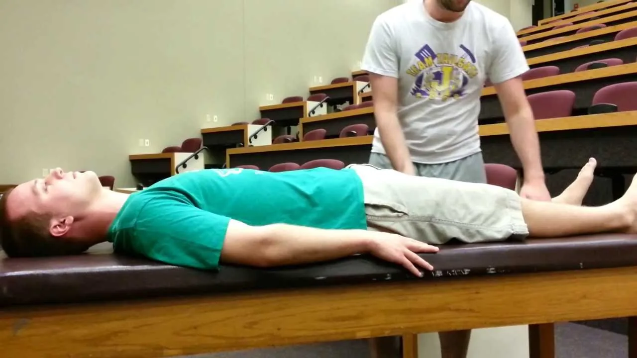
Bedside tests assess meningeal irritation. Kernig sign—described originally by Vladimir Kernig—is positive when knee extension causes resistance and back pain while the patient lies supine with the hip held at 90 degrees. Brudzinski sign, named after Jozef Brudzinski, occurs when passive neck flexion induces involuntary bending of the hips and knees. While both signs show high specificity, their low sensitivity means negative findings cannot rule out disease [6].
A non-blanching red or purple spotty rash provides a key clue in meningococcal disease, signaling capillary damage and leaking blood vessels from circulating endotoxins. The rash does not fade when pressed firmly with a clear drinking glass. Patients also develop photophobia, experiencing eye pain when looking at bright lights. Infants rarely show neck stiffness, displaying poor feeding, continuous crying, sleepiness, cold hands and feet, or a bulging soft spot. The National Health Service advises parents in England to call 999 or visit an A&E hospital emergency department immediately if these signs appear [3].
Why Meningitis Diagnosis Needs Rapid Care
Bacterial meningitis moves with alarming speed, with less than 24 hours separating first symptoms from hospital arrival in 47% of adult patients. Hasbun reported in JAMA that bacterial meningitis incidence ranges from 0.9 per 100,000 in high-income nations to 80 per 100,000 in low-income regions, where mortality reaches 54% [14].
Examining cerebrospinal fluid through an urgent lumbar puncture distinguishes bacterial attacks from viral syndromes, revealing opening pressures elevated above 20 to 30 centimeters of water as swollen brain tissue compresses surrounding fluid compartments. White blood cells surge with neutrophils. Impaired glucose transport and bacterial consumption drop fluid glucose below 2.2 millimoles per liter, while damaged vascular walls allow serum proteins to leak into the spinal canal until protein concentrations climb above 1.5 grams per liter. By contrast, viral meningitis yields clear fluid with normal glucose, mildly elevated protein between 0.5 and 2 grams per liter, and a lymphocyte count that dominates after 48 hours [13].
Before doing a spinal tap, doctors check whether a computed tomography scan of the head is needed to prevent brain herniation. Scans are advised for patients with focal neurological deficits, new seizures, papilledema, or impaired immunity. However, if imaging or spinal taps are delayed, doctors must give antibiotics right away. Guidelines from the Infectious Diseases Society of America emphasize that diagnostic imaging or lumbar puncture must never delay antibiotic treatment when bacterial infection is suspected [6].
Hospital Treatment Protocols
Speed dictates hospital survival. Initial management of acute bacterial meningitis demands rapid intravenous antimicrobial therapy adjusted according to patient age and specific clinical risk factors. For children and adults under 50 years of age, clinical guidelines recommend combining vancomycin plus ceftriaxone to defeat resistant pneumococcal and meningococcal strains, whereas clinicians add ampicillin for patients older than 50 years to ensure coverage against Listeria monocytogenes [6].
Steroid therapy with intravenous dexamethasone curbs brain harm from the body’s own immune reaction. When antibiotics burst bacterial walls, the release of cell fragments triggers heavy inflammation in the subarachnoid space. Administering dexamethasone before or with the first antibiotic dose dampens cytokine release, limits swelling across fragile nervous tissues, and significantly lowers the risk of permanent sensorineural hearing loss. Cochrane reviews show that early dexamethasone cuts hearing loss and improves survival in Streptococcus pneumoniae disease. In tuberculous meningitis, dexamethasone reduces death from stroke and blood vessel inflammation, as shown in clinical trials conducted in Vietnam by Guy Thwaites and colleagues [5].

Intensive care units manage blood pressure, fluids, and complications. Crystalloid fluids support circulation without worsening brain edema, while anti-seizure drugs control fits. For viral meningitis, care is supportive, but intravenous acyclovir is given for herpes simplex virus or varicella-zoster virus. Survivors of bacterial disease need hearing tests within weeks of discharge [8].
Vaccines and Public Health
Vaccines remain the most effective weapon against bacterial meningitis worldwide. Conjugate vaccines against Haemophilus influenzae type b (Hib) cut invasive childhood illness by more than 90% in countries with routine programs. Pneumococcal conjugate vaccines shield infants and older adults from invasive Streptococcus pneumoniae strains. Quadrivalent conjugate vaccines (MenACWY) and serogroup B vaccines (MenB) protect teenagers and university students. In England, the National Health Service offered MenB vaccines to young people born between 1 September 2007 and 31 August 2008. In 2023, the World Health Organization prequalified the pentavalent Men5CV formula, which shields against strains A, C, W, Y, plus X across African regions [4].
When meningococcal illness occurs, public health workers give preventive oral antibiotics like rifampin or ciprofloxacin to close household contacts. In maternity wards across Europe and North America, screening pregnant women for Group B streptococcus and giving penicillin during labor cuts early infant meningitis by 80% in developed nations [6].
Global health groups now coordinate action to stamp out epidemics. In 2020, the Seventy-third World Health Assembly approved the Defeating Meningitis by 2030 road map, targeting epidemic elimination, a 50% cut in cases, and a 70% drop in deaths. In the United Kingdom, the Meningitis Trust, formed in 1985 in Stroud, Gloucestershire, merged in 2013 with Meningitis UK—founded by Steve Dayman after losing his son Spencer Dayman in 1982—to build Meningitis Now. The charity previously funded the Spencer Dayman Meningitis Laboratories at the University of Bristol [4].
- WEBSITE Centers for Disease Control and Prevention. (2025). About Meningitis. [Article Link]
- WEBSITE Centers for Disease Control and Prevention. (2025). About Viral Meningitis. [Article Link]
- WEBSITE National Health Service. (2026). Meningitis. [Article Link]
- WEBSITE World Health Organization. (2026). Meningitis. [Article Link]
- ACADEMIC JOURNAL van de Beek, D., de Gans, J., Spanjaard, L., Weisfelt, M., Reitsma, J. B., & Vermeulen, M. (2004). Clinical Features and Prognostic Factors in Adults with Bacterial Meningitis. New England Journal of Medicine, 351(18), 1849-1859. [Article Link]
- ACADEMIC JOURNAL Mount, H. R., & Boyle, S. D. (2017). Aseptic and Bacterial Meningitis: Evaluation, Treatment, and Prevention. American Family Physician, 96(5), 314-322. [Article Link]
- WEBSITE MedlinePlus. (2024). Meningitis: MedlinePlus Medical Encyclopedia. [Article Link]
- WEBSITE Medscape. (2025). Meningitis: Practice Essentials, Background, Pathophysiology. [Article Link]
- WEBSITE Medscape. (2025). Aseptic Meningitis: Background, Etiology, Epidemiology. [Article Link]
- WEBSITE Medscape. (2025). Viral Meningitis: Background, Epidemiology, Etiology. [Article Link]
- WEBSITE Patient.info. (2024). Meningitis: Professional Reference. [Article Link]
- WEBSITE Merck Manual Consumer Version. (2026). Viral Meningitis. [Article Link]
- WEBSITE Pathophys.org. (2012). Meningitis. [Article Link]
- ACADEMIC JOURNAL Hasbun, R. (2022). Progress and Challenges in Bacterial Meningitis. JAMA, 328(21), 2147-2147. [Article Link]
- WEBSITE Wikipedia contributors. (2026). Meningitis. [Article Link]


