



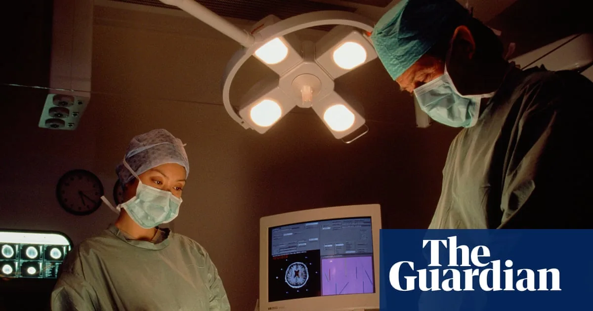
A pioneering brain tumour test trialled across the United Kingdom is slashing diagnostic waiting times from up to eight weeks down to just two hours while patients remain on the operating table [2]. Developed by researchers at the University of Nottingham and validated through collaborative clinical work at Newcastle University, this rapid genomic method analyzes tumor DNA directly during surgery [1]. NHS England has now initiated a multi-centre pilot across five specialist trusts to bring immediate genetic classification into neurosurgical theatres [2].
How Does the Rapid Brain Tumour Test Work?
The rapid brain tumour test operates by feeding small intraoperative tissue samples into a portable sequencer that decodes cancer DNA in real time rather than relying on delayed histological stains. When surgeons extract abnormal brain tissue during an operation, staff transfer the sample straight to a pathology laboratory where specialized sequencing hardware reveals the patient’s unique genomic architecture within hours. Stuart Smith, a consultant neurosurgeon at Nottingham University Hospitals NHS Trust and co-lead of the university’s research centre of excellence, highlighted that having precise genetic data while decisions occur inside the operating theatre is fundamentally gamechanging. Primary brain tumours represent the single largest cancer killer of children and adults under 40 in the United Kingdom [2].
Standard clinical protocols require pathologists to slice, stain, and inspect tissue under traditional light microscopes before dispatching specimens to regional facilities for extensive molecular profiling. Although standard laboratory analysis remains highly accurate, the logistical journey and multifaceted chemical assays routinely take several weeks to return actionable answers to clinical teams. Dr. Jonathan Coxhead, head of the Genomics Core Facility at Newcastle University, explained that partnering with the Newcastle Hospitals team allows scientists to drive the adoption of this rapid brain tumour test directly into the National Health Service [1].
Because oncological treatment hinges entirely on the exact biological category of the malignancy, delayed test results force patients and families into weeks of agonizing uncertainty. Clinicians cannot finalize radiotherapy schedules or specialized chemotherapy regimens without identifying whether a growth is a low-grade lesion or an aggressive malignancy [1]. The new test compresses that diagnostic lag [2].
Oxford Nanopore Sequencing in the Operating Theatre
The hardware driving this diagnostic shift relies on an innovative desktop sequencing device engineered by Oxford Nanopore. Instead of employing massive, room-sized sequencing machinery that demands days of chemical preparation, medical staff insert extracted tumour fragments into a compact unit roughly the size of a standard shoebox. Inside the device, individual DNA molecules pass through a microscopic aperture known as a nanopore (an ultra-fine opening across an electrically resistant membrane). As each nucleotide thread moves through the nanopore, it produces characteristic fluctuations in electrical current that instantly disclose the unique genetic code of the underlying tumour [2].
Traditional microscopy provides only structural snapshots of preserved tissue. Nanopore sequencing decodes the live genome. By assessing real-time genomic signals, the sequencing system distinguishes between subtle cellular abnormalities that appear visually identical to pathologists under conventional illumination [2]. This technical distinction proves crucial when dealing with complex central nervous system cancers, where visual inspection alone frequently fails to separate benign margins from infiltrative borders [1].
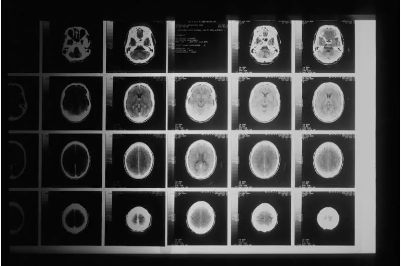
Earlier oncology investigations examining glioma cell activity and aggressive nervous system tumours have shown how vital metabolic and genetic markers are during surgical resection. In Newcastle, scientists combine the physical Oxford Nanopore sequencers with sophisticated bioinformatics software known as ROBIN to interpret raw biological data without manual delays. Project lead Angharad Goodman, a clinical scientist at the Newcastle Genetics Laboratory, explained that current practices require running several separate genetic tests that consume weeks before delivering a comprehensive diagnosis [1].
How Does the Brain Tumour Test Guide Surgical Care?
The brain tumour test guides surgical care by providing surgeons with precise tumour classification while the patient remains anesthetized on the table, allowing the operating team to adjust their surgical margins immediately. If the molecular read-out reveals an aggressive form of cancer that requires extensive resection, neurosurgeons can safely extend tissue removal without subjecting the individual to a second invasive operation weeks later [2]. Conversely, if the test classifies the lesion as a subtype that responds poorly to aggressive surgery but well to targeted medical therapies, surgeons can avoid removing critical brain tissue. The Brain Tumour Charity notes that because brain tumours remain the deadliest cancer for young patients, immediate clarity substantially improves clinical decision-making [1].
Medical classifications currently catalog more than 100 distinct types of brain tumors, each carrying radically different prognoses and therapeutic vulnerabilities. Determining whether a sample represents an astrocytoma, an ependymoma, or an aggressive glioblastoma (a high-grade and highly aggressive central nervous system malignancy) cannot be achieved with visual tissue staining alone. As Angharad Goodman highlighted, the overriding aspiration is to establish a verified provisional diagnosis before the surgical wound is closed [1]. Such intraoperative precision prevents redundant cranial interventions.
Prof Frankie Swords, the NHS medical director, described the diagnostic innovation as a huge leap forward that holds the potential to completely transform how healthcare services manage brain tumours across the country. Beyond immediate surgical tactics, the brain tumour test allows oncologists to enroll qualifying individuals into specialized clinical trials within days rather than waiting two months for laboratory confirmation. For patients suffering from rapid disease progression, accessing experimental therapies without administrative delays represents a meaningful survival advantage [2].


Data Analysis and the ROBIN Software Pipeline
Generating raw sequencing reads represents only the first phase of rapid intraoperative diagnosis; the biological signals must be converted into an unambiguous medical classification. In Newcastle, the multidisciplinary team utilizes specialized analytical software designated as ROBIN to process the continuous stream of nanopore data. ROBIN evaluates complex methylation profiles and specific genetic alterations across the tumour genome, cross-referencing findings against extensive reference databases. This automated computational pipeline bypasses the time-consuming manual interpretations that typically occupy pathology teams for weeks [1].
The Newcastle development cohort brings together clinical geneticists, pathologists examining cellular tissue, surgical oncologists, and neurology specialists. While unconventional diagnostic experiments such as experimental diagnostic models like nematode worm cancer screening explore alternative biomarker methods, nanopore sequencing focuses strictly on direct molecular sequencing of human tumour DNA. Clinical scientist Angharad Goodman stressed that having this unified testing platform replaces fragmented, multi-step assays with a single, highly integrated workflow [1].
Software automation ensures rapid, reproducible sequencing calls. Dr. Jonathan Coxhead noted that observing cutting-edge genomic infrastructure directly translate into improved bedside care provides immense motivation for the laboratory team. The software generates provisional classifications within minutes of sequence acquisition, creating an audit trail that clinical scientists review before transmitting findings to theatre [1]. Rigorous automated algorithms minimize diagnostic divergence.
Why Are NHS Hospitals Adopting the Brain Tumor Test?
NHS hospitals are adopting the brain tumor test to eliminate life-threatening delays in cancer treatment for vulnerable neurological patients across the country [2].
The clinical rollout expands upon an initial regional initiative funded by Newcastle Hospitals Charity, which successfully deployed the test across 40 distinct varieties of brain tumours in northeastern England. That charitable pilot included pediatric patients, adolescents, and mature adults, verifying that the brain tumour test maintains high fidelity across diverse age demographics. Simon Bailey, professor of pediatric neuro-oncology at Newcastle University, emphasized that the technology dramatically improves clinicians’ ability to deliver rapid diagnoses, allowing young children to commence optimal therapy immediately [1].
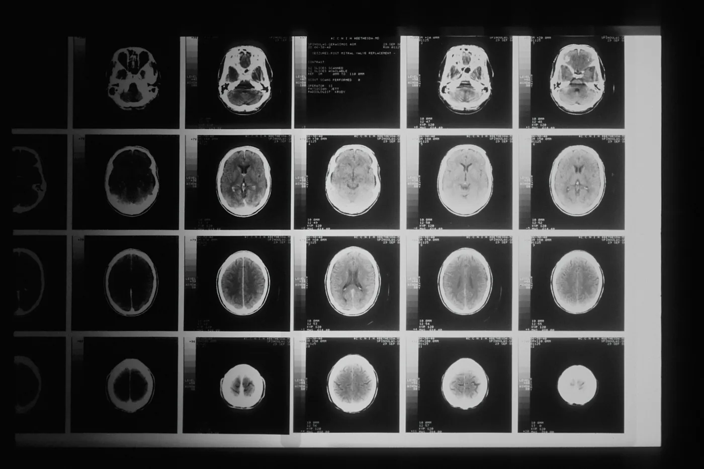
Professor Bailey noted that securing prompt diagnostic certainty enables affected children and their families to plan their lives during an emotionally traumatic period [1]. Conventional delays of six to eight weeks leave parents and oncologists trapped in limbo, unsure whether radiotherapy should begin or whether specialized targeted inhibitors are warranted. Stuart Smith from Nottingham reiterated that saving valuable weeks provides vital relief for individuals who are confronting aggressive malignancies and simply do not have time on their side [2].
Expansion Plans for Specialist Centres Across England
Under the supervision of NHS England, the intraoperative diagnostic platform is currently operational within five pioneer health trusts across the nation (Nottingham, Birmingham, London, and Newcastle). These frontline adopter sites comprise Nottingham University Hospitals NHS Trust, University Hospitals Birmingham NHS Foundation Trust, Great Ormond Street Hospital NHS Foundation Trust in London, King’s College Hospital, and Newcastle Hospitals NHS Foundation Trust [2]. Over the next two years, clinicians will gather detailed operational metrics to evaluate whether the rapid sequencing method delivers dependable outcomes inside demanding surgical environments [1].
Five specialist centres lead the initial rollout. Following the primary validation phase at these initial five trusts, healthcare authorities plan to expand the genomic sequencing pipeline to additional regional treatment centres in Bristol, Oxford, Leeds, and Manchester [2]. Health administrators are examining the logistical feasibility, laboratory equipment costs, and computational infrastructure necessary to support routine clinical deployment across all major neurosurgical units. Dr. Jonathan Coxhead emphasized that building this network of excellence is vital to ensure that the new brain tumour test reaches regional treatment centres across every territory with equal precision [1].
By transforming a diagnostic protocol that once demanded two months into a focused two-hour procedure, British researchers have demonstrated how bedside genomics can reshape cancer care in real time [2]. The two-year evaluation will determine whether rapid intraoperative sequencing transitions into a standard protocol across the entire National Health Service. As clinical trials gather patient outcomes over the coming months, surgical teams stand ready to turn rapid genetic sequencing into routine neurosurgical practice [1].
- ONLINE NEWS Clark, G., & Zinin, A. (2026, September 25). New brain tumor test could transform diagnosis and treatment. Medical Xpress. [Article Link]
- ONLINE NEWS Horton, H. (2026, September 25). ‘Gamechanging’ brain tumour test reduces diagnosis from eight weeks to two hours. The Guardian. [Article Link]
APA 7: PerEXP Teamworks. (2026, September 26). Rapid Brain Tumour Test Cuts NHS Diagnosis to Two Hours. PerEXP Teamworks.


