




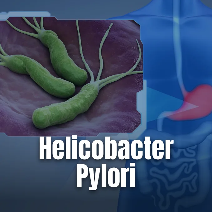
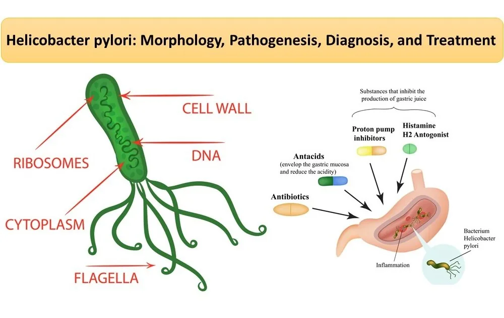
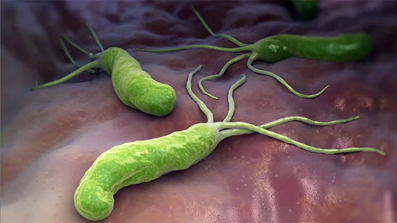
Helicobacter pylori is a spiral-shaped microbe that lives in the human stomach, infecting more than half of all people worldwide. While most carriers show no symptoms, chronic colonization damages the mucosal lining of the stomach and duodenum. Chronic gastritis, peptic ulcers, and stomach cancer often trace back to this bug. The organism measures 0.5 to 1 µm wide and 2 to 4 µm long [2]. By making urease enzymes, the germ neutralizes stomach acid in its local spot and sets up a lifelong home within the gut [16].
Discovery of Helicobacter Pylori
Italian pathologist Giulio Bizzozero first saw spiral microbes inside the stomach of dogs in 1892. Doctors long held that living germs could not survive inside human gastric acid, assuming that ulcers came from emotional stress and spicy food. In 1983, Australian physician-scientists Barry Marshall and Robin Warren challenged that old view by growing the spiral bug from stomach biopsies at Royal Perth Hospital in Australia [2].
Warren and Marshall first named the bug Campylobacter pyloridis because its curved body looked like other campylobacter species, but later taxonomist C. S. Goodwin and his team in 1989 grouped it into the new genus Helicobacter due to its corkscrew shape and its habit of living in the pylorus [16]. To prove that the newly isolated germ caused gastritis, Marshall drank a broth of cultured microbes, falling sick with acute nausea and vomiting within days. Endoscopy showed that the germs had colonized his stomach lining. Marshall drank the culture. He then took antibiotics and bismuth salts to cure his illness, offering clear proof of bacterial disease [1].
Marshall and Warren received the Nobel Prize in Physiology or Medicine in 2005. The Nobel Committee noted that peptic ulcer disease was no longer a chronic illness, but a condition curable with short courses of pills. In 1997, Tomb and a group of geneticists sequenced the complete genome of reference strain 26695, finding 1.7 million base pairs and roughly 1,566 genes [1].

Acid Survival in the Stomach
The human stomach keeps a harsh fluid bath with gastric acid pushing the pH below 2.0 (pH below 2.0). Strong acid quickly destroys common gut microbes that enter from food or drink. Helicobacter pylori grows best in neutral conditions between pH 6.5 and 7.5, making its survival in acidic fluid a remarkable biological trick [11].
Urease production powers this acid defense system. Urease makes up 10% of total protein. The microbe uses a gated inner membrane channel called UreI to run this survival mechanism. When external acidity climbs, UreI opens and lets urea from gastric juice flood into the cytoplasm, where urease quickly splits urea into carbon dioxide and ammonia. Ammonia then moves out into the periplasmic space, soaking up free hydrogen ions to yield ammonium. The buffering process keeps the periplasmic pH close to 6.2, shielding delicate metabolic enzymes from destruction while the bug swims across the gastric cavity to reach the epithelial wall [4].
Polar flagella supply the physical drive to flee acidic fluid. The microbe uses two to six sheathed flagellar strands, built from FlaA and FlaB proteins, to move its body through thick mucus toward gastric cells. Guided by pH chemotaxis, it swims away from acid and nests beneath the mucus blanket where conditions remain near neutral [3].
How Helicobacter Pylori Spreads
Global surveys show that about 4.4 billion people carry Helicobacter pylori, meaning more than half the world harbors the bug. Rates differ widely between regions and social groups, with developing countries reporting rates over 70%, while richer nations stay below 30%. In the United States, Cleveland Clinic notes that about 5% of children under age 10 carry the germ. National health surveys by the National Cancer Institute found that seroprevalence reached 21% in non-Hispanic white adults, 52% in African American adults, and 64% in Mexican American adults, reflecting gaps in household crowding, sanitation, and clean water access during childhood [7].
Most people catch the bug in early childhood, usually before turning five. The germ passes from person to person through close home contact between parents and young siblings. Poor sanitation spreads the microbe through contaminated food and water, while shared saliva and dental plaque serve as the main path in cleaner urban homes [5].
Once the bug settles in, it stays for life unless cleared by drugs. Natural loss of the microbe is rare in adults, with annual reinfection in developed nations holding below 2%. In Turkey, a 2010 study by Uyanıkoğlu evaluated 1,298 antrum biopsies, finding positive rates of 71.5% between ages 31 and 45, 68.6% between ages 46 and 60, and 70.4% above age 60, showing that adult carrier rates stay high throughout life [2].
Toxins and Cell Damage
Sticking tightly to stomach cells lets the bug resist gastric emptying. The adhesin BabA docks onto Lewis b blood group antigens on host cells, while another adhesin, SabA, binds sialyl-Lewis x sugars that show up during inflammation. In addition, bacterial lipopolysaccharide mimics human Lewis antigens to slip past immune patrols [11].
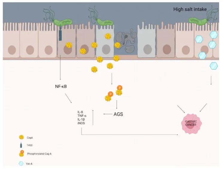
Harmful strains carry the cag pathogenicity island to inject toxic proteins straight into stomach cells. The genetic cluster builds a type IV secretion system that works like a tiny needle. It injects the oncoprotein CagA into host cells. Once inside, host enzymes phosphorylate CagA, causing it to scramble the cell skeleton, break tight junctions between neighbors, boost cell growth, and trigger the release of interleukin-8. The surge in chemical signals pulls white blood cells into the mucosal wall, causing chronic gastritis that wears down the stomach lining over decades of persistent colonization [11].
A second weapon is the vacuolating toxin VacA, an autotransporter made by nearly all strains. VacA drills anion pores into host cell membranes, forming large fluid vacuoles that cause cells to burst. VacA also punctures mitochondrial walls to trigger cell death and stops local T cells from multiplying, blunting the immune counterattack [6].


How Helicobacter Pylori Causes Ulcers
Peptic ulcer disease strikes about 10% to 15% of people infected with Helicobacter pylori. The microbe causes over 75% of duodenal ulcers and most non-drug stomach ulcers. Where the germ settles in the stomach dictates the clinical outcome. When the bug gathers in the lower antrum, inflammation blocks somatostatin release from D cells. Free from regulation, antral G cells pour excess gastrin into the blood, driving parietal cells in the upper fundus to flood the stomach with hydrochloric acid. The massive acid flow spills into the duodenum, wearing down the delicate intestinal surface and paving the way for ulceration [16].
Heavy acid loads induce gastric metaplasia in the duodenum, creating patches that Helicobacter pylori can colonize. Bacterial proteases and ammonia then eat away the protective mucus film. Without mucus, stomach acid and digestive pepsin bore through the raw tissue, opening painful peptic sores [10]. Taking nonsteroidal anti-inflammatory drugs (NSAIDs) such as aspirin or ibuprofen multiplies the risk of bleeding ulcers [8].
Patients with peptic ulcers usually feel a dull or burning gnawing pain below the ribs, especially on an empty stomach. Eating food or taking antacids brings quick relief by buffering acid. Severe ulcers can eat into blood vessels, triggering life-threatening bleeding. Bleeding ulcers produce melena (dark, tarry stools) or vomit resembling coffee grounds. These sores bleed easily. Perforation can also tear through the stomach wall, requiring emergency surgery [10].
Gastric Cancer Risk
Epidemiological studies investigated the link between chronic gastritis and stomach tumors in 1991 and 1994, prompting the World Health Organization and the International Agency for Research on Cancer to classify the germ as a Group 1 human carcinogen, a rating reaffirmed in 2009 [2]. Chronic colonization is the top cause of non-cardia gastric adenocarcinoma worldwide. Cancer growth follows Correa’s cascade, moving slowly from superficial gastritis to atrophic gastritis, intestinal metaplasia, dysplasia, and full invasive cancer. About 1% to 3% of carriers develop stomach cancer, which claimed roughly 769,000 lives globally in 2020. Bugs bearing the CagA toxin carry the greatest malignant danger [7].
The bug also causes gastric mucosa-associated lymphoid tissue lymphoma. Continuous immune stimulation from bacterial antigens sparks runaway B cell proliferation in the mucosa. Clearing the bug with antibiotic therapy leads to complete tumor disappearance in 75% to 80% of early localized MALT cases, making drugs the first-line cure for this lymphoma [13].
Infection also reaches outside the digestive tract. The microbe hinders nutrient absorption, causing unexplained iron deficiency anemia and low vitamin B12 levels. It can also spark immune thrombocytopenia (ITP) when anti-CagA antibodies mistake human platelets for the bug. Curiously, childhood colonization lowers the risk of esophageal adenocarcinoma, as long-standing stomach atrophy reduces acid reflux into the gullet over a lifetime [7].
Breath and Stool Testing
Doctors advise non-invasive testing for young patients who have indigestion without alarming red flags like sudden weight loss. The urea breath test is among the most accurate diagnostic tools in gastrointestinal medicine. During the test, the patient drinks a small cup of liquid containing urea labeled with non-radioactive carbon-13 or carbon-14. If live germs reside in the stomach, bacterial urease breaks the urea into ammonia and labeled carbon dioxide, which enters the blood and exits through the lungs. Breath bags filled ten to twenty minutes later capture the labeled gas with high precision, confirming an active infection [4].
The stool antigen test provides another fast, reliable choice for spotting active germs. Stool tests detect bacterial proteins directly in bowel movements using monoclonal antibodies or PCR. Blood antibody tests, by contrast, stay positive long after bugs vanish, making blood tests useless for checking if medical treatment successfully eradicated the infection [9].

Invasive testing uses upper endoscopy with tissue biopsies for patients over 50 or those vomiting blood. Biopsy tissue goes into a rapid urease test (CLO test), where urea turns pink as pH rises. Pathologists also stain samples with Giemsa to spot the corkscrew bugs directly under a microscope [12]. Patients must stop taking proton pump inhibitors (PPIs) for two weeks and antibiotics for four weeks before testing to prevent false negative results [9].
Antibiotic Regimens Facing Drug Resistance
Eradicating the germ cures ulcers and shields against stomach cancer. For years, standard first-line care paired a proton pump inhibitor (PPI) with clarithromycin and amoxicillin for 14 days. But rising drug resistance has weakened that triple regimen across North America and Europe. In areas where clarithromycin failure tops 15%, Maastricht VI guidelines recommend bismuth quadruple therapy for 14 days as the best first choice. The regimen bundles a PPI, bismuth subsalicylate, metronidazole, and tetracycline, curing over 90% of cases in clinical trials and overcoming macrolide resistance [17].
Resistance rates fluctuate by prior drug use and local prescribing trends. In French clinical samples tested by Raymond and colleagues, resistance stood at 26% for clarithromycin and 61% for metronidazole, with prior macrolide use driving clarithromycin resistance up to 68% among previously treated patients, highlighting why doctors must consult local resistance data before prescribing antibiotics [15].
In 2022, the Food and Drug Administration approved two vonoprazan combinations (Voquezna) based on the 1,046-patient PHALCON-HP trial. William D. Chey from the University of Michigan said, “I believe the added flexibility of having two additional effective therapies, including a dual therapy option that does not contain clarithromycin, offers the potential to improve clinical outcomes in patients with H. pylori infection” [14]. Cure must be confirmed with a breath or stool test four weeks after antibiotics end [9].
- ACADEMIC JOURNAL Marshall, B., & Adams, P. C. (2008). Helicobacter Pylori: A Nobel Pursuit?. Canadian Journal of Gastroenterology, 22(11), 895-896. [Article Link]
- ACADEMIC JOURNAL Öztekin, M., Yılmaz, B., Ağagündüz, D., & Capasso, R. (2021). Overview of Helicobacter pylori Infection: Clinical Features, Treatment, and Nutritional Aspects. Diseases, 9(4), 66. [Article Link]
- ACADEMIC JOURNAL Ansari, S., & Yamaoka, Y. (2017). Survival of Helicobacter pylori in gastric acidic territory. Helicobacter, 22(4), e12386. [Article Link]
- ACADEMIC JOURNAL Graham, D. Y., & Miftahussurur, M. (2018). Helicobacter pylori urease for diagnosis of Helicobacter pylori infection: A mini review. Journal of Advanced Research, 13, 51-57. [Article Link]
- ACADEMIC JOURNAL Brown, L. M. (2000). Helicobacter Pylori: Epidemiology and Routes of Transmission. Epidemiologic Reviews, 22(2), 283-297. [Article Link]
- ACADEMIC JOURNAL Kusters, J. G., van Vliet, A. H. M., & Kuipers, E. J. (2006). Pathogenesis of Helicobacter pylori Infection. Clinical Microbiology Reviews, 19(3), 449-490. [Article Link]
- REPORT National Cancer Institute. (2013). Helicobacter pylori (H. pylori) and Cancer. National Institutes of Health. [Article Link]
- WEBSITE Cleveland Clinic. (2023). What Is an H. Pylori Infection?. Cleveland Clinic Health Library. [Article Link]
- WEBSITE MedlinePlus. (2026). Helicobacter pylori (H. pylori) Tests. National Library of Medicine. [Article Link]
- WEBSITE WebMD. (2025). H. pylori: Causes, Symptoms, Treatment. [Article Link]
- WEBSITE Medscape. (2026). Helicobacter Pylori Infection: Background, Etiology, Pathophysiology. Medscape Drugs & Diseases. [Article Link]
- ONLINE NEWS Raffier, N. (2022). The Changes Made in the New Expert Consensus on H pylori. Medscape. [Article Link]
- WEBSITE Patient.info. (2024). Helicobacter pylori. [Article Link]
- ONLINE NEWS Hamza, Z. (2022). Two New Regimens Win FDA Approval for H. Pylori Infection. MedPage Today. [Article Link]
- ACADEMIC JOURNAL Raymond, J., Lamarque, D., Kalach, N., Chaussade, S., & Burucoa, C. (2010). High Level of Antimicrobial Resistance in French Helicobacter pylori Isolates. Helicobacter, 15(1), 21-27. [Article Link]
- WEBSITE Wikipedia. (2026). Helicobacter pylori. Wikimedia Foundation. [Article Link]
- WEBSITE Wikipedia. (2026). Helicobacter pylori eradication protocols. Wikimedia Foundation. [Article Link]


