






Synapses are microscopic gaps where neurons pass voltage pulses or chemical cues to communicate with partner cells in the nervous system. Understanding how synapses work clarifies how brain circuits coordinate movement, process sensory input, and preserve memories over time. In the human brain alone, roughly 86 billion neurons form between 1,000 and 5,000 trillion synaptic connections [2, 4]. Most of these connections rely on chemical messengers called neurotransmitters that diffuse through a fluid gap to trigger or inhibit nerve firing in the receiving cell [1].
- Discovery of the Synaptic Junction
- Structural Architecture of the Junction
- How Synapses Work to Release Transmitters
- Receptor Activation in Postsynaptic Membranes
- Signal Termination Through Reuptake Mechanisms
- Spatial Summation in Synaptic Transmission
- Differences in Electrical Synapses
- Neural Plasticity in Synaptic Transmission
Discovery of the Synaptic Junction
Early anatomists believed that the nervous system formed an unbroken reticular network. Spanish histologist Santiago Ramón y Cajal disproved that view, demonstrating that nerve cells remain separate units that communicate across narrow borders, establishing the foundational neuron doctrine [10].
British physiologist Charles Sherrington introduced the term synapse in 1897 in Michael Foster’s Textbook of Physiology, selecting a name suggested by classical scholar Arthur Woollgar Verrall from the Greek word synapsis, which means to clasp together [10]. Researchers argued for decades over whether signals crossed this boundary through sparks or chemicals. Pharmacologist Henry Dale settled the debate in 1936 by showing that chemical agents transmit signals across the gap between nerves and muscle, identifying acetylcholine as the active agent [3]. Light microscopes could not resolve the gap [10]. In 1952, Alan Hodgkin and Andrew Huxley used voltage-clamp tests on the squid giant axon to map how sodium and potassium currents produce the action potential, an achievement recognized with the 1963 Nobel Prize [1].
Direct visual proof arrived in the 1950s when electron microscopes entered laboratories. Neurobiologist Sanford Palay used electron microscopy to view tissue ultrastructure, showing that cell surfaces remain separated by a fluid gap of 20 to 40 nanometers. Scientists named this gap the synaptic cleft [3, 5]. Palay’s photographs confirmed Dale’s chemical model, proving that nerve cells communicate without fused cytoplasm [5].
Structural Architecture of the Junction
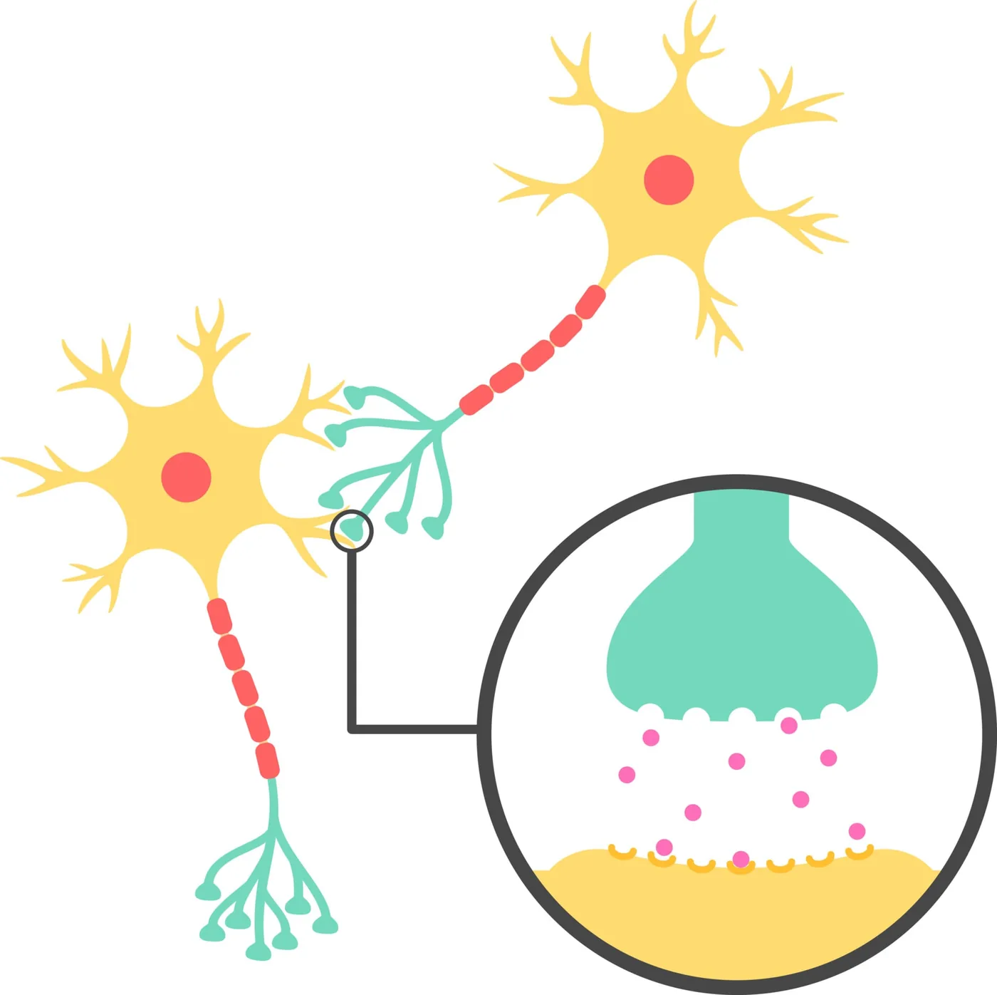
Brain tissue organizes synapses into different anatomical layouts based on which cell parts touch. The most common cortical layout is the axodendritic synapse, where an axon terminal meets a dendritic spine on an adjacent neuron [6].
Other anatomical patterns include axosomatic contacts touching the cell soma, axoaxonic junctions ending on neighboring axons, and dendrodendritic contacts connecting two dendrites [3, 6]. In 1966, researchers Wilfrid Rall, Gordon Shepherd, Thomas Reese, and Milton Brightman identified dendrodendritic synapses in the mammalian olfactory bulb, showing that dendrites can send signals directly without an axon [10]. Both sides of the junction rely on protein scaffolds that align release zones with receptor fields across the synaptic cleft. Synaptic adhesion molecules project from both cell surfaces, binding each other within the cleft to hold structural alignment and guide wiring. In the Encyclopedia of Neuroscience from Springer, Masahito Yamagata wrote that synaptic adhesion molecules guide junction assembly and transmit cues into the cytoplasm [8]. Sensory cells in the Organ of Corti also form specialized synapses with auditory nerves [10, 12].
Presynaptic nerve terminals store signaling agents in distinct vesicle pools. Small agranular vesicles of 35 to 50 nanometers hold glutamate and glycine, while large granular vesicles of 80 to 160 nanometers store neuropeptides [3, 5]. Actin filaments and synapsin hold vesicles near the active zone [5]. Mitochondria cluster inside the terminal bouton, producing adenosine triphosphate (ATP) to power ongoing vesicle recycling [6, 7].
How Synapses Work to Release Transmitters
Studying how synapses work at the molecular scale begins when an action potential invades the nerve terminal. At rest, a neuron holds an internal charge roughly 70 millivolts more negative than fluid outside the cell, an electrical balance kept by sodium and potassium gradients. When incoming dendritic inputs push membrane voltage past the threshold of -55 mV, voltage-gated sodium channels open in fast waves, propagating an action potential along the axon until the positive wave enters the terminal bouton to flip open voltage-gated calcium channels. Extracellular calcium ions (Ca²⁺) flood into the axoplasm down a steep concentration gradient. Free calcium acts as an intracellular command signal [1, 3].
Entering calcium mobilizes vesicles to the active zone [1, 3]. Calcium binds sensor proteins, prompting vesicular synaptobrevin to coil with syntaxin 1 and SNAP-25 on the plasma membrane, forming the SNARE complex that forces lipid bilayers to fuse [12].
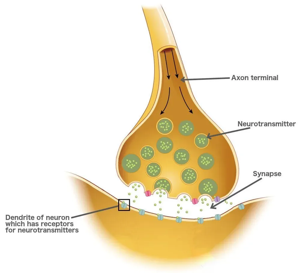
Opening a fusion pore lets stored transmitters spill into the 20 to 40 nanometer synaptic cleft through exocytosis in less than a millisecond [3, 5]. Thousands of chemical messenger molecules diffuse across the narrow fluid gap to bind selective receptors on the receiving membrane, a process central to decoding neurotransmitters and their signaling roles in the central nervous system. Transmitter diffusion takes under a millisecond [4, 5].
Receptor Activation in Postsynaptic Membranes
Chemical messengers diffusing through the cleft bind docking proteins clustered on the target cell surface [4, 5]. These target proteins operate through two main formats: ionotropic receptors and metabotropic receptors [1, 6]. Binding triggers distinct electrical responses [1, 7].
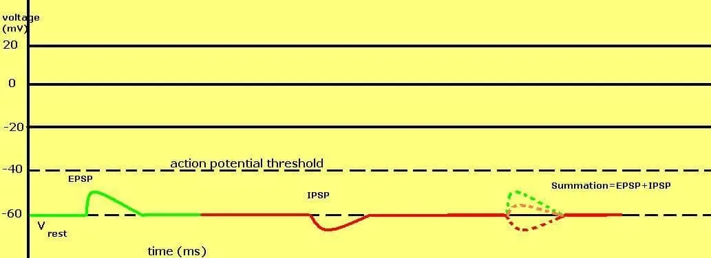
Ionotropic receptors contain built-in ion channels that snap open when bound by a ligand, producing voltage shifts within a single millisecond, whereas metabotropic receptors couple to G-proteins that launch enzymatic cascades to modulate cell states over longer periods [1, 6]. The functional outcome depends on receptor properties rather than the chemical messenger alone [1, 7]. When glutamate binds to ionotropic AMPA receptors or NMDA receptors, non-selective cation channels open, driving an influx of sodium ions. This inward current produces an excitatory postsynaptic potential (EPSP), nudging membrane voltage toward the firing threshold. If multiple excitatory inputs cooperate, cumulative depolarization prompts the receiving cell to fire an impulse [1, 7]. Receptors dictate the functional response across the nervous system [1].
Inhibitory signaling uses mechanisms that pull membrane voltage away from the firing threshold. Gamma-aminobutyric acid (GABA) acts as the main inhibitory transmitter in the brain, binding to ionotropic GABAA receptors to open selective chloride channels. Negatively charged chloride ions enter the cell, generating a hyperpolarizing inhibitory postsynaptic potential (IPSP). This negative charge stabilizes membrane potential, making action potential firing far less likely [7, 10].


Signal Termination Through Reuptake Mechanisms
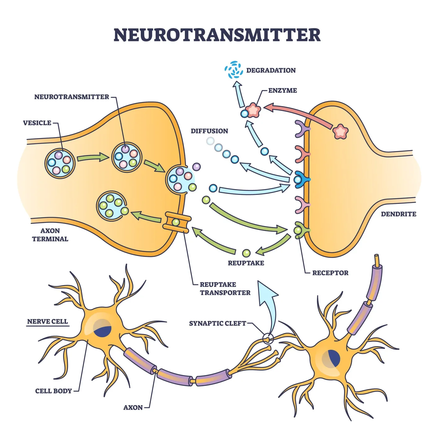
Examining how synapses work during signal clearance reveals why transmission must shut off swiftly [1, 4]. If transmitter molecules stayed bound to receptors, receiving cells would remain locked in constant stimulation, blinding neural circuits to new incoming messages [4].
Signal clearance restores resting membrane potential, preparing the junction for subsequent signaling cycles through three cooperating pathways: reuptake, enzymatic degradation, and diffusion [1, 6, 10]. In presynaptic reuptake, transporter proteins use sodium gradients to pump released transmitters back into the terminal bouton, where cells repackage them into vesicles or break them down with internal enzymes [1, 10]. At cholinergic junctions, enzymatic breakdown takes precedence: the enzyme acetylcholinesterase anchored in the cleft hydrolyzes acetylcholine into choline and acetate within microseconds. Presynaptic terminals reabsorb choline to synthesize fresh transmitter, preventing sustained muscle contraction [4, 7]. In Betz cells of the cerebral cortex, transmitter depletion can lead to synaptic fatigue during prolonged motor activity [3]. Without fast clearance, neuromuscular transmission halts under persistent depolarization block [4, 7].
Surrounding glial cells provide an essential line of defense, wrapping around synapses to form an astroglial cradle. Astrocytes express high-affinity transporter proteins that soak up excess glutamate and potassium ions from the cleft, protecting neurons from excitotoxicity. Selective serotonin reuptake inhibitors (SSRIs) block serotonin transporters to prolong signaling in mood circuits, while toxins like strychnine block inhibitory glycine receptors, producing violent convulsions and death [1, 10].
Spatial Summation in Synaptic Transmission
Understanding how synapses work as computing nodes shows that individual synaptic inputs rarely trigger an action potential in isolation [1, 4]. A single EPSP produces only a minor membrane depolarization of a fraction of a millivolt, which decays along the dendrite [1, 3].
Because a typical central neuron receives thousands of inputs across its dendritic tree, the receiving cell integrates competing signals through summation before deciding whether to fire. Spatial summation occurs when multiple presynaptic terminals fire at distinct dendritic locations at the same moment. If several excitatory synapses release glutamate simultaneously, their individual EPSPs combine into an additive depolarizing current that spreads to the axon initial segment. However, if an inhibitory synapse fires concurrently, its hyperpolarizing IPSP cancels the incoming positive charge, preventing depolarization [1, 7]. Through spatial summation, the neuron acts as an analog calculator, weighing inputs from multiple circuits before firing [4, 7]. Summation turns the synapse into a voting system [4].
Temporal summation operates along a chronological axis, combining inputs that arrive at the same synaptic site in rapid succession. Because postsynaptic potentials persist for several milliseconds before decaying, a second impulse arriving before the first fades adds to the remaining depolarization [1, 7]. When the summed depolarization reaches the threshold potential of -55 mV at the axon hillock, voltage-gated sodium channels fire an all-or-none action potential down the axon [1, 3].
Differences in Electrical Synapses
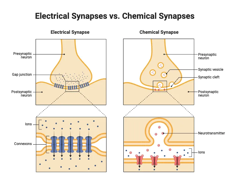
Contrasting how synapses work in chemical and electrical modes highlights how nervous systems match structure to physiological demand. While chemical synapses carry most signals across mammalian nervous tissue, electrical synapses provide direct electrical coupling between neighboring cells [1, 6].
At an electrical synapse, the gap separating adjacent cell membranes measures only 3.5 nanometers, compared to the 20 to 40 nanometer cleft found at chemical junctions [1, 5]. Membranes at electrical junctions are bridged by protein assemblies termed gap junctions [1, 6]. Each gap junction consists of paired hexameric protein complexes called connexons, which join across intercellular spaces to form an open aqueous pore. Ions and small signaling molecules such as ATP pass through these pores from one cytoplasm to the next without requiring vesicle exocytosis [6]. Because ions diffuse through open channels, electrical transmission occurs virtually without delay, bypassing the multi-millisecond synaptic delay characteristic of chemical synapses [1, 3]. Impulses pass in either direction [5, 6]. Direct coupling welds adjacent cells into functional syncytia [6].
Despite their high speed, electrical synapses lack the regulatory versatility of chemical transmission. Electrical junctions cannot convert an excitatory signal into an inhibitory response, nor can they produce long-lasting modulatory changes through secondary messenger cascades. Nervous systems deploy electrical synapses in circuits requiring instantaneous synchronization, such as cardiac muscle fibers, intestinal smooth muscle, and escape reflex pathways in invertebrates [3, 5]. Retinal alpha-ganglion cells also employ dendritic gap junctions to coordinate contrast adjustments [10].
Neural Plasticity in Synaptic Transmission
Neuroscientists studying how synapses work have discovered that these connections are dynamic structures rather than static anatomical relays. Synapses exhibit synaptic plasticity, the capacity to modify transmission strength in response to changing patterns of neural activity [4].
Plasticity manifests primarily through two opposing processes: long-term potentiation (LTP) and long-term depression (LTD). When connected neurons fire repeatedly in close synchrony, the synapse undergoes LTP, increasing postsynaptic AMPA receptor density and strengthening synaptic responses for hours or weeks [4, 7]. Conversely, low-frequency stimulation triggers LTD, internalizing receptors and reducing synaptic efficacy [4]. In sensory organs, neurons utilize unique ribbon synapses to maintain tonic transmission without exhausting vesicle stores. Ribbon synapses in retinal photoreceptors, retinal bipolar cells, and cochlear hair cells hold thousands of vesicles docked along protein ribbons anchored by Bassoon, Piccolo, and RIBEYE scaffolds [11, 12]. Saaid Safieddine and Robert Wenthold showed that cochlear hair cell ribbons express core SNARE machinery, including syntaxin 1 and SNAP-25, to drive fast auditory neurotransmission [12].
Recent discoveries continue to reveal novel synaptic architectures in mammalian brain tissue. In 2022, researchers Shu-Hsien Sheu and David Clapham at the Janelia Research Campus of HHMI discovered axo-ciliary synapses, where brainstem serotonergic axons form functional junctions with primary cilia on hippocampal CA1 pyramidal neurons. Stimulation of ciliary 5-HTR6 serotonin receptors activates signaling pathways that alter nuclear chromatin accessibility and gene transcription [9]. Beyond chemical diffusion, mechanical tension on hippocampal membranes modulates synaptic firing properties, providing a cellular parallel to light-induced cell movements and rapid cellular responses observed in biophysics [10].
- WEBSITE Guy-Evans, O. (2026). What Happens at The Synapse? Simply Psychology. [Article Link]
- WEBSITE Mukherjee, S. (2020). Synapse – Definition, Types, Structure, Functions, and Diagram. Science Facts. [Article Link]
- WEBSITE GeeksforGeeks. (2023). Synapse | Anatomy, Function & Types. GeeksforGeeks. [Article Link]
- WEBSITE Jalan, M. (2018). What Would Happen If Our Neurotransmitters Stopped Working? ScienceABC. [Article Link]
- WEBSITE Pan, S. (2024). Synapse – Definition, Structure, Principles, Types & Function. Biology Notes Online. [Article Link]
- WEBSITE Kenhub. (2024). Synapse. Kenhub. [Article Link]
- WEBSITE Lasica, A., & Brewer, C. (2023). Excitatory and Inhibitory Synaptic Signalling. TeachMePhysiology. [Article Link]
- BOOK CHAPTER Yamagata, M. (2009). Synaptic Adhesion Molecule. In M. D. Binder, N. Hirokawa, & U. Windhorst (Eds.), Encyclopedia of Neuroscience. Springer. [Article Link]
- ONLINE NEWS Howard Hughes Medical Institute. (2022). Scientists discover new kind of synapse in neurons’ tiny hairs. Phys.org. [Article Link]
- WEBSITE Wikipedia contributors. (2026). Synapse. Wikipedia, The Free Encyclopedia. [Article Link]
- WEBSITE Wikipedia contributors. (2026). Ribbon synapse. Wikipedia, The Free Encyclopedia. [Article Link]
- ACADEMIC JOURNAL Safieddine, S., & Wenthold, R. J. (1999). SNARE complex at the ribbon synapses of cochlear hair cells: analysis of synaptic vesicle- and synaptic membrane-associated proteins. European Journal of Neuroscience, 11(3), 803-812. [Article Link]
- ACADEMIC JOURNAL Sheu, S., et al. (2022). A serotonergic axon-cilium synapse drives nuclear signaling to alter chromatin accessibility. Cell, 185(18), 3390-3407.e18. [Article Link]


