





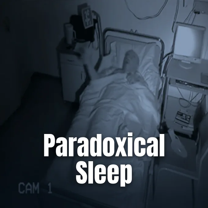
Understanding what is REM sleep begins with knowing how the brain shifts from quiet rest into an active state that mirrors daytime wakefulness. During this night phase, which alternates with non-rapid eye movement (NREM) rest in ninety-minute cycles, cerebral oxygen and sugar use rises to levels seen during busy waking hours. Mammals and birds enter this recurring state about four times during seven hours of rest. Neural networks fire rapidly, but brainstem pathways keep limb muscles limp so the sleeper cannot act out unfolding dreams [1].
What Is REM Sleep?
Sleep researchers Nathaniel Kleitman and Eugene Aserinsky first identified rapid ocular bursts at the University of Chicago in 1953, linking those physical movements to vivid dream reports. Their work proved that sleep is not a passive mental blackout. Later investigators William Dement and Michel Jouvet built upon these Chicago trials, tracking how human sleep cycles through distinct phases across each night [1].
Adult humans cycle between non-rapid eye movement rest and rapid eye movement episodes roughly every ninety minutes. Early cycles feature brief REM intervals of ten minutes, but later morning cycles expand to thirty minutes or more. Healthy adults cycle four times nightly. When subjects are woken whenever this phase starts, they show a rebound effect the next night by entering the state much faster. Waking imagery fades quickly because norepinephrine levels drop to zero, leaving short-term dream scenes unrecorded [1].
Modern sleep research tracks these night patterns through digital tools and video cameras. In an open sleep trial hosted by LSDBase, investigators tested the CGX Patch, a single-channel forehead EEG sensor, next to an automated camera system called the INSPEC. The setup ran machine vision models like BlazeFace in TensorFlow to log eye movements through the night. The CGX Patch verified these eye movements. Dream logs in Lucid Scribe showed that camera readings matched frontal EEG peaks [5].
Brain Waves During Paradoxical Sleep
French neurobiologist Michel Jouvet popularized the label paradoxical sleep because EEG traces during this phase mimic an alert, waking brain. While limb muscles stay limp, cortical electrodes register fast, low-voltage, desynchronized waves instead of slow delta patterns. Neurons in the thalamus and cortex fire in quick rhythms, creating rich mental activity while the sleeper rests quietly in bed [1].
This lively state features 3 to 10 Hz theta waves in the hippocampus alongside bursts of 40 to 60 Hz gamma waves in the cerebral cortex. Cortical neurons remain depolarized, so they fire impulses as readily as they do during waking daytime work. Brain energy use matches waking states. Oxygen and glucose consumption in the brain equals conscious levels, but deep NREM sleep cuts this metabolic rate by 11 to 40 percent. Furthermore, posterior brain areas stay coherent with one another across the cortex, especially when dreamers gain lucid control of their visions, showing that sleeping circuits can sustain organized mental tasks [1].
PET brain scans show that forebrain systems activate in unique patterns during these episodes. The anterior paralimbic REM activation area (APRA), which includes networks tied to memory, emotion, and fear, lights up far more than in quiet waking rest. The amygdala fires actively and helps drive brainstem bursts. In contrast, the frontal gyrus and parietal areas work at levels similar to daytime thought, organizing sensory fragments into narrative scenes [1].

Chemical Signals in the Brain Stem
Rhythms that launch this stage originate in lower brainstem centers, specifically the pontine tegmentum and locus coeruleus. Just as the brain transitions out of deep slow-wave rest, electrical bursts known as PGO waves (ponto-geniculo-occipital waves) travel from the pons into the lateral geniculate nucleus and the occipital cortex. These signals arrive in clusters about every six seconds for up to two minutes, driving rapid eye twitches under closed eyelids [1].
Neurotransmitters shift abruptly when these pontine centers take charge of cerebral circuits. Acetylcholine surges through the pons and cerebral cortex, generating fast brain waves. In contrast, monoamine messengers drop to near zero. Serotonin, norepinephrine, and histamine vanish from synapses during the entire episode. Laboratory injections of carbachol or acetylcholinesterase inhibitors induce paradoxical sleep in resting animals, but waking animals only respond if monoamine stores are drained beforehand [1].
Harvard researchers Robert McCarley and Allan Hobson proposed the reciprocal interaction model in 1975 to explain these cyclical shifts. Their model used formulas based on Lotka–Volterra equations to show how cholinergic REM-on cells activate aminergic REM-off cells, which then inhibit the cholinergic drivers. Neuroscientist Kayuza Sakai and Michel Jouvet refined this concept in 1981, adding gamma-aminobutyric acid (GABA) and orexin to explain how brainstem switches cycle smoothly through the night [1].
Mechanisms of Muscle Atonia
To keep sleepers from thrashing during dreams, the brainstem imposes a total motor block called REM atonia. Somatic motor neurons enter a state of hyperpolarization, where their resting membrane potential falls by an additional 2 to 10 millivolts. This drop lifts the stimulus threshold needed to cause muscle firing, leaving voluntary limbs flaccid while sparing autonomic muscles such as the diaphragm. Motor cells hyperpolarize by several millivolts [1].
Neural pathways running from the locus subcoeruleus in the brain stem project downward through the medulla to silence spinal motor neurons. Scientists McKenna and Peever at the University of Toronto found that glycinergic and GABAergic transmitters drive this motor inhibition, preventing cortical commands from reaching the limbs. Animal studies show that lesions in the locus subcoeruleus wipe out muscle atonia, prompting sleeping subjects to leap and run around their cages while remaining soundly asleep [7].
This active motor block separates rapid eye movement sleep from non-REM phases where muscle tone is merely relaxed. Sleepwalking and confusional arousals occur during deep NREM slow-wave sleep because somatic motor pathways remain unlocked. Similar motor gating issues appear in sleep disorders like narcolepsy, where sudden muscle collapse interrupts ordinary waking activities during daytime emotional reactions [2].



Autonomic Shifts and Temperature Loss
As voluntary muscles go limp, autonomic regulation enters an erratic state that diverges from ordinary body control. Heart rate, blood pressure, and cardiac output fluctuate widely from minute to minute, accompanied by irregular breaths and shallow breathing spells. Autonomic homeostasis stops during paradoxical sleep. Protective reflexes against low oxygen weaken, meaning the brain reduces its automatic governance over respiration compared to waking or slow-wave rest [1].
Internal thermoregulation halts during these periods, leaving the body unable to defend core temperature against chilly or warm rooms. Small mammals and humans stop shivering when cold and stop sweating when hot while in paradoxical sleep. Research on cats reveals that even when brainstem cuts remove muscle atonia, the animals still do not shiver during REM sleep. Warm-sensing and cold-sensing neurons in the hypothalamus simply do not fire. Sleepers wake if bedroom temperatures swing. If thermal indicators drift outside a tolerable range at the close of deep sleep, the brain skips REM sleep to prevent hypothermia [1].
Blood flow shifts also create recurring genital changes that unfold regardless of dream themes or erotic thought. Men undergo nocturnal penile tumescence (NPT), while women experience clitoral enlargement and increased blood supply, lasting between one and three hours across an average night. In medical clinics, doctors evaluate these nocturnal erections to discover if male sexual problems arise from psychological stress or physical nerve damage [1].
Comparing NREM and REM Sleep Stages
Allan Rechtschaffen and Anthony Kales established the modern staging of sleep in their 1968 R&K system, sorting human rest into four NREM stages and one REM stage. In 2007, an AASM update by the American Academy of Sleep Medicine combined stages 3 and 4 into a single deep sleep category [2]. Healthy sleep alternates between these three quiet phases and active rapid eye movement periods [4].
Stage 1 marks the initial drift from wakefulness, lasting five to ten minutes with theta waves and hypnic twitches [2, 4]. Stage 2 brings sleep spindles and K-complexes, which are single delta waves that last for one second and react to outside sounds like a door tap. Slow spindles run from 11 to 13 Hz in the superior frontal gyrus, while fast spindles run from 13 to 15 Hz to recruit sensorimotor circuits and the hippocampus. K-complexes can be triggered voluntarily by transient noises, helping researchers test how external auditory events enter the sleeping mind [2]. Stage 3 slow-wave sleep follows, using delta waves to repair tissues before the brain cycles back toward REM [4].
Circadian transitions between these stages are guided by the suprachiasmatic nucleus (SCN), a master neural pacemaker located in the hypothalamus. Clinical specialists at Rex MD note that morning light synchronizes the SCN, helping the body cycle cleanly through deep rest and REM sleep. Poor habits such as evening caffeine or erratic schedules disrupt this rhythm, cutting down on restorative rapid eye movement time [4].
Why Vivid Dreams Occur in REM Sleep
Why do dreamers encounter such intense storylines during this sleep phase? Sleep records show that 90 to 95 percent of people woken from rapid eye movement sleep recall rich, emotional dreams. In contrast, only 5 to 10 percent of people woken from NREM sleep recall clear plots, though 70 percent mention vague feelings [2]. Eye movements during this phase move in loops roughly seven times per minute, matching visual scene shifts [1]. Dreamers recall vivid plots upon waking [2].
Beyond generating dreams, this stage supports brain development and synaptic growth. In a 2015 study published in Science Advances, researchers Bridi, Aton, Seibt, Renouard, Coleman, and Frank showed that rapid eye movement sleep drives cortical plasticity in young brains. Using an ocular dominance model in cats, the Michigan team found that losing REM sleep blocked the activation of extracellular signal-regulated kinase (ERK), a key enzyme needed to cement waking experience into the brain [8].
Even though dreams feel vivid, the brain seldom saves them for long. A lack of norepinephrine in the cortex stops short-term dream scenes from moving into long-term memory circuits. While night REM cycles process emotional skills, simple quiet periods of daytime wakeful rest offer an easy way for the brain to stabilize fragile episodic memories without needing sleep [1].
Clinical Features of REM Sleep Disorders
When brainstem systems that cause muscle limpness fail, patients develop REM sleep behavior disorder (RBD), a condition first described by Carlos Schenck and Mark Mahowald in 1986 [3, 6]. Instead of resting quietly, people with RBD scream, punch, kick, and leap out of bed to fight off attackers in bad dreams. These night actions frequently injure both the patient and their sleep partner. Sleeping individuals often recall vivid attack scenarios when woken, which match the movements observed by doctors. Melatonin helps patients manage sleep disruptions. Secondary RBD can also stem from drug withdrawal or head injuries [3].
Clinical reports reveal that RBD is often an early warning sign of severe brain disease. In a study published in JAMA Neurology, researchers Howell and Schenck in Minnesota found that dream enactment is often the first clue of an impending alpha-synuclein disorder [6]. A subsequent paper by McKenna and Peever in Movement Disorders confirmed that about 80 percent of patients with idiopathic RBD eventually develop Parkinson’s disease, dementia with Lewy bodies, or multiple system atrophy [7].
Neuroimaging uncovers the physical roots of this motor failure. Brain scans by García-Lorenzo and Ehrminger in Brain using 3T MRI showed loss of signal in the locus coeruleus and subcoeruleus of RBD patients, confirming localized tissue decay in the brainstem [9, 10]. In Movement Disorders, Heintz-Buschart and Wilmes showed that Parkinson’s and RBD patients share distinct changes in their gut bacteria, such as Akkermansia, offering biological markers years before motor symptoms begin [11].
- WEBSITE Wikipedia. Rapid eye movement sleep. [Article Link]
- WEBSITE Wikipedia. Non-rapid eye movement sleep. [Article Link]
- WEBSITE Wikipedia. Rapid eye movement sleep behavior disorder. [Article Link]
- WEBSITE Rex MD. (2022, March 28). Types of Sleep: Everything You Need To Know. [Article Link]
- WEBSITE LSDBase. (2026, June 13). REM Sleep. [Article Link]
- ACADEMIC JOURNAL Howell, M. J., & Schenck, C. H. (2015). Rapid Eye Movement Sleep Behavior Disorder and Neurodegenerative Disease. JAMA Neurology, 72(6), 707. [Article Link]
- ACADEMIC JOURNAL McKenna, D., & Peever, J. H. (2017). Degeneration of rapid eye movement sleep circuitry underlies rapid eye movement sleep behavior disorder. Movement Disorders, 32(5), 636-644. [Article Link]
- ACADEMIC JOURNAL Bridi, M., Aton, S. J., Seibt, J., Renouard, L., Coleman, T., & Frank, M. G. (2015). Rapid eye movement sleep promotes cortical plasticity in the developing brain. Science Advances, 1(6), e1500105. [Article Link]
- ACADEMIC JOURNAL García-Lorenzo, D., Santos, C. L., Ewenczyk, C., Leu‐Semenescu, S., Galléa, C., Quattrocchi, G., Lobo, P. P., Poupon, C., Benali, H., Arnulf, I., Vidailhet, M., & Lehéricy, S. (2013). The coeruleus/subcoeruleus complex in rapid eye movement sleep behaviour disorders in Parkinson’s disease. Brain, 136(7), 2120-2129. [Article Link]
- ACADEMIC JOURNAL Ehrminger, M., Latimier, A., Pyatigorskaya, N., García-Lorenzo, D., Leu‐Semenescu, S., Vidailhet, M., Lehéricy, S., & Arnulf, I. (2016). The coeruleus/subcoeruleus complex in idiopathic rapid eye movement sleep behaviour disorder. Brain, 139(4), 1180-1188. [Article Link]
- ACADEMIC JOURNAL Heintz‐Buschart, A., Pandey, U., Wicke, T., Sixel‐Döring, F., Janzen, A., Sittig, E., Trenkwalder, C., Oertel, W. H., Mollenhauer, B., & Wilmes, P. (2017). The nasal and gut microbiome in Parkinson’s disease and idiopathic rapid eye movement sleep behavior disorder. Movement Disorders, 33(1), 88-98. [Article Link]
Cover image: Uguccioni G, Pallanca O, Golmard J, Dodet P, Herlin B, Leu-S / Wikimedia Commons, CC BY 4.0


