






Nighttime light exposure may leave a measurable mark on the heart’s structure. In a UK Biobank analysis of 11,071 adults, Professor Lu Qi and colleagues at Tulane University in New Orleans linked light exposure at night with differences in cardiac MRI (magnetic resonance imaging). Their study, published in the European Heart Journal, identifies an association rather than a cause: participants exposed to brighter nights tended to have thicker left ventricular walls and smaller chamber volumes. [1, 3]
Nighttime Light Exposure: What Counts?
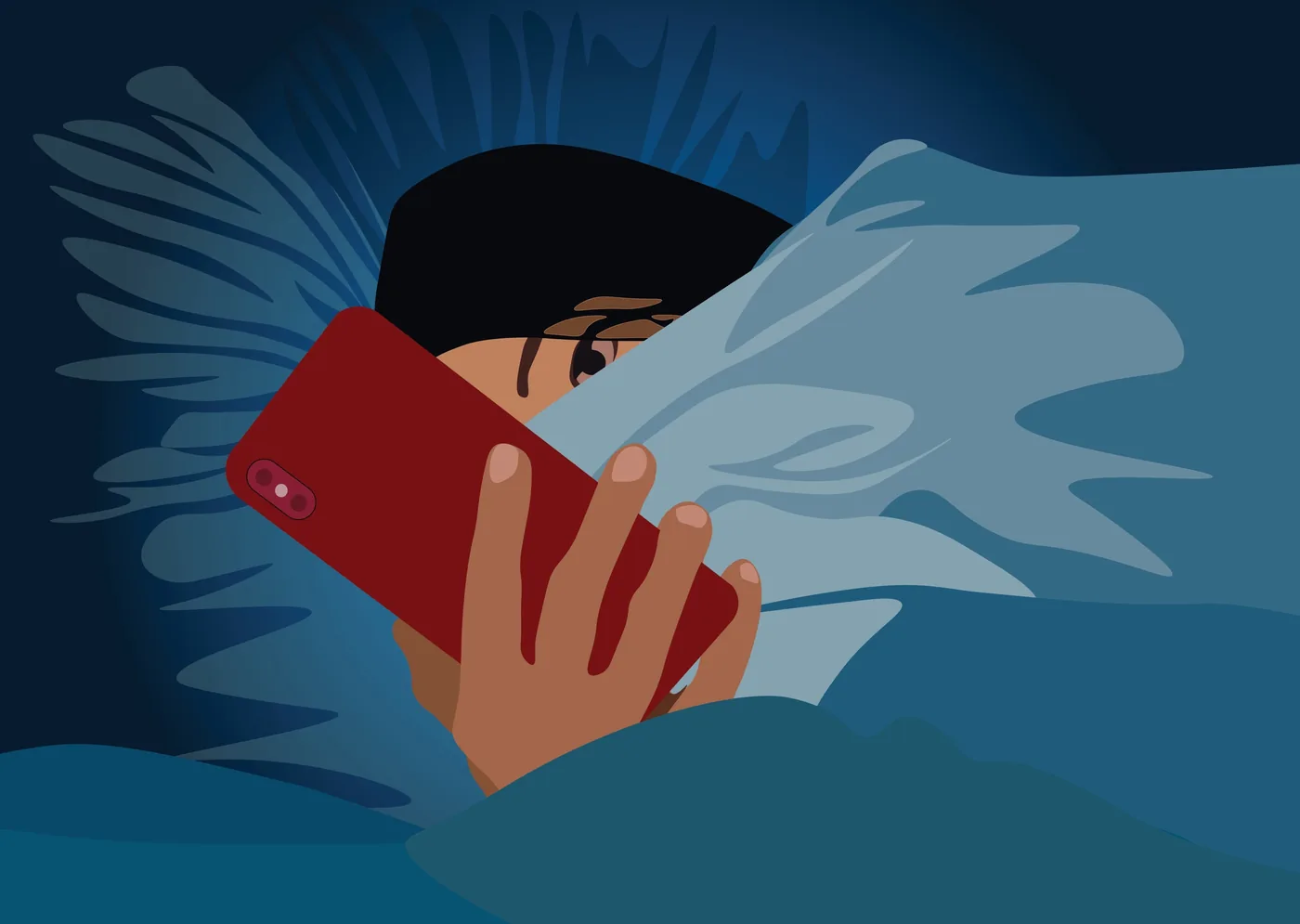
Nighttime light exposure means encountering light during the night, including illumination from streetlamps, televisions, phone screens and electronic equipment. Brightness can remain low enough that a room feels dark without reaching near-total darkness. For the Tulane investigation, participants wore wrist sensors for seven days, allowing the researchers to compare recorded light levels with later measurements of the heart. They measured personal exposure. Satellite maps were not the study’s exposure measure. [2, 3]
The team compared exposure above three lux with almost complete darkness. Lux measures light reaching a surface. The comparison concerns relatively dim conditions, extending beyond sleeping under a bright ceiling lamp, but three lux does not establish a boundary between safe and dangerous bedrooms. The reports do not identify a level below which cardiac effects disappear, or a brightness above which everyone develops them. [2, 3]
A wrist sensor records light at the wrist, while the eyes may receive a different amount depending on the person’s position and surroundings during the night. Nautilus identifies this mismatch as a possible source of either overestimation or underestimation. The instrument supplies useful data, but a week of wrist measurements cannot fully describe the light someone encounters over several years. [2]
What Did the MRI Reveal?
Qi’s colleagues examined cardiac structure and function using MRI, with Knowridge reporting scans about three years after the light measurements in the 11,071 participants drawn from UK Biobank. The paper lists four authors. J. Dai, W. Dai, Y. Heianza and L. Qi titled their study Nighttime Light Exposure and Cardiac Structure and Function. The central result concerns the left ventricle (the chamber that pumps oxygen-rich blood around the body): participants with greater nighttime light exposure tended to have thicker walls and less room inside. [1, 3]
The researchers also found signs of reduced muscle deformation, meaning that the heart muscle did not flex as well during each beat, and Knowridge reports differences in the right ventricle and left atrium too. Several chambers showed associations. However, Knowridge gives less detail about those additional findings than about the left ventricle. The Tulane analysis extends beyond an isolated wall measurement to several features of cardiac structure and performance. [3]
Nautilus describes the average thickening as less than 3 percent, a measurable difference that does not establish visible heart damage in everyone who sleeps near a light source. Existing cardiovascular problems may matter. Nautilus raises particular concern for people who already have them, without establishing how much additional risk the imaging finding represents for an individual. Researchers would need further evidence to connect the size of these anatomical differences with specific clinical outcomes. [2]
Why Does Heart Shape Matter?
Clinicians use cardiac remodeling (changes in heart shape and function) to describe adjustments that can accompany repeated stress on the heart. A chamber can change its wall thickness or internal volume as the muscle adapts to demands, with some adaptations initially helping it cope and others eventually contributing to stiffness or less efficient pumping. Knowridge places the Tulane results within this broader concept, while emphasizing that the study cannot show that a bedside lamp will cause heart failure. The researchers’ imaging findings concern differences in anatomy and performance, which are distinct from a diagnosis or a prediction of an individual’s future health. [3]
Heart failure was not an inevitable outcome of the MRI findings. The study reports associations with structural and functional measurements. [3]
Earlier observational research had already connected nighttime light with cardiovascular disease, according to Qi’s explanation quoted by Nautilus. What could imaging add? Qi said, “little is known about the related cardiac structure and functional changes.” By examining the heart itself, the Tulane team addresses that gap between environmental exposure and disease associations. The scans provide anatomical evidence for further investigation, but they do not establish which biological process produced the differences or whether reducing light would reverse them. [2, 3]


How Might Sleep Connect Them?
The body’s circadian rhythms (roughly 24-hour biological cycles) offer one possible connection between light and cardiovascular function. Light helps set the internal clock. That clock influences sleep, hormones, blood pressure and metabolism, so illumination during the normal rest period could affect several processes relevant to the heart at once. The reports describe this pathway as a biological explanation worth investigating; the MRI associations do not independently establish every step in it. [2, 3]
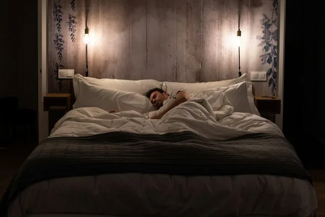
Qi’s team estimated that shorter sleep could statistically explain roughly 24 to 49 percent of the structural associations, according to Nautilus, which describes sleep durations below five or six hours. The sleep analysis does not mean that light directly caused that share of heart changes. Statistical explanations depend on the relationships measured in the participants. Nor does the remaining proportion identify a single additional mechanism; other exposures, health differences and measurement limitations may contribute. [2]
Sleep research also examines pathways involving stress, including the neurons associated with stress and poor sleep in mice. That is a separate investigation. Similarly, research on light-induced cellular responses asks questions at a different biological scale. Neither supplies a direct explanation for the UK Biobank result; the Tulane study links personal light measurements and human cardiac imaging without isolating a cellular pathway. [1, 3]
Can Light Explain the Differences?
Tulane’s researchers did not randomly assign participants to bright or dark bedrooms. They observed existing exposures. Night-shift work, neighborhood conditions, sleep habits, underlying illness and lifestyle may influence both the amount of light people encounter during the night and the condition of their cardiovascular system. Knowridge identifies these as possible alternative explanations, which matter because an association can persist even when the exposure is not the direct cause. A large cohort offers more observations without automatically removing those differences. [3]
The wrist measurements covered seven days. How well did that week represent the following years? Neither supplied report settles that question, and Nautilus explicitly warns that a short recording period may be a poor proxy for habitual exposure. A participant’s routine could differ from the measurement week; the study’s interpretation depends partly on whether those brief readings capture a meaningful longer-term pattern. The same concern applies when interpreting a later scan against an earlier environmental measurement. [2, 3]
The two news accounts describe the MRI schedule differently, so they do not support a confident claim here that repeated scans demonstrated progressive thickening within the same individuals. Knowridge describes imaging about three years after exposure measurement. The clearest shared result is an association between nighttime light exposure and cardiac measurements. Future work must establish whether changing the exposure changes the heart. [2, 3]
Which Nighttime Lights Need Attention?
The accompanying editorial, as described by Knowridge, suggests attention to unnecessary illumination: streetlight entering bedrooms, bright bedside lamps and lights on electronic equipment. Its proposals include blackout curtains, warmer and dimmer lighting, and covering unwanted equipment lights. Nautilus also discusses red alarm-clock displays and reducing phone use before bed. These suggestions address the nighttime environment. The study did not test whether a particular bulb color prevents cardiac remodeling, and the reports provide no validated color prescription for heart protection. [2, 3]
Cities enter the discussion too. Knowridge reports editorial proposals for shielded lamps, warmer bulbs and lighting that dims or activates only when needed, extending the question beyond individual bedrooms to shared outdoor environments. Those are proposals, not interventions evaluated in this cohort. Researchers still need to test whether reducing nighttime light exposure prevents or reverses the imaging differences, with measurements long enough to distinguish lasting changes from a single recorded week. [3]
- ACADEMIC JOURNAL Dai, J., Dai, W., Heianza, Y., & Qi, L. (2026). Nighttime light exposure and cardiac structure and function. European Heart Journal. [Article Link]
- ONLINE NEWS Currie, J. (2026, September 11). Nighttime light may change the shape of your heart. Nautilus. [Article Link]
- ONLINE NEWS Knowridge Science Report. (2026, September 11). Sleeping with light may change heart structure and function. [Article Link]
APA 7: PerEXP Teamworks. (2026, September 12). Nighttime light exposure linked to changes in heart shape. https://perexpteamworks.com/en/nighttime-light-exposure-heart-shape/


