





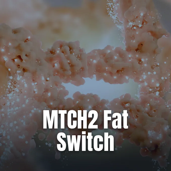
Biologists seeking better treatments for obesity face a persistent problem: weight-loss regimens frequently reduce lean muscle as well as fat. Researchers at the Weizmann Institute of Science in Israel have identified a cellular pathway that could change this metabolic equation. By silencing a gene regulator called the MTCH2 protein, nicknamed Mitch, researchers at the Weizmann Institute of Science in Israel prompted human cells to burn stored lipids and prevented precursor cells from forming new fat [1]. The intervention also stopped immature precursor cells from developing into new fat stores.
The Muscle Loss Dilemma in Obesity Care
Obesity affects roughly 40 percent of adults in the United States and drives cardiovascular disease and metabolic illness across industrial societies. Modern commercial GLP-1 medications like semaglutide and tirzepatide help patients reduce total body mass by mimicking glucagon-like peptide-1, an intestinal hormone that signals satiety directly to neural appetite centers in the brain. Pharmaceutical developers originally synthesized these therapeutic peptide compounds using biological insights gained from Exendin-4, an active peptide hormone first isolated by endocrinologists from the venom of the Southwestern Gila monster [4]. Brand-name drugs include Wegovy and Ozempic. Appetite suppression carries a biological trade-off. Because active skeletal muscle tissue expends energy continuously even when a patient rests, shedding substantial lean mass impairs physical strength, joint stability, and long-term metabolic health throughout life. When people lose muscle, maintaining a stable weight becomes far more difficult over time.
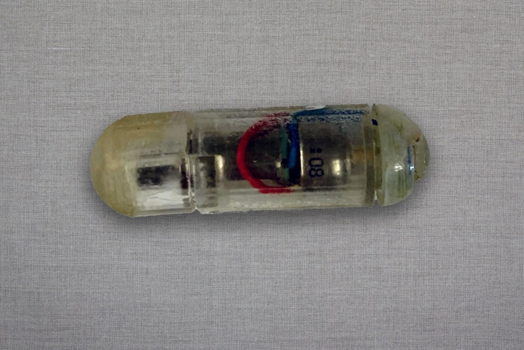
In 2026, clinical researchers reviewed comprehensive trial records from 20 separate pharmaceutical studies covering 15,782 participants across medical centers located throughout Europe and North America. Their statistical synthesis showed that lean tissue represented between 25% and 39% of all weight lost under GLP-1 treatments. That loss rate closely mirrored the muscle declines seen in standard calorie-restricted diets without exercise, although regular resistance training helped patients preserve more functional muscular tissue. Sarcopenia poses severe threats for older adults by increasing the incidence of physical frailty and falls. Biomedical engineers have developed innovative mechanical devices like a vibrating pill designed to fight obesity by mechanically stimulating gastric mechanoreceptors to induce early feelings of satiety. Discovering targeted biological mechanisms that selectively burn lipid deposits without sacrificing skeletal muscle remains the central therapeutic goal for clinical endocrinologists treating chronic metabolic disease worldwide [1, 6].
How MTCH2 Protein Influences Mitochondria
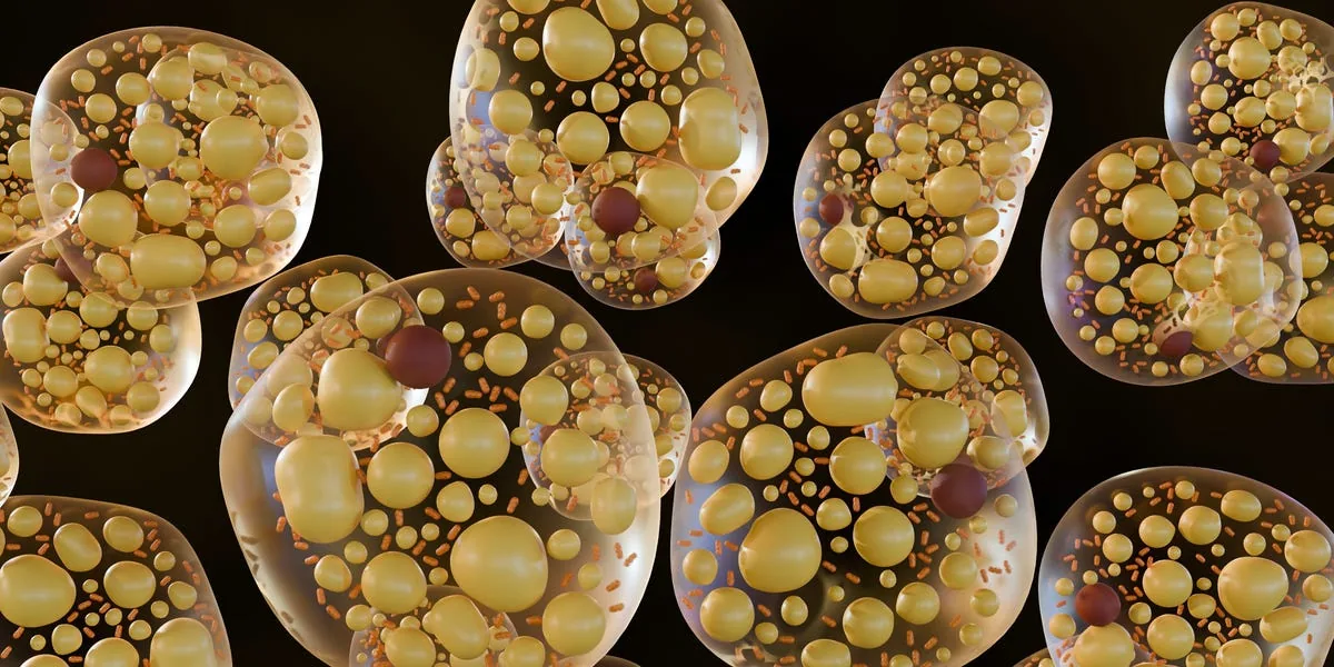
Mitochondria produce adenosine triphosphate, or ATP, the biochemical energy currency that fuels muscular movement and cellular growth. These organelles do not stay isolated inside human cells. Instead, they constantly fuse and split through a dynamic biological process known as mitochondrial dynamics. When individual mitochondria merge into extensive interconnected networks, they produce adenosine triphosphate with peak efficiency and consume smaller quantities of nutrient fuel to sustain basic cellular viability. Professor Atan Gross, Yehudit Zaltsman, and Limor Regev in the Weizmann Immunology and Regenerative Biology Department discovered that the MTCH2 protein directly coordinates this structural merging process. Without Mitch in the outer mitochondrial membrane, the organelle web fragments into tiny pieces that produce energy inefficiently [1, 2].
Inefficient mitochondria force an organism to consume greater quantities of fuel to survive. In 2016, Gross and his Weizmann team deleted MTCH2 from the skeletal muscle of laboratory mice. The modified mice ate standard quantities of high-fat chow. Yet they never became obese. Their muscles developed extra oxygen-rich endurance fibers. These fibers improved stamina during treadmill stress tests. The mice also demonstrated improved cardiac performance [1, 4]. Genetically altered mice lacking MTCH2 in skeletal muscle consumed normal amounts of high-fat food yet stayed lean because their fragmented mitochondria burned incoming calories at accelerated rates.

Accelerated Fuel Use in Human Cells
To determine whether human tissue behaves like mouse models, doctoral researcher Sabita Chourasia silenced Mitch in cultured human cells. Working with Christopher Petucci and Clarissa Shoffler at the University of Pennsylvania, alongside Hu Wang and Xianlin Han at the University of Texas at San Antonio, the researchers evaluated HeLa cultures. Chourasia and Gross tracked more than 100 metabolic substances with Alexander Brandis, Tevie Mehlman, Sergey Malitsky, Maxim Itkin, Ayala Sharp, and Ron Rotkopf to monitor energetic shifts [2]. The human cells accelerated cellular respiration to counteract a persistent ATP deficit. Chourasia said that oxygen consumption surged as the human cells rapidly broke down fatty acids, amino acids, and carbohydrates to compensate for low ATP levels. “This explains the increase in muscular endurance,” Chourasia said, connecting the cellular observations to earlier animal trials [1].
The altered human cells did more than speed up their metabolic engine; they completely restructured their choice of fuel. Normal cells rely primarily on carbohydrates and protein substrates. But human cells lacking MTCH2 protein turned toward stored fatty acids. Gross and his co-workers discovered that the cells broke down lipids inside their own cellular membranes to feed the energy-demanding mitochondria. Membrane lipids dropped sharply, whereas storage lipids temporarily increased. Detailed lipidomics confirmed that cells deprived of MTCH2 broke down membrane phospholipids to feed high mitochondrial respiration, creating a permanent catabolic state focused on burning fatty acids [1, 2]. “Mitch determines the fate of fat in human cells,” Gross said, describing how the protein controls lipid partitioning [1, 6].

Blocking the Formation of New Fat Cells
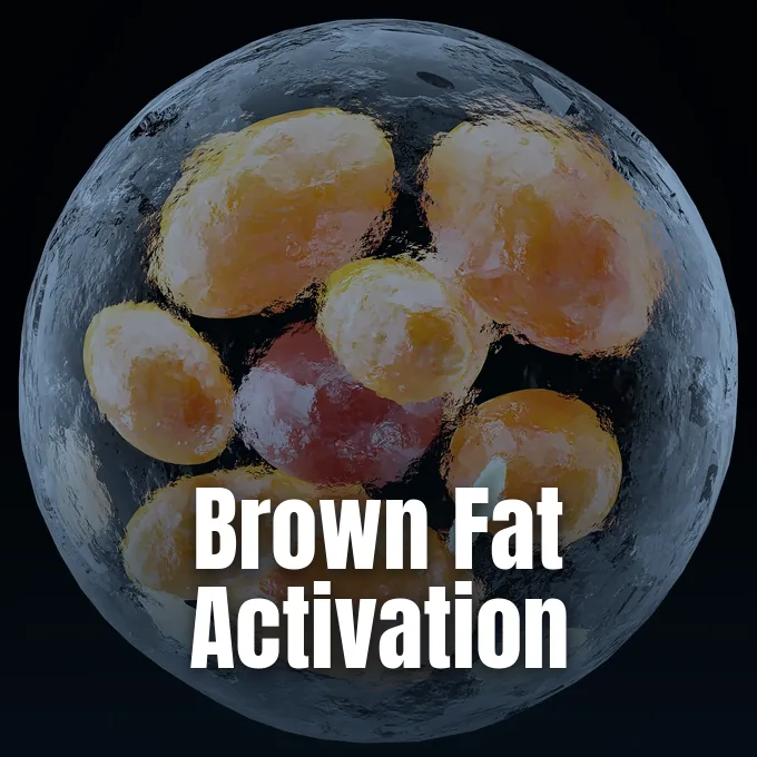
Earlier clinical studies observed that women with obesity possess higher baseline concentrations of MTCH2 within their subcutaneous fat tissue. That clinical clue led Gross, supported by the Marketa & Frederick Alexander Professorial Chair and philanthropist Amnon Shoham, to examine how the protein influences adipogenesis in progenitor cells. Adipogenesis is the biological process through which immature preadipocytes mature into specialized, lipid-storing adipocytes. Building a mature fat cell requires massive amounts of reductive energy to assemble fresh cellular membranes and accumulate large storage droplets of neutral lipid [1, 2].
Gross and Chourasia tested this differentiation pathway by deleting MTCH2 from mouse NIH3T3L1 preadipocytes. Under normal laboratory conditions, 80% to 90% of standard precursor cells mature into functional, fat-storing cells within six days. But when researchers knocked out MTCH2, only 5% to 10% completed that transition. The altered cells accumulated virtually no lipid droplets. Because the cells suffered from a chronic energy deficit, they could not generate the building blocks required for lipid storage. When researchers deleted MTCH2 from mouse NIH3T3L1 preadipocytes, only 5% to 10% matured into adipocytes, compared to 80% to 90% of normal control cells after six days [1, 5]. “There is a shortage of energy,” Gross said [1]. He said that suppressing Mitch silenced key adipogenic genes [5].

Regulation of CPT1 by MTCH2 Protein
In October 2025, an international research team published follow-up findings in Nature Communications that detailed the precise molecular machinery of Mitch. An international consortium led by C. Wu and M. Blüher analyzed human and murine adipose tissue samples to identify regulators governing fatty acid entry into the mitochondrial matrix. Working with Gross and Chourasia, the researchers screened mitochondrial outer membrane proteins to understand lipid oxidation. Their screen identified MTCH2 as a direct regulator of carnitine palmitoyltransferase 1, or CPT1. CPT1 serves as the main enzymatic gatekeeper controlling the influx of free fatty acids into mitochondria for energy extraction [3].
In healthy cells, a metabolic substance called malonyl-CoA binds CPT1 to shut off fat oxidation when ample fuel exists. Wu and Blüher showed that MTCH2 binds directly to CPT1. This physical contact sensitizes CPT1 to malonyl-CoA signals, keeping fat burning tightly restricted. When MTCH2 is deleted, CPT1 loses that sensitivity. Fatty acids then enter mitochondria freely, accelerating cellular respiration. This heightened respiratory activity occurs independently of uncoupling protein 1, the protein responsible for non-shivering heat generation during brown fat activation in cold thermal environments [3]. Mitch acts as a biochemical brake on fat consumption.

Challenges Facing Cellular Metabolic Therapies
Discovering a cellular switch that burns fat while blocking new adipocyte development offers fresh hope, but clinical therapies remain far away. The adipogenesis experiments relied on mouse cell lines, and the metabolic data came from cultured human cells rather than living patients. Health journalism vertical First Check, an initiative of DataLEADS, pointed out that systemic inhibition of mitochondrial carrier proteins could induce severe off-target toxicities in cardiac or neural tissues [5]. Mitochondria generate energy for every major organ, including the heart and central nervous system. Indiscriminate, whole-body suppression of MTCH2 could trigger toxicities in healthy tissues [6, 7].
In our evolutionary past, the MTCH2 protein protected early humans by conserving calories during periods of famine and starvation. In the 21st century, with abundant processed food and sedentary desk jobs, that energy-saving mechanism drives chronic obesity [4]. To make Mitch a viable drug candidate, medicinal chemists must design selective small molecules that target adipose depots or skeletal muscles without disturbing energy metabolism in the heart. Such selective treatments could work with appetite suppressants, helping patients eliminate stubborn fat while safeguarding healthy muscle mass [1, 6].
- ONLINE NEWS ScienceDaily. (2026, October 24). Scientists discover a hidden switch that burns fat and blocks new fat cells. [Article Link]
- ACADEMIC JOURNAL Chourasia, S., Petucci, C., Shoffler, C., Abbasian, D., Wang, H., Han, X., Sivan, E., Brandis, A., Mehlman, T., Malitsky, S., Itkin, M., Sharp, A., Rotkopf, R., Dassa, B., Regev, L., Zaltsman, Y., & Gross, A. (2025). MTCH2 controls energy demand and expenditure to fuel anabolism during adipogenesis. The EMBO Journal, 44(4). [Article Link]
- ACADEMIC JOURNAL Wu, C., Wang, T., Ghosh, A., Long, F., Sharma, A. K., Dahlby, T., Noé, F., Severi, I., Colleluori, G., Cinti, S., Giordano, A., Ding, L., Khandelwal, R., Kostidis, S., Giera, M., Balazova, L., Gardeux, V., Abu-Nawwas, L., Deplancke, B.,. Blüher, M. (2025). MTCH2 modulates CPT1 activity to regulate lipid metabolism of adipocytes. Nature Communications, 16(1). [Article Link]
- ONLINE NEWS Orf, D. (2026, July 17). Scientists Just Found a Secret Switch That Turns Off Fat Storage—And Turns On Fat Burning. Popular Mechanics. [Article Link]
- ONLINE NEWS Gupta, A. (2026, July 8). Scientists find the ‘off switch’ that could stop your body from storing fat. THE WEEK Magazine. [Article Link]
- WEBSITE VitaBasix. (2026, July 8). Scientists Discover a Protein Switch That Burns Fat and Prevents the Formation of New Fat Cells. [Article Link]
- ONLINE NEWS Patrick, G. (2026, July 3). Fat-Burning Protein Called ‘Mitch’ Could Transform Future Weight Loss Treatments. Science Times. [Article Link]
APA 7: PerEXP Teamworks. (2026, October 25). Weizmann Scientists Disable MTCH2 Protein to Boost Fat Burning. PerEXP Teamworks. https://perexpteamworks.com/en/mtch2-protein-fat-burning-switch/


1 comment