




Tau protein misfolding has long mirrored the destructive progression of fatal prion disorders, sparking intense debate over whether common dementias share identical molecular propagation mechanics. In a landmark investigation published in Nature, researchers demonstrated that prion-like transmission of human tau strains occurs directly inside living wild-type mice without requiring artificial genetic manipulation [1]. Structural biologist Sjors Scheres and neuroscientist Michel Goedert at the MRC Laboratory of Molecular Biology observed that ordinary rodent tau copied the exact atomic folds of introduced human assemblies, proving that structural strains faithfully self-propagate through templated seeding [2].
- How Tau Folds Drive Prion-Like Transmission
- Can Tau Exhibit Prion-Like Spreading in Mice?
- Cryo-EM Resolves Exact Folds of Tau Seeds
- Cellular Tropism Separates Alzheimer’s from Corticobasal Degeneration
- Does Human to Human Prion Transmission Apply to Tau?
- Translational Frontiers in Blocking Tau Strain Transmission
How Tau Folds Drive Prion-Like Transmission
Prions represent one of the most destructive phenomena in biology, arising when normally folded peptides twist into aberrant, self-replicating conformations [2]. When a pathological template contacts a native monomer, it forces that healthy molecule into its own corrupted architecture, establishing a self-sustaining cascade known formally as templated seeding [1]. ScienceAlert science writer Michelle Starr noted that prions behave like snaggled cogs in complex machinery, where a damaged tooth systematically deforms adjacent gears across healthy neural tissue [2]. Mounting evidence suggests similar seeding mechanics in Parkinson’s disease and diverse tauopathies, including Alzheimer Disease, Pick Disease, and Corticobasal Degeneration [1]. Medical Research Council investigators confirmed that distinct folds produce unique clinical strains.
Under ordinary physiological conditions, tau protein operates as a vital structural component within the Central Nervous System. It stabilizes internal microtubules, supports nutrient transport along elongated axons, and facilitates the conversion of short-term memories into enduring storage [2]. In pathological states, hyperphosphorylated tau detaches from cellular scaffolding and coalesces into insoluble intracellular fibrils. The prion-like transmission of these abnormal assemblies enables pathological seeds to recruit disordered monomeric tau into growing filaments. Scheres verified identical core folds [1]. Prior to this MRC Laboratory investigation, structural biologists lacked definitive proof that host aggregates copied the exact molecular geometry of donor seeds [2].

Can Tau Exhibit Prion-Like Spreading in Mice?
Extracted human tau seeds trigger progressive, prion-like spreading throughout recipient mouse brains without requiring foreign transgenic proteins [1]. Structural biologist Sjors Scheres and neuroscientist Michel Goedert led an international team at the MRC Laboratory of Molecular Biology in Cambridge in the UK to resolve this transmission puzzle [2]. Working with collaborators Sofia Lövestam, Aki Shimozawa, Airi Tarutani, Reiko Ohtani, and Kazuko Hasegawa, the researchers harvested filamentous tau aggregates from Frontal Cortex autopsy tissue donated by patients diagnosed with Alzheimer Disease or Corticobasal Degeneration. They stereotaxically injected these human extracts into the striatum of Wild Type mice aged 6 to 18 weeks [1]. Unlike earlier studies relying on animals engineered to overexpress human mutations, these experimental subjects possessed only native murine tau [2].
Tracking experiments established that the injected human seeds disappeared rapidly from recipient brain tissue. Human seeds cleared within 7 days. Staining with the human-specific HT7 antibody became completely negative after one week, ensuring that downstream aggregates could not be attributed to lingering inocula. Monitored at 1, 3, 6, and 9 months post-injection, endogenous murine tau developed extensive insoluble inclusions that spread outward from the striatal injection site into the Cerebral Cortex and Corpus Callosum. This sustained progression confirmed active prion-like transmission across interconnected circuits. By connecting these cellular pathways with wider research into elaborate brain cell maps, investigators traced how regional anatomical connectivity dictates neuroanatomical vulnerability [1].
Cryo-EM Resolves Exact Folds of Tau Seeds
Cryo-electron microscopy confirmed that endogenous mouse filaments copied the three-dimensional atomic structure of human disease seeds with near-perfect fidelity. In mice inoculated with Alzheimer Disease preparations, 83% of extracted filaments formed Paired Helical Filaments. The researchers calculated a high-resolution reconstruction at 3.6 Å resolution, demonstrating that the mouse fold matched the human Alzheimer core with a root mean square deviation of 0.906 Å across all non-hydrogen atoms. The remaining 17% displayed 2D class averages matching Straight Filaments or single protofilaments. Cryo-EM reconstructions resolved 3.6 Å details [1]. The molecular resemblance was extraordinary, proving that native murine peptides folded into precise copies of human pathological conformers during prion-like transmission [2].
Extracts from animals receiving Corticobasal Degeneration seeds revealed a distinct structural distribution comprising 74% single protofilaments and 26% double protofilaments. The team reconstructed the singlets to 3.4 Å resolution, revealing an atomic fold identical to human type I CBD filaments with an RMSD of just 0.576 Å, while the doublets reconstructed to 10 Å consistent with type II arrangements. Advanced image processing workflows for resolving amyloid filaments in cryo-EM were developed using RELION-5.1 software, an algorithmic protocol described by Sofia Lövestam and Sjors Scheres in a methodological preprint that has not undergone peer review. Sofia Lövestam confirmed structural match. This atomic confirmation established that distinct tau folds act as genuine conformational templates in mammalian brains [1].

Cellular Tropism Separates Alzheimer’s from Corticobasal Degeneration
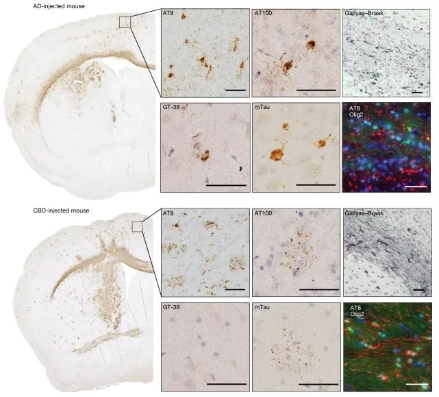
Inoculation with distinct human tau strains produced radically divergent neuropathological lesion profiles in the recipient brain tissue. Mice injected with Alzheimer Disease tau accumulated hyperphosphorylated inclusions confined strictly to neuronal cell bodies and neuropil threads. In sharp contrast, brains receiving Corticobasal Degeneration tau developed extensive pathology across both nerve cells and surrounding glial cells. Collaborators Andrew Robinson, Yuko Saito, Shigeo Murayama, and Mari Yoshida noted that these CBD-injected animals displayed abundant oligodendrocytic coiled bodies and astrocytic plaque-like inclusions identical to hallmark lesions documented in human clinical cases [1]. Strain identity dictates cellular tropism directly in vivo, demonstrating that the physical fold itself governs which cell types become vulnerable to prion-like transmission [2].
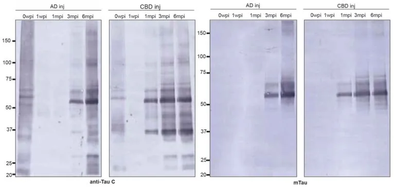
Western blots revealed a 37 kDa band. Tau filaments from Corticobasal Degeneration brains also exhibited significantly longer helical cross-over distances than Alzheimer Disease fibrils [1].
Biochemical discrimination was further confirmed through epitope accessibility testing with the GT-38 antibody. In adult humans, alternative splicing of the MAPT transcript generates six tau isoforms divided evenly into three-repeat (3R) and four-repeat (4R) variants. Adult mice express exclusively 4R tau. The GT-38 antibody specifically recognizes an epitope inside the second Microtubule Binding Repeat (R2), which is naturally absent in 3R isoforms. Tissue from Alzheimer-seeded mice bound GT-38 robustly because R2 remains partially accessible on the filament exterior. In CBD tau filaments, R2 is buried within the tightly packed core, rendering the antibody completely unable to bind [1].
Does Human to Human Prion Transmission Apply to Tau?
Human to human prion transmission does not occur in Alzheimer Disease or Corticobasal Degeneration under ordinary clinical or everyday conditions [2]. Genuine prion diseases such as kuru, Creutzfeldt-Jakob disease, scrapie, Chronic Wasting Disease, and Bovine Spongiform Encephalopathy are notorious for their infectious transmission across individuals or between species [1]. In sharp contrast, common human tauopathies show no epidemiological signature of contagious transmission. Although misfolded tau proteins clearly spread through templated molecular seeding from cell to cell inside an individual brain, they lack the environmental stability and infectivity required to transmit spontaneously between people. Tau aggregates behave like prions within brain tissue, yet they do not constitute infectious pathogens in social environments [2].
The experimental animals exhibited another unexpected clinical outcome during the nine-month monitoring timeline. The subjects showed no motor decline. Despite accumulating dense phosphorylated tau assemblies across cortical layers, the rodents failed to develop noticeable behavioral impairments or motor decline. ScienceAlert writer Michelle Starr emphasized that researchers cannot yet distinguish whether the mice did not live long enough for overt neurodegeneration to manifest, whether accumulated pathology remained below a critical threshold, or whether filament propagation and tissue destruction constitute functionally dissociated disease stages. Rebecca Dyer, who fact-checked the ScienceAlert analysis, verified that the asymptomatic presentation highlights ongoing questions regarding how structural assemblies translate into symptomatic dementia [2].
Translational Frontiers in Blocking Tau Strain Transmission
Demonstrating that Wild Type mice faithfully replicate human amyloid folds provides researchers with a powerful physiological platform for preclinical therapeutics. Previous drug discovery pipelines frequently depended on transgenic overexpression strains whose aberrant filament architectures failed to mirror human autopsy specimens. By revealing that endogenous murine proteins replicate human strain geometry without genetic tampering, the MRC Laboratory study validates ordinary rodents as tractable biological testbeds. While parallel initiatives develop rodent models carrying human neural cells to mimic developmental disorders, wild-type seeding allows direct evaluation of how native mammalian cells process human seeds [1].
Unraveling the molecular checkpoints of prion-like transmission offers immediate targets for disease-modifying medicine. Future investigations seek to determine how cellular membranes internalize seed filaments, which cytoplasmic chaperones facilitate conformational conversion, and why specific folds selectively invade neurons or glia. Whereas earlier therapeutic paradigms struggled with the therapeutic targeting of neurodegenerative proteins due to their intrinsically disordered structures, locking onto strain-specific conformations offers unprecedented precision. Monoclonal antibodies designed to shield unique interfacial epitopes could halt spreading before widespread cortical damage takes root [1]. Preventing seed propagation before irreversible neuronal loss occurs represents a vital objective in modern translational neuropathology [2].
Resolving whether structural differences explain clinical variability among diverse tauopathies remains an urgent objective for molecular medicine. Co-authors Hisaomi Suzuki, Mitsumoto Onaya, and Masato Hasegawa emphasized that identifying strain-specific conformational barriers will clarify why different tauopathies progress along distinct anatomical trajectories [1]. Clarifying these kinetic barriers will ultimately establish whether interrupting prion-like transmission can halt disease progression in human patients [2].
- ACADEMIC JOURNAL Lövestam, S., Shimozawa, A., Tarutani, A., Ohtani, R., Masuda-Suzukake, M., Hasegawa, K., Robinson, A. C., Saito, Y., Murayama, S., Yoshida, M., Suzuki, H., Onaya, M., Hasegawa, M., Goedert, M., & Scheres, S. H. W. (2026). Prion-like transmission of human tau strains in the mouse brain. Nature. [Article Link]
- ONLINE NEWS Starr, M. (2026, September 30). Scientists watched human dementia proteins spread through mouse brains like prions. ScienceAlert. [Article Link]
APA 7: TWs Editor. (2026, October 1). How Human Tau Strains Drive Prion-Like Transmission in Mice. PerEXP Teamworks.


