





In a significant neurological advance reported in Nature, researchers have engineered mice with human brains by replacing missing rodent neural structures with expanding human cellular grafts. Can living animal hosts finally allow scientists to inspect human psychiatric disorders that laboratory glassware cannot replicate? The resulting xenocortical mice (animals carrying human-derived cortical tissue) developed functional connections with host circuitry. Stanford University psychiatrist Sergiu Pașca announced in reports covered by The Guardian that transplanted cellular structures expanded until human cells occupied roughly half the rodent brain volume [2].
Why Create Mice with Human Brains?
Neurology and psychiatry have historically lagged behind oncology and cardiology in discovering curative treatments. Stanford University researcher Sergiu Pașca pointed out that therapies for psychiatric conditions remain scarce because human brain tissue is largely inaccessible for direct experimental manipulation during development. While other medical fields examine living biopsies or target well-defined peripheral tissues, neuroscientists cannot easily sample healthy developing human brains. The search for viable alternatives led scientists to cultivate miniature three-dimensional tissues known as neural organoids (clusters of stem cells that self-assemble into brain-like structures) to study cellular specialization. These lab-grown clumps provide accessible cellular platforms. Yet isolated dishes lack blood vessels, sensory inputs, and the complex physiological cues that guide normal biological maturation [3].
The underlying technology began taking shape during the early 2010s when researchers in Austria demonstrated that pluripotent stem cells could self-organize into multi-layered cerebral structures [3]. **Translating dish-based organoids into whole animals became the logical next phase for neurobiology.** Earlier comparative studies had already begun cataloguing structural variations across mammalian species, detailed in earlier work that developed elaborate brain cell maps for human and nonhuman primates. Stanford University scientists noted in PerEXP Teamworks coverage that moving from glass dishes into living rodents transforms static cell colonies into dynamic functional networks [2].
By grafting human tissue into rodent hosts, researchers sought to create faithful living testbeds for disorders such as schizophrenia, epilepsy, and intellectual disability [2]. Many fundamental questions about developmental brain disorders cannot be answered through computer simulations or static cellular assays alone. Why is mouse biology so frequently used as the starting canvas? Rodent models share conserved mammalian genetic pathways, offering a compressed temporal window for observation [3].
Genetics Behind the Xenocortical Model
In 2022, Sergiu Pașca and his colleagues at Stanford University took an initial step by transplanting human neural organoids into newborn rats. The grafted human neurons successfully survived and formed functional synapses with host circuitry. However, rat brain tissue grew so rapidly that it constrained the expanding human graft to only one-third of a single cortical hemisphere. **The physical skull of the animal left inadequate room for substantial tissue growth.** Furthermore, rat neurons mature on a much faster biological timetable than human neurons, preventing the transplanted human tissue from reaching more advanced stages of functional and structural maturation [3].
To overcome spatial limitations, the Stanford University team led by Sergiu Pașca engineered genetically modified mice born without a cerebral cortex or hippocampus. These two brain structures govern sensory processing, voluntary movement, and memory storage, comprising roughly half of the rodent brain volume. Under normal biological circumstances, such extensive congenital cranial deficits would be immediately fatal, yet the genetically altered rodents survived and adapted to their missing brain regions. The animals survived [2]. Remaining subcortical structures adapted to take over essential autonomic tasks, leaving a cavernous cranial space ready to host donor cells [3].
The resulting biological vacancy transformed the skull into an open vessel. In total, each genetically altered rodent lacked about 14m mouse brain cells, opening physical space for human cellular colonization in mice with human brains [2]. Sergiu Pașca observed that the animals functioned despite substantial congenital brain differences. **Genetically silencing endogenous cortical expansion created the biological niche required for xenocortical integration.** The procedure established a live physiological scaffold where human neural precursors could expand, migrate, and develop without encountering mechanical compression or physical resistance from surrounding host tissue [1].
How Donor Organoids Expand Inside Rodents
The transplantation protocol developed at Stanford University relied on human organoids generated by reprogramming donated human skin cells into induced pluripotent stem cells. After cultivating the stem cells into three-dimensional neural tissue, researchers delivered several microinjections into the empty cranial cavity of newborn pups. Each injection contained approximately 100,000 human brain cells suspended in specialized nutrient medium [2]. Across the experimental cohort of neonatal rodents, the delicate surgical transplantation took hold successfully in 25 of 29 attempts, confirming that early developmental timing was critical for graft survival [3].
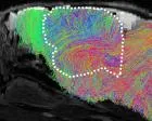
Over the following three months, the grafted human tissue expanded nearly fivefold in volume inside the rodents’ skulls [3]. Host blood vessels penetrated the human neural clusters, establishing an active circulatory supply that nourished deep cellular layers. **The expanding human tissue ultimately occupied more than 90 percent of the cortical cavity, yielding roughly 4m human neurons in each animal.** Writing in Nature, Sergiu Pașca explained that the expanding human cells wired extensively into host subcortical regions and extended functional axonal projections reaching deep toward the mouse spinal cord [1].
Despite this dramatic volume growth, the resulting brain was not an identical miniature copy of an adult human cortex. The human neural tissue remained developmentally immature, resembling the cerebral structure found halfway through human pregnancy [2]. Furthermore, the human cells did not organize into the strict six-layered anatomical laminate characteristic of a mature human cortex. The technique was bold [3]. Microscopic examination confirmed that human neurons developed elaborate dendritic branches and established synaptic contacts with mouse tissue, demonstrating functional integration into an alien mammalian architecture without losing human identity [1].


Modeling Cerebral Palsy and Neurological Damage
To demonstrate that mice with human brains can model clinical disease, researchers investigated cerebral palsy (a motor disability often triggered by perinatal oxygen deprivation) [2]. The Stanford University team subjected grafted animals to five hours of low oxygen, mimicking hypoxic birth complications. **Low oxygen exposure produced cellular injury across the human graft, mirroring perinatal complications.** Afterward, microscopic analysis revealed acute cellular injury across the human graft, mirroring neuropathological changes seen in human infants [3]. The damage confirmed that grafted human neurons respond to systemic insults with their own distinct biological vulnerability [1].
Following the hypoxic exposure, the grafted rodents developed noticeable defects in gait and limb coordination [3]. Sergiu Pașca explained that the physical symptoms directly resembled the motor impairments seen in human clinical presentations of cerebral palsy. Because dish-grown cell cultures cannot walk, run, or demonstrate physical reflex deficits, observing living animals allowed the team to evaluate directly how cellular damage translates into physical disability. Similar methodologies have expanded into neurovascular investigations, complementing earlier rodent studies examining how mice reveal the neurons that cause stress and poor sleep. Oxygen deprivation proved decisive [2].
Beyond hypoxic injury, the xenocortical model yielded rare cellular discoveries in Stanford University laboratories. Within the expanding human grafts, researchers identified von Economo neurons (spindle-shaped nerve cells previously observed almost exclusively in postmortem brain tissue) [2]. In humans, these specialized cells are among the earliest to degenerate in frontotemporal dementia, an aggressive neurodegenerative condition that degrades language and social behavior. Because von Economo neurons have proved nearly impossible to generate and maintain in standard culture dishes, the living rodent model provides an unprecedented platform for testing candidate dementia therapeutics [3].

What Behavioral Tests Reveal About Enhancement
A central question raised by observers is whether introducing human brain tissue confers augmented intelligence or unnatural cognitive enhancement. Stanford University researchers placed the chimera rodents through a battery of standardized behavioral assays testing fine motor coordination, spatial navigation, and short-term memory [3]. Animals lacking a cortex and hippocampus displayed severe unsteadiness, trembling gaits, and profound memory impairments. **Transplanting human brain organoids partially rescued these deficits without producing cognitive super-abilities.** The grafted mice exhibited noticeable improvements in limb stability and navigational performance, yet their scores consistently remained below those of healthy rodents with intact native brains during all testing phases [2].
Bioethicist Insoo Hyun, director at the National University of Singapore Yong Loo Lin School of Medicine, reviewed the behavioral outcomes carefully. Hyun observed that ethical boundaries fundamentally depend on whether foreign tissue elevates cognitive capacity beyond natural biological baselines. On motor coordination tasks and spatial memory challenges, performance fell squarely into an intermediate zone between completely decorticated animals and healthy control animals with fully intact native brains. Insoo Hyun pointed out that secular bioethics focuses on functional capability rather than tissue origin. The animals showed no evidence of human-like self-awareness or enhanced reasoning [3].
The mice remained forgetful [2]. Pop-culture fears of hyper-intelligent rodents proved groundless; Sergiu Pașca’s animals remain far more Pinky than Brain in Animaniacs lore. World domination was absent [3].
Where Bioethicists Draw the Line
Despite the absence of heightened cognitive abilities, creating mice with human brains sparks significant philosophical and regulatory debate. Legal and bioethical authorities emphasize that animal welfare must remain the central operational consideration as neural transplants become more sophisticated. Emily Jackson, a professor of law at the London School of Economics and chair of a report on neural organoids for the Nuffield Council on Bioethics, emphasized the necessity of persistent animal welfare monitoring. Emily Jackson stressed that regulatory bodies must closely monitor transplanted rodents to detect hidden distress throughout their lifespan [2].
Academic perspectives within the neurobiology community differ regarding the necessity of whole-animal chimera models. Madeline Lancaster, a group leader at the MRC Laboratory of Molecular Biology in Cambridge, expressed reservations about whether xenocortical animals accurately reflect natural human neurodevelopment. Lancaster pointed out that because transplanted tissue develops within a highly artificial foreign environment, it cannot fully replicate authentic embryonic growth. She argued that international laboratories should prioritize fully in vitro alternatives (petri dish cultures without living animal hosts) to minimize animal experimentation while improving culture conditions to support tissue maturation [2].
At the University of Pennsylvania, neurosurgeon Isaac Chen praised Sergiu Pașca for actively initiating ethical oversight rather than treating bioethics as an afterthought. **Ethical frameworks must evolve in parallel with experimental capabilities as cross-species neural grafting expands.** H. Isaac Chen noted that transplanting human brain organoids into rodents raises legitimate boundaries that require transparent international oversight [3]. As researchers prepare to model frontotemporal dementia and autism using patient-derived cells, continuing scientific scrutiny will ensure that therapeutic progress proceeds with strict ethical safeguards, opening new medical possibilities without transgressing humane standards [1].
- ACADEMIC JOURNAL Kaganovsky, K., Kelley, K. W., Gschwind, T., Harary, P. M., Kochalka, J., White, A. D., Lerma-Usabiaga, G., Chen, X., Revah, O., Gore, F., Aoyama, A., Shadrach, J. L., Yoon, S., Valencia, A., Ogawa, S., Reis, N., Vogel, H., Wandell, B., Kaltschmidt, J. A.,. Pașca, S. P. (2026). Developmental xenocortication using human-derived organoids in mice. Nature. [Article Link]
- ONLINE NEWS Sample, I. (2026, September 16). Scientists create mice with part-human brains. The Guardian. [Article Link]
- ONLINE NEWS Dolgin, E. (2026, September 16). These mice have human (nerve cells) on the brain. Science News Magazine. [Article Link]
APA 7: TWs Editor. (2026, September 17). Growing mice with human brains to model rare disorders. PerEXP Teamworks. https://perexpteamworks.com/en/mice-with-human-brains/


