





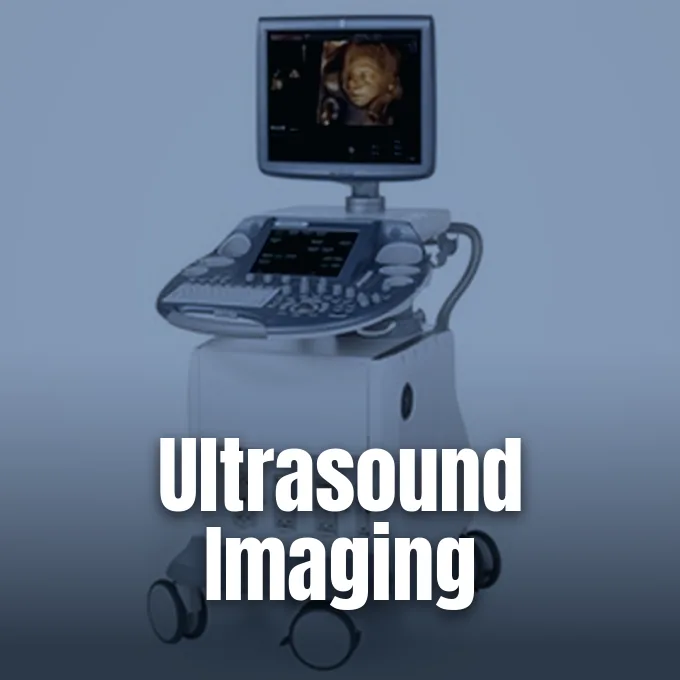
Ultrasound imaging maps body organs and blood flow by sending high-frequency sound waves into soft tissue and recording returning echoes. These sound pulses vibrate at frequencies greater than 20,000 Hz, which sit far above the hearing range of the human ear. Without exposing patients to ionizing radiation or requiring surgical cuts, sonography delivers real-time clinical pictures. Doctors place small handheld probes directly against lubricated skin to check heart valves, monitor fetal growth during pregnancy, and trace blood clots inside deep veins during routine hospital visits. It works without invasive needles [9].
Acoustic Principles in Medical Sonography
Acoustic physics began when Swiss researcher Jean-Daniel Colladon measured sound speed in Lake Geneva in 1826, while Lord Rayleigh formulated sound equations in England in 1877 [7]. Sound waves move through human tissue as longitudinal pressure waves that squeeze and release cellular material along their propagation path. When a transmitted acoustic pulse encounters the boundary between two biological structures with differing acoustic impedance values, a measurable fraction of the pressure wave reflects back to the probe while the remaining acoustic energy travels deeper into underlying tissues. Scanners compute tissue depth by using an assumed sound speed of 1540 meters per second [9].
How do sound pulses turn into electric signals? The answer lies in the piezoelectric effect discovered by Pierre Curie and Jacques Curie in Paris in 1880. They observed that pressing quartz crystals or Rochelle salt produced an electric charge on the crystal face. Gabriel Lippman deduced the reverse reaction in 1881, confirming through thermodynamic principles that applying an alternating voltage to quartz or Rochelle salt crystals causes them to expand and contract to produce high-pitched sound waves [7]. Modern ultrasound probes use engineered ceramic discs that send acoustic pulses into tissue and convert returning echoes into voltage spikes [2]. Sound echoes bounce off organs within microseconds [9].
Diagnostic ultrasound imaging relies on these returning acoustic echoes to build clinical images. Soft organs differ slightly in density, so sonograms show echo travel times rather than true geometric slices. Air scatters sound waves. For this reason, sonographers apply water-based acoustic gel to patient skin to seal out air gaps before scanning [9].
How Transducers Form Sound Beams
A modern probe houses an array of tiny crystal elements that fire in timed patterns controlled by digital beamformers. J. Powers and F. W. Kremkau reported in Interface Focus that by delaying electric pulses sent to individual crystal elements, the scanner steers the sound beam and focuses acoustic energy at chosen depths inside the patient [2]. Shallow targets like thyroid tissue, testes, and breasts need high frequencies between 7 and 18 MHz for sharp detail, whereas deeper organs like the liver and kidneys require 1 to 6 MHz for sound to penetrate dense tissue without losing signal strength [9].
Most routine exams use curved or flat probes placed on lubricated skin. When skin exams lack clarity, doctors turn to internal probes like transvaginal, endorectal, and transesophageal devices. These specialized tools place crystal arrays next to target organs [9].
Cardiologists also thread miniaturized IVUS catheters inside coronary arteries to inspect plaque buildup on vessel walls [9]. Matrix array probes use two-dimensional grids of crystal elements to steer beams across a pyramid-shaped volume rather than a single flat slice. This design captures broad tissue areas [11]. Systems also blend frequencies to smooth out grain and sharpen organ borders [2].
Primary Modes in Ultrasound Imaging
Clinicians choose distinct scanning modes based on whether they need single depth readings, flat organ slices, or dynamic heart valve tracings during ultrasound imaging. Scanners rely on three classic display formats known as A-mode, B-mode, and M-mode. In A-mode, or amplitude mode, returning echoes produce vertical spikes along a baseline, where spike height indicates echo strength [9]. Neurologist Karl Theo Dussik and his brother Friederich Dussik tested early one-dimensional transmission at the University of Vienna during the 1940s [7]. Today, A-mode assists specialized eye tests and precise distance checks [9].
B-mode, or brightness mode, serves as the workhorse of clinical sonography by building two-dimensional grey-scale slices of living tissue. In this mode, an array of transducer elements fires sequential sound beams through a body plane, converting the voltage amplitude of each returning echo into a bright dot on a digital monitor. M-mode, or motion mode, fires rapid pulses along a single acoustic line to record moving tissue boundaries over time. This fast recording behaves like a streak video recording in high-speed cameras, allowing cardiologists to measure heart valve opening velocity and track ventricle wall contraction throughout the cardiac cycle [9].
Harmonic imaging filters echoes to isolate signals that vibrate at twice the transmitted frequency, wiping out surface clutter to improve image clarity. B-flow displays flowing red blood cells while erasing still tissue. It works without Doppler color bleed [9].
Doppler Measurement of Blood Flow
Medical scanners assess vascular health by using the Doppler effect, which shifts returning sound frequencies when acoustic waves bounce off moving red blood cells traveling through arteries and veins. Duplex sonography merges two-dimensional B-mode anatomical pictures with spectral Doppler traces to show vessel structure alongside numeric blood flow velocities. Power Doppler measures the total energy of backscattered acoustic waves from moving erythrocytes, giving doctors high sensitivity to detect slow flow in tiny vessels even though it provides no velocity direction. These tools spot leg clots and carotid artery narrowing [9].
Functional ultrasound imaging, or fUS, applies Doppler methods to track brain activity by monitoring tiny changes in cerebral blood volume. When brain neurons fire, nearby blood flow surges through a vascular process called neurovascular coupling. The response begins within 0.3 seconds [10].
Sensory-evoked cerebral blood volume peaks around 1 second after neural stimulation, giving neuroscientists an accurate blood-based readout of brain activity. Researcher Mickael Tanter and his team at ESPCI in Paris created ultrasensitive functional ultrasound using ultrafast plane-wave transmissions. While standard clinical scanners send focused acoustic lines sequentially, ultrafast systems transmit tilted plane waves at thousands of frames per second, compounding echoes across multiple angles to boost signal-to-noise ratios without contrast agents [10].


Origins From Early Echo Sounding
The history of diagnostic ultrasound grew from early physics experiments and maritime navigation gear. In 1794, Italian biologist Lazzaro Spallanzani proved that bats fly through dark rooms by listening to reflected echoes of inaudible high-frequency sounds. English scientist Francis Galton built the Galton whistle in 1876 to produce high-frequency acoustic waves beyond human hearing. Lord Rayleigh published his mathematical equations on wave motion in his 1877 book titled “The Theory of Sound” in England. Jean-Daniel Colladon had already measured underwater sound speed in Lake Geneva in 1826 [7].
Naval sonar leaped forward after the Titanic sank in 1912, prompting Alexander Belm in Vienna and Lewis Richardson at the British Patent Office to patent echo-ranging designs. Canadian inventor Reginald Fessenden built a moving-coil sonar oscillator in the United States in 1914 that emitted low-frequency sound pulses and detected an iceberg underwater from two miles away. Paul Langevin and Constantin Chilowsky designed quartz hydrophones at 150 kHz in France to track submarines. In 1928, Sergei Y. Sokolov at the Electrotechnical Institute of Leningrad proposed finding metal flaws with ultrasound. Floyd A. Firestone patented the supersonic reflectoscope at the University of Michigan in 1941, while Donald Sproule developed the M1 pulse-echo flaw detector for Kelvin and Hughes in England. Josef Krautkrämer and Herbert Krautkrämer built flaw tools in Köln in 1949, Karl Deutsch worked in Wuppertal, and Siemens produced units in Erlangen. KretzTechnik in Austria also built early ultrasound flaw gear [7].
Medical diagnostic work followed in the 1940s when Karl Theo Dussik in Austria attempted to map brain ventricles with sound beams, a method he called hyperphonography. In the late 1950s and 1960s, Scottish physician Ian Donald developed clinical obstetric sonography at the University of Glasgow, proving that pulse-echo sound could evaluate fetal growth during pregnancy without surgical risks [7, 9]. Donald showed that sound scans could assess unborn infants safely [7].
Three-Dimensional and 4D Scanning
Volumetric scans give surgeons spatial views that standard two-dimensional ultrasound imaging can’t show on flat screens. Sonographers generate three-dimensional datasets by gathering a dense sequence of adjacent B-mode slices using freehand probe tilting, motorized mechanical probe sweeps, endoprobes, or matrix array transducers. Digital software merges these planar slices into a single digital block. Doctors can rotate this reconstructed volume on screen to inspect oblique planes from any viewing angle [11].
Four-dimensional ultrasound adds real-time motion to volumetric rendering, allowing doctors to watch moving structures inside the body. General Electric released the Voluson 730 system to display fetal motion and heart valve movement, later expanding the lineup with the Voluson Signature Series S6 and S8 models. According to Arvind Gopalratnam, GE Healthcare Global Communications Manager for Ultrasound, real-time scans offer vital fetal details during clinical care. “Ultrasound systems can be used to determine fetal age, evaluate multiple and/or high-risk pregnancies, detect fetal and placental abnormalities, identify structural problems with the uterus, and determine ectopic pregnancies and other abnormalities,” he said. Gopalratnam said that these clear diagnostic scans help expectant mothers feel confident [8].
Volumetric imaging aids delicate surgical interventions outside obstetrics. In cardiology, three-dimensional echocardiography measures heart chamber volumes and traces valve leaflet motion. Doctors at the Mayo Clinic in Jacksonville use real-time 3D ultrasound to map nerve clusters and guide needle paths for regional nerve blocks during knee and shoulder operations. It guides needles with high accuracy [11].
Clinical Safety and Energy Guidelines
Medical ultrasound enjoys a strong clinical safety record because acoustic pressure waves emit no ionizing radiation. But high acoustic power can produce physical bioeffects in soft tissues through mild temperature rises and acoustic cavitation bubbles [9, 11]. To keep patients safe, modern machines show live output readings known as the thermal index and mechanical index. The FDA establishes maximum regulatory limits for thermal and mechanical indices to prevent thermal and mechanical injury to fetal and maternal tissues during diagnostic examinations [11]. Sound energy can slightly warm soft organs, so trained staff follow clinical limits to protect patients [9].
Emergency doctors learn point-of-care sonography to guide swift decisions during trauma cases. The Focused Assessment with Sonography for Trauma, known as FAST, detects internal bleeding inside the abdomen within minutes of patient arrival. Emergency medicine teams also rely on extended EFAST scans to check for pericardial tamponade around the heart [9].
The European Committee of Medical Ultrasound Safety, known as ECMUS, advises that ultrasonic examinations should be performed exclusively by trained personnel who maintain current knowledge of acoustic bioeffects. Major medical societies discourage commercial keepsake scans that expose developing fetuses to sound waves merely for souvenir videos. Sonographers use the lowest acoustic output that yields clear diagnostic information [9].
Advanced Analysis in Ultrasound Imaging
Modern computing tools now improve ultrasound imaging by cleaning up acoustic noise and classifying tissue lesions [1, 2]. In the journal Engineering, S. Liu and T. Wang showed that deep learning architectures assist radiologists with lesion segmentation, tumor detection, and objective breast mass classification to reduce operator variability across clinical centers. Researchers use deep learning to analyze complex echo textures [1]. H. K. So and A. C. H. Yu demonstrated in IEEE Micro that high-performance graphic processing units accelerate synthetic aperture beamforming, enabling real-time image reconstruction on portable ultrasound hardware [4].
Acoustic wave propagation modeling and elastography offer deeper physical views into tissue health. In IEEE Transactions, J. Gu and Y. Jing showed that numerical wave propagation models help engineers simulate complex sound diffraction across tissue boundaries, improving probe design [5]. In the Royal Society Interface journal, P. N. T. Wells and H. Liang detailed how ultrasound elastography measures soft tissue strain and shear wave speed to quantify tissue stiffness without invasive needle biopsies. Doctors use shear wave elastography to grade liver fibrosis, check thyroid nodules, and evaluate breast masses [6]. Historical pioneers like William Fry at the University of Illinois and Russell Meyers at the University of Iowa used ultrasound in brain surgery, while Peter Lindstrom in San Francisco performed neural ablation for pain relief. Jerome Gersten also tested ultrasound at the University of Colorado in 1953, while Peter Wells in Bristol, Douglas Gordon in London, and Michele Arslan in Padua treated Meniere’s disease [7].
Sonographers master open psychomotor skills to guide probes across patient contours, combining visual and spatial coordination on scanner screens. D. Nicholls, L. P. Sweet, and J. A. Hyett wrote in the Journal of Ultrasound in Medicine that teaching these psychomotor skills requires standard visual exemplars to evaluate anatomical structures accurately [3]. Handheld devices from GE Healthcare and other makers now bring clinical ultrasound to rural clinics lacking large radiology suites [2].
- ACADEMIC JOURNAL Liu, S., Wang, Y., Yang, X., Lei, B., Liu, L., Li, S. X., Ni, D., & Wang, T. (2019). Deep Learning in Medical Ultrasound Analysis: A Review. Engineering, 5(2), 261-275. [Article Link]
- ACADEMIC JOURNAL Powers, J., & Kremkau, F. W. (2011). Medical ultrasound systems. Interface Focus, 1(4), 477-489. [Article Link]
- ACADEMIC JOURNAL Nicholls, D., Sweet, L. P., & Hyett, J. A. (2014). Psychomotor Skills in Medical Ultrasound Imaging. Journal of Ultrasound in Medicine, 33(8), 1349-1352. [Article Link]
- ACADEMIC JOURNAL So, H. K., Chen, J., Yiu, B. Y. S., & Yu, A. C. H. (2011). Medical Ultrasound Imaging: To GPU or Not to GPU?. IEEE Micro, 31(5), 54-65. [Article Link]
- ACADEMIC JOURNAL Gu, J., & Jing, Y. (2015). Modeling of wave propagation for medical ultrasound: a review. IEEE Transactions on Ultrasonics Ferroelectrics and Frequency Control, 62(11), 1979-1992. [Article Link]
- ACADEMIC JOURNAL Wells, P. N. T., & Liang, H. (2011). Medical ultrasound: imaging of soft tissue strain and elasticity. Journal of The Royal Society Interface, 8(64), 1521-1549. [Article Link]
- WEBSITE Woo, J. (2020). A Short History of the Development of Ultrasound in Obstetrics and Gynecology. OB-Ultrasound. [Article Link]
- ONLINE NEWS GE Healthcare. (2011). What Is 4D Ultrasound Technology? GE News. [Article Link]
- WEBSITE Wikipedia contributors. Medical ultrasound. Wikipedia, The Free Encyclopedia. [Article Link]
- WEBSITE Wikipedia contributors. Functional ultrasound imaging. Wikipedia, The Free Encyclopedia. [Article Link]
- WEBSITE Wikipedia contributors. 3D ultrasound. Wikipedia, The Free Encyclopedia. [Article Link]


