





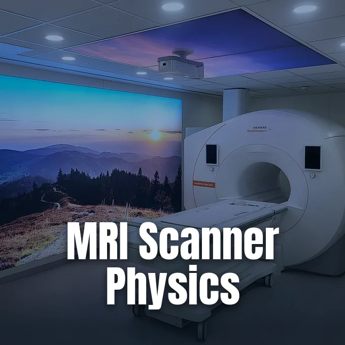
Understanding how MRI scans work begins with the physical behavior of hydrogen atoms inside human flesh. Unlike computed tomography or X-ray examinations, magnetic resonance imaging does not use ionizing radiation to generate pictures of living organs. Medical scanners rely on a static magnetic field, pulsed radio waves, and magnetic gradients to track how water and fat protons absorb and emit electromagnetic energy [1, 3]. By recording these faint radio signals through receiving coils, computers reconstruct cross-sectional views that differentiate healthy soft tissue from diseased regions in living human bodies [3].
How MRI Scans Work in Living Tissue
Human tissues consist mostly of water and fat molecules packed with hydrogen atoms [3]. Each hydrogen nucleus contains a single proton carrying an intrinsic quantum property known as nuclear spin with a value of one half. Because moving electrical charges produce magnetic dipoles, protons act like microscopic bar magnets in biological cells [1].
Patient bodies enter a hollow scanner bore where a primary magnet forces cellular protons to align along the main field axis [3]. Protons precess around these magnetic field lines at the Larmor precession frequency, an exact speed determined by field strength multiplied by the gyromagnetic ratio of hydrogen. Radiofrequency coils transmit pulses tuned to that exact resonance frequency, tipping the aligned protons into a transverse plane [1, 3].
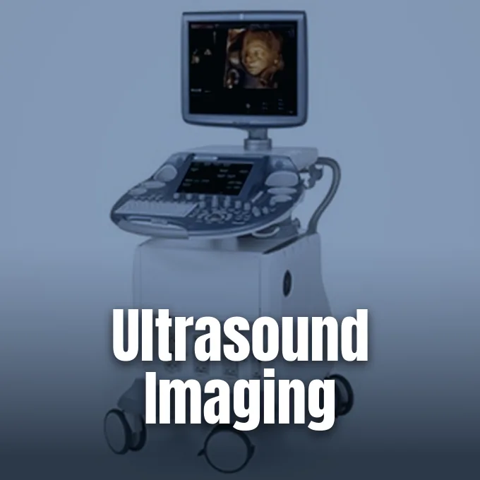
Relaxing protons release coherent radiofrequency signals that nearby antennas measure as soon as the radio pulse stops. Computers analyze these emitted radio waves to calculate proton density across living organs, revealing how MRI scans work in tissue without ionizing radiation [3]. Clinicians also evaluate soft structures alongside ultrasound imaging that maps human tissues through acoustic echoes to compare organ boundaries. At the antenna level, Slobozhanyuk reported in Advanced Materials that metallic wire metasurfaces placed under specimens strengthen radiofrequency fields to improve scan resolution [2]. Felix Bloch and H. E. Rorschach previously laid the physical groundwork for such radiofrequency measurements at Rice University. Researchers build upon these physical principles to image internal anatomical structures across multiple planes [1, 3].
Spatial Gradients
A uniform magnetic field cannot locate where a radio signal originates inside a patient. If every proton spun at the exact same frequency, detector antennas would register a single confused hum. To solve this localization challenge, engineers install gradient coils along three orthogonal axes within the scanner housing [3].
Adjustable electric currents running through gradient coils create small magnetic variations across the bore, causing protons at different physical positions to precess at distinct frequencies. Radio pulses selectively excite a single thin plane of tissue, allowing computers to decode position from signal phase and frequency shifts. Spatial encoding lets clinicians observe how MRI scans work when generating three-dimensional volume data from anatomical slices across diverse body planes [3].
Rapid switching of gradient coils produces the loud knocking and clicking sounds that patients hear during an examination. Magnetic forces push violently against wire windings, causing tiny mechanical vibrations through magnetostriction. Diagnostic scanners in modern hospitals typically use superconducting magnets operating at 1.5 teslas or 3 teslas, which need liquid helium cooling. Permanent magnets operate in low-field open scanners designed for patients who experience claustrophobia [3]. In ultra-low-field research, V. S. Zotev described a seven-channel SQUID system in an arXiv preprint that imaged sheep brains at 46 microteslas with pre-polarization at 40 milliteslas, though the preprint did not undergo peer review. Engineers continue refining gradient timing to reduce examination duration while maintaining high diagnostic accuracy [3, 4].
Relaxation Times T1 and T2
Visual contrast between organs depends on how excited protons relax back to thermal balance [1, 3]. Once a radio pulse tilts proton spins into the transverse plane, nuclei return to equilibrium through two separate processes. Longitudinal recovery, termed T1 relaxation, tracks how quickly magnetization regrows along the main field axis [1, 3].
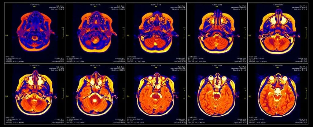
Operators capture T1-weighted images by selecting short repetition times between successive pulses. T1-weighted images show fatty structures as bright areas and watery fluids as dark regions, outlining brain cortex anatomy with high clarity. Transverse decay, designated as T2 relaxation, measures how quickly spinning protons lose phase coherence through local magnetic interactions [1, 3]. T2-weighted scans display fluids as bright white zones, highlighting inflammatory edema and white matter lesions across the central nervous system and soft organs [3].
Spin-echo sequences deliver a 90-degree pulse to create transverse magnetization, followed by a 180-degree pulse that inverts spins to generate a refocused echo signal [1]. Felix Bloch formulated the Bloch equations to describe mathematically how these nuclear vector components evolve over time. Clinicians frequently administer exogenous contrast agents like gadolinium to alter local relaxation rates, making abnormal blood vessels and lesions more conspicuous. Because water molecules bind differently to intracellular proteins in healthy muscle than in damaged tissue, varying pulse timing reveals subtle pathology across diverse anatomical structures [1, 3]. Radiologists examine both T1 and T2 contrasts to evaluate tissue health across the human body [3].
Magnetic Resonance Imaging Origins in Cancer Studies
The conceptual foundation of magnetic resonance imaging emerged from scientific debates over cellular water. In the 1960s, physical biochemists argued whether intracellular water acted like simple fluid in a test tube or formed structured macromolecular complexes. Biologists debated whether water inside cells possessed special physical properties [1].
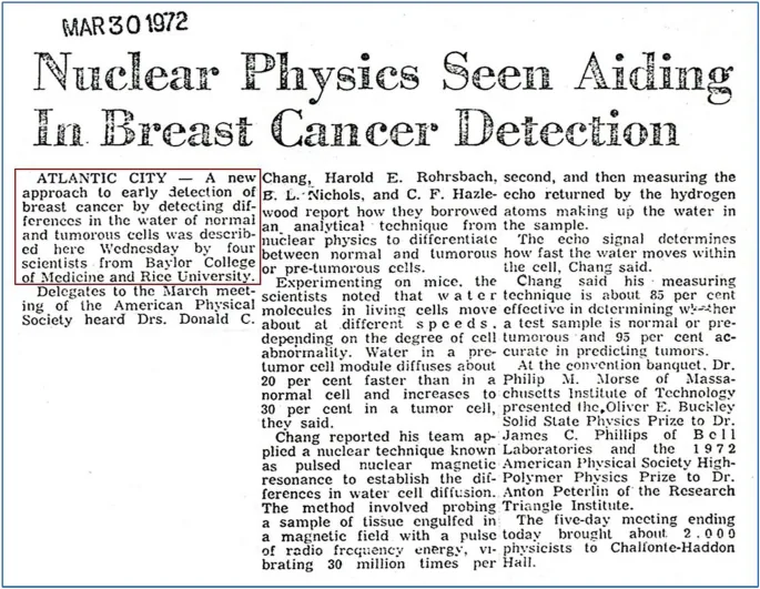
Donald C. Chang and C. F. Hazlewood investigated cellular water at Rice University and Baylor College of Medicine using spin-echo spectrometers. Chang and Hazlewood showed that water protons in biological tissues exhibit shorter relaxation times and slower diffusion coefficients than bulk water. Immature muscle cells in newborn rats displayed longer relaxation times than differentiated adult muscle fibers. In the early 1970s, physician Ray Damadian discovered that malignant tumors exhibited elevated relaxation values compared to healthy tissues, suggesting that nuclear resonance could detect cancer [1, 3].
Ray Damadian realized that nuclear magnetic resonance could identify cancer cells without invasive surgical biopsies. Chemist Paul Lauterbur soon proposed adding magnetic field gradients to encode spatial positions, creating two-dimensional NMR cross-sections [1, 3]. Paul Lauterbur received the Nobel Prize in 2003 for developing MRI, an award Damadian contested in newspaper advertisements. As Chang said in Discover Physics, practical scanning succeeded because multiple teams solved separate challenges, including relaxation measurements, biological cancer links, and spatial gradient encoding. Early medical prototypes confirmed that magnetic fields and radio waves could safely visualize internal pathology [1, 3].

High-Field Systems
Advanced medical research facilities build scanners with field strengths far exceeding standard hospital equipment. While ordinary diagnostic units operate at 1.5 teslas or 3 teslas, research systems run at 7 teslas, 9.4 teslas, and 11.7 teslas [3]. The FDA cleared 7-tesla scanners for clinical use in the United States in 2017 [6].
FDA radiologist Robert Ochs said that “the added field strength allows for better visualisation of smaller structures and subtle pathologies that may improve disease diagnosis.” [6] In Concepts in Magnetic Resonance, É. V. Reeth reviewed super-resolution algorithms that sharpen anatomical boundaries after scan acquisition. Researchers also examine optical imaging limits with the doughnut beam technique used to resolve nanometer-scale structures, but magnetic scanners uniquely penetrate deep internal organs. Medical software algorithms show how MRI scans work when reconstructing microstructural boundaries from raw data [5, 6].
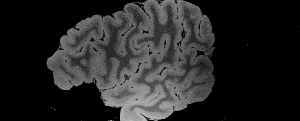
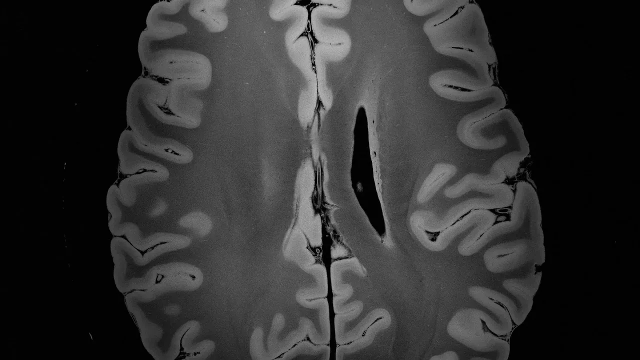
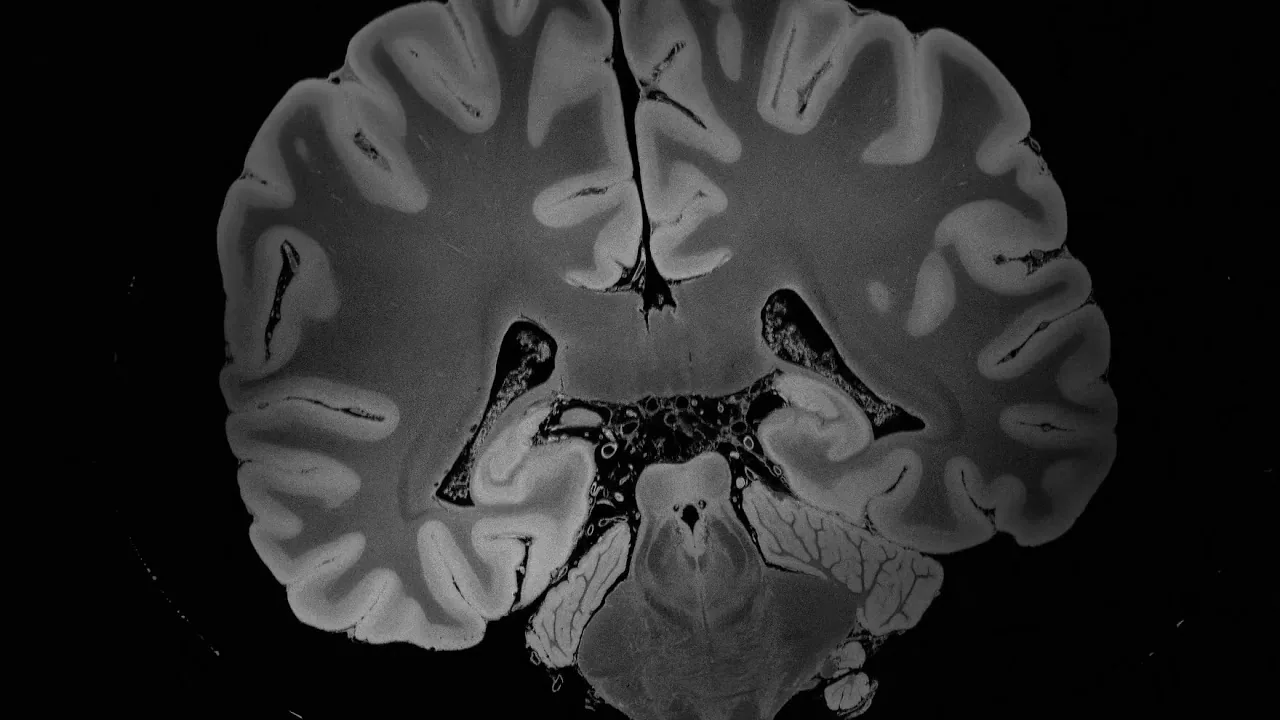
Neuroimaging scientist Brian L. Edlow led an experiment at Massachusetts General Hospital that tested high-field capabilities on motionless tissue. Researchers examined an ex vivo brain donated by a 58-year-old woman who died from viral pneumonia, securing the organ in a custom airtight urethane container. Continuous scanning over 100 hours inside a custom 7-tesla magnet yielded eight terabytes of raw imaging data at 100-micrometer resolution. Mount Sinai electrical engineer Priti Balchandani told Science News: “It’s definitely unprecedented.” The findings appeared on the preprint platform bioRxiv before peer review, and engineers integrated the dataset into the Lead DBS brain stimulation toolbox [6].
Functional Mapping with Blood Oxygen
Functional neuroimaging maps working brain activity rather than static anatomical structures. Active neurons consume metabolic energy, triggering local blood vessels to supply oxygenated blood. Seiji Ogawa discovered BOLD contrast in 1990, establishing blood-oxygen-level dependent imaging as the dominant technique for mapping human cognition [7].
Red blood cells carry oxygen bound to hemoglobin proteins throughout cerebral circulation. Deoxygenated hemoglobin contains unpaired electrons that make the molecule paramagnetic, causing local magnetic field disruptions that attenuate the magnetic resonance signal across surrounding blood vessels. Oxygenated hemoglobin is diamagnetic and produces negligible magnetic interference. When active brain regions fire, incoming oxygenated blood displaces deoxygenated blood within two seconds, reaching peak signal strength between four and six seconds later. Detectors register stronger radio signals from working brain zones [7].
The biological connection between cerebral blood flow and neuronal activity dates back to 1890, when Charles Roy and Charles Sherrington tested brain circulation at Cambridge University. Around that time, Italian physiologist Angelo Mosso invented the human circulation balance to weigh cerebral blood shifts during emotional and intellectual tasks. William James mentioned Mosso’s balance in 1890, and a modern replication by David T. Field confirmed that the balance detected blood volume shifts. Stefano Sandrone also recovered Mosso’s original scientific manuscripts. Since 1998, resting-state fMRI has allowed neuroscientists to map the default mode network while patients rest quietly without performing active tasks inside the scanner bore [7].
Can Brain Scans Produce False Signals?
Functional brain imaging does not read conscious thoughts or display raw electrical spikes [7, 8]. Computer algorithms divide three-dimensional brain scans into thousands of cubic volume elements called voxels. As Richard Chirgwin reported for The Register, “Software, rather than humans … scans the voxels looking for clusters” across three-dimensional neural maps [8].
In 2016, Swedish researcher Anders Eklund from Linköping University evaluated common fMRI software packages, including SPM, FSL, and AFNI. Eklund and his colleagues gathered resting-state data from 499 healthy participants and executed three million random comparisons. The statistical software generated false-positive rates reaching 70 percent, far exceeding the expected five percent rate. A software bug that remained uncorrected for fifteen years questioned findings in roughly 40,000 published papers, as journalist Kate Lunau reported for Motherboard [8].
Equipment operating fees of 600 dollars per hour had historically limited neuroimaging experiments to small cohorts of twenty or thirty volunteers [8]. Structural imaging also faces calibration challenges [9, 10]. Shinohara showed in NeuroImage Clinical that raw MRI signal intensities lack absolute physical units, varying widely across hospital sites, scanner models, and patient visits [9]. Chandra said in Journal of Neurology that while diffusion tensor imaging tracks Alzheimer’s disease, conventional structural imaging remains the recommended clinical standard for patient diagnosis [15]. Radiologist Bekiesińska-Figatowska cautioned in Polish Journal of Radiology that patient motion, vascular pulsation, clothing clips, and surgical implants can easily mimic spinal cord pathology [10].
Magnetic Resonance Imaging Safety in Hospital Suites
Magnetic resonance imaging avoids ionizing radiation, making it generally safer than computed tomography for routine diagnosis [11]. Around 50,000 scanners operate worldwide across hospitals and outpatient imaging centers [3]. However, the powerful main magnet generates a persistent projectile hazard because the magnetic field remains fully energized at all hours, even when no patient is undergoing an active scan [11, 12].
Ferromagnetic objects brought near the scanner room fly into the bore at high speeds between 20 and 30 feet per second, posing severe blunt trauma risks. At Westchester Medical Center in Valhalla, six-year-old Michael Colombini of Croton-on-Hudson underwent an examination after brain surgery. A 6.5-pound metal oxygen tank entered the room, became magnetized, and struck the child with fatal force [12, 13]. David Hebert, speaking for District Attorney Jeanine F. Pirro, called the occurrence a tragic accident [12]. A formal hospital review cited staff training deficits and absent written procedures for administering oxygen as critical factors behind the fatal accident [13].
Medical devices are now classified into MR Safe, MR Conditional, and MR Unsafe categories. Titanium alloys resist magnetic attraction, but ferromagnetic aneurysm clips and older cardiac pacemakers pose severe hazards in the magnetic environment. The American Board of Magnetic Resonance Safety credentials supervising physicians as MRMD, safety officers as MRSO, and consulting physicists as MRSE. In 2026, the credentialing board introduced an MR Safety Tech certification to educate technologist aides in the fundamental safety knowledge needed to manage scan environments. Knowing how MRI scans work helps clinical teams maintain safe facilities while acquiring high-quality diagnostic images [11].
- ACADEMIC JOURNAL Chang, D. C. (2025). A new understanding on the development of MRI for cancer detection. Discover Physics. [Article Link]
- ACADEMIC JOURNAL Slobozhanyuk, A. P., Poddubny, A. N., Raaijmakers, A. J. E., Berg, C. A. T. v. d., Kozachenko, A. V., Dubrovina, I. A., Melchakova, I. V., Kivshar, Y. S., & Belov, P. A. (2016). Enhancement of magnetic resonance imaging with metasurfaces. Advanced Materials, 28(9), 1832-1838. [Article Link]
- WEBSITE Wikipedia. Magnetic resonance imaging. [Article Link]
- PREPRINT Zotev, V. S., Matlashov, A. N., Volegov, P. L., Urbaitis, A. V., Espy, M. A., & Kraus Jr, R. H. (2007). SQUID-based instrumentation for ultra-low-field MRI. arXiv. [Article Link]
- ACADEMIC JOURNAL Reeth, É. V., Tham, I. W. K., Tan, C. H., & Poh, C. L. (2012). Super-resolution in magnetic resonance imaging: A review. Concepts in Magnetic Resonance Part A, 40A(6), 306-325. [Article Link]
- ONLINE NEWS Dockrill, P. (2019). 100-Hour-Long MRI of Human Brain Produces Most Detailed 3D Images Yet. ScienceAlert. [Article Link]
- WEBSITE Wikipedia. Functional magnetic resonance imaging. [Article Link]
- ONLINE NEWS Crew, B. (2016). A Bug in FMRI Software Could Invalidate 15 Years of Brain Research. ScienceAlert. [Article Link]
- ACADEMIC JOURNAL Shinohara, R. T., Sweeney, E. M., Goldsmith, J., Shiee, N., Mateen, F. J., Calabresi, P. A., Jarso, S., Pham, D. L., Reich, D. S., & Crainiceanu, C. M. (2014). Statistical normalization techniques for magnetic resonance imaging. NeuroImage Clinical, 6, 9-19. [Article Link]
- ACADEMIC JOURNAL Bekiesińska-Figatowska, M. (2015). Artifacts in magnetic resonance imaging. Polish Journal of Radiology, 80, 93-106. [Article Link]
- WEBSITE Wikipedia. Safety of magnetic resonance imaging. [Article Link]
- ONLINE NEWS Chen, D. W. (2001). Boy, 6, Dies Of Skull Injury During M.R.I. The New York Times. [Article Link]
- ONLINE NEWS Archibold, R. C. (2001). Hospital Details Failures Leading to M.R.I. Fatality. The New York Times. [Article Link]
- ACADEMIC JOURNAL Heim, B., Krismer, F., Marzi, R. D., & Seppi, K. (2017). Magnetic resonance imaging for the diagnosis of Parkinson’s disease. Journal of Neural Transmission, 124(8), 915-964. [Article Link]
- ACADEMIC JOURNAL Initiative, A. D. N., Chandra, A., Dervenoulas, G., & Politis, M. (2018). Magnetic resonance imaging in Alzheimer’s disease and mild cognitive impairment. Journal of Neurology, 266(6), 1293-1302. [Article Link]
Cover image: FbrG / Wikimedia Commons, CC BY-SA 4.0


