




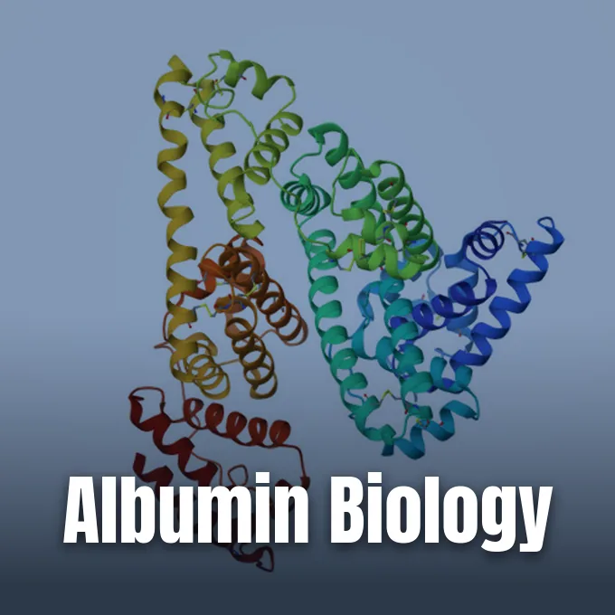
Understanding what is albumin begins with blood plasma, where this water-soluble globular protein makes up roughly half of all serum protein mass in healthy adults. Synthesized by liver cells, the monomeric molecule lacks attached sugars and circulates at normal adult levels between 3.5 and 5.0 g/dL [5]. Albumin belongs to an ancient family of vertebrate transport proteins that includes alpha-fetoprotein and vitamin D-binding protein [4]. Its presence keeps bodily fluids balanced and lets insoluble drugs travel safely through blood.
Identity of Human Serum Albumin
The word comes from Latin albumen, meaning egg white [4]. The mature monomer weighs about 66.5 kDa across a chain of 585 amino acids folded into three homologous domains [5]. Bridging these loops, seventeen disulfide bonds maintain three-dimensional stability, and an unpaired cysteine at Cys34 provides a free sulfhydryl group that supplies broad antioxidant defense against reactive oxygen species [1]. Because its isoelectric point is 4.7, the molecule carries a net negative electrical charge at normal blood pH of 7.4 [4]. Electrical repulsion stops filtration across kidney glomeruli [6].
Genetically, the ALB gene spans 16,961 nucleotides on human chromosome 4 at locus 4q13.3, split into 15 exons that mirror the domain architecture of the protein [5]. Rare mutations cause congenital analbuminemia, documented in OMIM entry 103600 alongside dysalbuminemic hyperthyroxinemia and dysalbuminemic hyperzincemia, where individuals survive through compensatory shifts in other blood globulins [14].
Within each eukaryotic cell in hepatic tissue, genetic control maintains balanced protein output [5]. Protein databases like SMART document over 2,800 related albumin domains across more than 1,100 animal proteins, demonstrating the evolutionary preservation of this molecular blueprint [14]. Though small variations exist among mammals, the core globular design stays constant across vertebrate biology [4].
Hepatic Synthesis in Liver Cells
Liver hepatocytes synthesize roughly 15 grams of protein daily in a healthy adult. Production begins in the nucleus when the ALB gene undergoes transcription into messenger RNA, which migrates into the cytoplasm where membrane-bound ribosomes translate the transcript into preproalbumin, a nascent polypeptide containing a 24-amino-acid leader sequence [3]. This sequence routes the molecule into secretory pathways.
Inside the rough endoplasmic reticulum, signal peptidases cleave eighteen amino acids off preproalbumin to form proalbumin. Retaining a six-amino-acid extension as the principal intracellular form, proalbumin moves forward through transport vesicles to the Golgi apparatus, where specific endoproteases cleave this remaining hexapeptide just before cellular release into tissue fluid. The liver never stores mature albumin. Hepatocytes discharge every completed molecule into circulation immediately after synthesis finishes [3].
Newly formed protein enters the blood through two anatomical routes. Most molecules pass from liver cells into sinusoids after crossing the microscopic Space of Disse, and a smaller fraction enters lymphatic vessels to join the thoracic duct. After two hours, about 90% of secreted molecules stay inside vascular channels, and the colloid level in hepatic interstitial fluid acts as an osmotic sensor to govern output [3].
Fluid Balance in Capillaries
The main job of the protein is generating intravascular colloid oncotic pressure, also known as colloid osmotic pressure [3]. Because large serum proteins cannot pass freely across healthy capillary endothelial walls, they exert a persistent osmotic pull that draws water from interstitial spaces back into circulating blood [6]. The molecule supplies 75% to 80% of normal plasma oncotic pressure and accounts for half of total plasma protein mass [3]. Without this steady inward pull, hydrostatic pressure generated by the beating heart would force liquid out of blood vessels into surrounding tissues, causing profound fluid accumulation [6].
Total body albumin spans 300 to 500 grams in a healthy adult. Between 30% and 40% of this total pool, amounting to approximately 210 grams, circulates within the vascular channels of muscle, skin, liver, and gut, and the remaining extravascular fraction permeates connective tissues with skin serving as the primary exchangeable reservoir. Lymphatic vessels return daily vascular losses back to the blood via the thoracic duct. The protein persists in human blood with a mean lifespan of three weeks, as liver enzymes clear roughly 4% each day [3].
When blood levels fall, declining oncotic pressure upsets microvascular fluid balance. Water moves outward into loose connective tissues, causing dependent peripheral edema in lower limbs or fluid accumulation in the abdominal cavity [3]. Smaller mammals operating at lower arterial pressures need less oncotic force and maintain lower baseline protein levels than humans [6].
Molecular Architecture of Albumin Domains
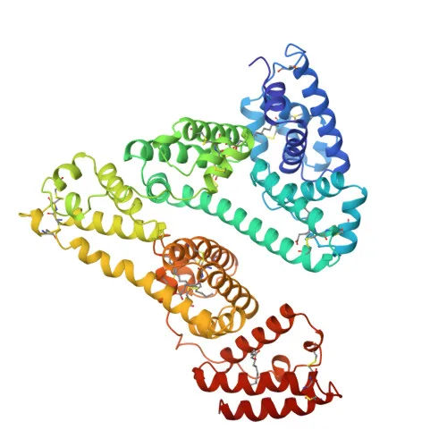
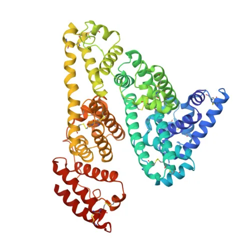
The three-dimensional fold was solved at Osaka Laboratories within Yoshitomi Pharmaceutical Industries by researchers Sugio, Kashima, Mochizuki, Noda, and Kobayashi at 2.5 Å resolution using molecular replacement from triclinic crystals (PDB entry 1AO6), establishing that the 585-residue chain forms a heart-shaped assembly of three alpha-helical domains designated I, II, and III [1]. Each domain divides into subdomains A and B [4].
The chief drug-binding pockets sit inside deep nonpolar cavities in subdomains IIA and IIIA. Historically designated as Sudlow site I and Sudlow site II, these pockets feature nonpolar walls lined with positively charged lysine and arginine side chains. This biochemical arrangement lets each pocket hold a broad collection of small drugs, ranging from diagnostic dyes to anticoagulants and anti-inflammatory compounds. Recombinant protein grown in Pichia pastoris yeast matches human plasma albumin with a root-mean-square deviation of only 0.24 Å for alpha-carbon atoms, proving that recombinant expression preserves natural tertiary folding [1]. In structural databases including SCOPe 2.08, the 1AO6 entry was released in 1998 and updated in 2009, confirming its role as an alpha-helical carrier protein [13].
Fatty acid handling is another central task handled by this modular fold. At Blackett Laboratory within Imperial College London, investigators Curry, Mandelkow, Brick, and Franks mapped five fatty acid binding sites at 2.5 Å resolution using PDB entry 1BJ5. Asymmetric nonpolar pockets lock long hydrocarbon chains securely [2].


Hormone and Metabolite Transport
Beyond fatty acids, the protein serves as an unspecialized vehicle for lipophilic hormones and organic waste products. It reversibly binds thyroid hormones, including thyroxine, and steroid hormones to keep them soluble in blood plasma and shields them from kidney clearance. The protein also carries unconjugated bilirubin, an insoluble breakdown product of red blood cell hemoglobin, transporting it from spleen and bone marrow to the liver for glucuronidation [5]. Without binding, toxic free bilirubin crosses the blood-brain barrier and causes permanent neurological damage [3].
Inorganic ions bind to albumin. Negatively charged surface residues coordinate calcium, zinc, copper, and cobalt, making the protein the chief reservoir for non-ionized calcium in blood [4]. Because half of serum calcium binds to the protein, measured calcium drops during hypoalbuminemia. Clinicians adjust measured calcium upward by 0.8 mg/dL for every 1.0 g/dL drop in serum albumin below 4.0 g/dL to evaluate true calcium status [6]. The molecule also binds free hemin and trace redox-active metals, preventing these compounds from catalyzing destructive oxidation reactions in vascular beds [12]. Its presence safeguards endothelial health.
Drug binding shapes the pharmacological behavior of many therapeutic agents [5]. Barbiturates, warfarin, and nonsteroidal anti-inflammatory agents bind strongly to subdomains IIA and IIIA [4]. When two highly bound medications compete for identical binding cavities, displacement increases the unbound free drug fraction in circulation, potentially elevating pharmacological potency or toxicity. The protein also protects circulating folic acid from photodegradation in light-exposed skin vessels [5].
Why Does Hypoalbuminemia Develop?
Hypoalbuminemia develops whenever liver output drops, breakdown accelerates, vascular permeability expands, or protein spills into urine. Defined as serum albumin below 3.5 g/dL, it affects around 20% of patients admitted to hospitals. Why does this decline occur so frequently in clinical practice? In most acute hospital settings, systemic inflammation drives the decline rather than inadequate dietary intake, as inflammatory cytokines including tumor necrosis factor and interleukin-6 suppress transcription of the ALB gene and speeds up capillary leakage into interstitial tissue [3].
In chronic liver disease, such as cirrhosis, loss of functional hepatocyte mass reduces the total output of new protein. Distorted microcirculation impairs the delivery of oxygen and amino acid substrates needed for ongoing protein assembly [3]. In advanced decompensated cirrhosis, clinical studies published in Gut by Bernardi, Angeli, Clària, and Moreau showed that circulating albumin undergoes irreversible oxidative modifications that impair its antioxidant, toxin-scavenging, and endothelial-protective properties [11]. When cirrhosis causes ascites, the enlarged fluid volume dilutes serum levels even if liver production stays partially active [3]. Peripheral edema and circulatory dysfunction worsen as effective oncotic pressure declines.
Excessive protein loss across damaged epithelial surfaces forms another major cause of depressed blood levels. In nephrotic syndrome, glomerular barrier failure allows over 3.5 grams of protein to spill into urine each day. Protein-losing enteropathy causes similar losses through the gut wall, and deep dermal burns weep massive amounts of extravascular fluid [3]. Severe malnutrition also halts protein synthesis.
Laboratory Measurement and Clinical Value
Clinical pathology laboratories measure serum albumin levels using automated dye-binding spectrophotometric tests. The two standard diagnostic reagents are bromocresol green and bromocresol purple, which undergo distinct light absorbance shifts upon binding selectively to circulating protein molecules. In healthy adult humans, the normal reference range is 3.5 to 5.0 g/dL, or 35 to 50 g/L. In young children under three years of age, normal physiology displays a wider reference range of 2.9 to 5.5 g/dL [5]. Measurements outside these boundaries signal underlying disease states.
Serum albumin level is a recognized prognostic indicator across varied acute and chronic illnesses. Hospital analyses cited in Medscape by Sans-García and Gerstmeyer showed that lower albumin predicts mortality in dialysis and spondylodiscitis cohorts, and pooled cohort data showed that each 10 g/L reduction in serum albumin raised inpatient deaths by 137% and complication risks by 89% [3]. In neurology, studies by LeVine in BMC Neurology revealed that multiple sclerosis involves blood-brain barrier leakage that permits circulating albumin to enter neural tissue beds, where it alters astrocyte potassium clearance and promotes neuroinflammation [12]. The cerebrospinal fluid to serum ratio tracks this barrier leakage.
Elevated serum levels, termed hyperalbuminemia, occur almost exclusively because of fluid depletion rather than increased liver production. When plasma water volume shrinks during dehydration, protein concentration rises artificially through hemoconcentration. Retinol deficiency can also produce higher values near 4.9 g/dL because altered vitamin A dynamics cause cellular swelling, but intravenous rehydration quickly restores standard baseline readings [5].
Albumin in Drug Delivery Systems
In clinical therapeutics, human serum albumin solutions extracted from donor plasma are prepared at concentrations of 5% and 25% for intravenous infusion. These solutions serve as volume expanders during trauma surgery, deep burn management, and cirrhosis complications [5]. In a randomized trial reported in Hepatology by Cavallin, Kamath, Merli, Fasolato, and Angeli, terlipressin combined with intravenous albumin reversed hepatorenal syndrome significantly better than midodrine and octreotide combined with albumin [10]. However, routine infusion in acute inpatient care is limited, as Cochrane systematic reviews found no survival benefit over crystalloid fluids in routine hypovolemia [5].
The natural properties of albumin have turned it into an attractive carrier for drug delivery in pharmacology [8]. Structural work led by Sand, Bern, Nilsen, Sandlie, and Andersen in Frontiers in Immunology showed how the neonatal Fc receptor, known as FcRn, shields albumin from cellular breakdown, explaining its long circulatory half-life of 19 days. Cellular endosomes take up albumin, where acidic pH promotes binding to FcRn, diverting the protein away from lysosomal degradation and recycling it back into the bloodstream [7]. Protein reviews by Sleep in Expert Opinion on Drug Delivery documented how albumin binds cell receptors including gp60, gp18, gp30, cubilin, and megalin [9]. Tumors and inflamed tissues consume large amounts of extracellular protein to fuel cellular growth, causing albumin to accumulate naturally at disease sites [8].
Nanoparticle albumin-bound formulation stands as the most successful clinical implementation of this carrier concept. Pharmaceutical evaluations published in Molecular Pharmaceutics by Spada, Emami, Tuszyński, and Lavasanifar tracked how Nab-Paclitaxel (Abraxane) binds paclitaxel into albumin nanoparticles, replacing CrEL-paclitaxel (Taxol) to reduce solvent toxicity. This formulation reduces hypersensitivity reactions and improves intratumoral delivery. Clinical trials are exploring further conjugates to prolong peptide therapeutics [8].
- ACADEMIC JOURNAL Sugio, S., Kashima, A., Mochizuki, S., Noda, M., & Kobayashi, K. (1999). Crystal structure of human serum albumin at 2.5 Å resolution. Protein Engineering, Design and Selection, 12(6), 439-446. [Article Link]
- ACADEMIC JOURNAL Curry, S., Mandelkow, H., Brick, P., & Franks, N. (1998). Crystal structure of human serum albumin complexed with fatty acid reveals an asymmetric distribution of binding sites. Nature Structural Biology, 5(9), 827-835. [Article Link]
- WEBSITE Medscape. (2025). Hypoalbuminemia: Background, Pathophysiology, Etiology. [Article Link]
- WEBSITE Wikipedia. (2026). Albumin. [Article Link]
- WEBSITE Wikipedia. (2026). Human serum albumin. [Article Link]
- WEBSITE Wikipedia. (2026). Serum albumin. [Article Link]
- ACADEMIC JOURNAL Sand, K. M. K., Bern, M., Nilsen, J., Noordzij, H. T., Sandlie, I., & Andersen, J. T. (2015). Unraveling the Interaction between FcRn and Albumin: Opportunities for Design of Albumin-Based Therapeutics. Frontiers in Immunology, 5, 682-682. [Article Link]
- ACADEMIC JOURNAL Spada, A., Emami, J., Tuszyński, J. A., & Lavasanifar, A. (2021). The Uniqueness of Albumin as a Carrier in Nanodrug Delivery. Molecular Pharmaceutics, 18(5), 1862-1894. [Article Link]
- ACADEMIC JOURNAL Sleep, D. (2014). Albumin and its application in drug delivery. Expert Opinion on Drug Delivery, 12(5), 793-812. [Article Link]
- ACADEMIC JOURNAL Cavallin, M., Kamath, P. S., Merli, M., Fasolato, S., Toniutto, P., Salerno, F., Bernardi, M., Romanelli, R. G., Colletta, C., Salinas, F., Giacomo, A. D., Ridola, L., Fornasiere, E., Caraceni, P., Morando, F., Piano, S. S., Gatta, A., & Angeli, P. (2015). Terlipressin plus albumin versus midodrine and octreotide plus albumin in the treatment of hepatorenal syndrome: A randomized trial. Hepatology, 62(2), 567-574. [Article Link]
- ACADEMIC JOURNAL Bernardi, M., Angeli, P., Clària, J., Moreau, R., Ginès, P., Jalan, R., Caraceni, P., Fernández, J., Gerbes, A. L., O’Brien, A. J., Trebicka, J., Thévenot, T., & Arroyo, V. (2020). Albumin in decompensated cirrhosis: new concepts and perspectives. Gut, 69(6), 1127-1138. [Article Link]
- ACADEMIC JOURNAL LeVine, S. M. (2016). Albumin and multiple sclerosis. BMC Neurology, 16(1), 47-47. [Article Link]
- WEBSITE SCOPe. (2026). Structural Classification of Proteins: PDB entry 1ao6. [Article Link]
- WEBSITE SMART. (2025). ALBUMIN domain annotation (SM00103). [Article Link]
- WEBSITE RCSB Protein Data Bank. (1997). Crystal Structure of Human Serum Albumin (PDB 1AO6). [Article Link]


