



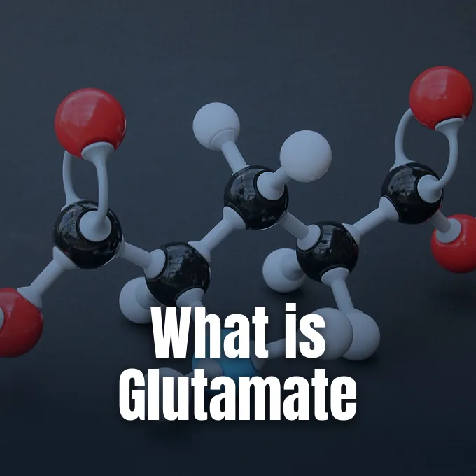
Understanding what is glutamate begins with identifying it as the chief excitatory neurotransmitter operating in the vertebrate nervous system [6]. In living tissue and neutral water solutions, the molecule exists as an anion that accounts for an estimated 90% of excitatory synaptic connections in the human brain [2]. Beyond its signalling role, the compound functions as a versatile metabolic fuel involved in nitrogen removal and protein synthesis [5]. Astrocytes and cellular transport systems keep extracellular fluid levels low to prevent brain cell damage [8].
Chemical Identity of Glutamic Acid
German chemist Karl Heinrich Ritthausen discovered and identified the compound in 1866 in Germany while treating wheat gluten with sulfuric acid during laboratory work on cereal grains. He named the substance after gluten. The molecule is an α-amino acid with the formula C5H9NO4, designated by Glu and the single letter E. Biochemical charts use E because it follows D for aspartate, reflecting its extra carbon group [2].
When the chemical dissolves in water, its electrical charge changes with acidity levels. In strong acid below pH 2.10, both carboxyl groups hold protons to create a positive cation. Within the mild range between pH 2.5 and 4.1, the carboxyl group nearest the amino terminus surrenders its proton, yielding a neutral zwitterion with both positive and negative charges. At pH 4.07, the second carboxyl group loses its proton as well, leaving a singly negative anion. Neutral blood at pH 7.35 to 7.45 holds this negative form almost exclusively, whereas alkaline solutions above pH 9.47 strip another proton from the amino group to create a doubly negative ion [2].
The molecule forms two optical enantiomers with mirror symmetry. Living cells use the l-form to build proteins. But bacteria also make the d-form with the enzyme glutamate racemase, placing it into their cell walls and outer capsules to resist host attacks and survive hostile environmental conditions [2]. In the ChEBI database run by the European Bioinformatics Institute, the compound carries the code CHEBI:18237. The catalog lists it as a polar amino acid and a fundamental metabolite in cellular life [1].
Discovery and Culinary Uses of MSG
In 1908, Japanese researcher Kikunae Ikeda of Tokyo Imperial University in Japan evaporated large volumes of kombu seaweed broth to identify the chemical source of savory taste. He isolated brown crystals of glutamic acid. Their taste matched the savory flavor of dashi broth. Ikeda named this flavor umami [2]. He patented a method to make monosodium glutamate, launching sales in 1909 [3].
Making the seasoning changed in the 1950s when factories moved from wheat extraction to bacterial fermentation [2]. Facilities cultured the soil bacterium Corynebacterium glutamicum, also known as Brevibacterium flavum, feeding it ammonia and sugars from molasses, sugar cane, or starch to yield high commercial volumes. The aerobic fermentation runs like the steps used to make vinegar [3]. By 2006, annual world output of glutamic acid reached 1.5 million tons [2]. Trade officials track the product under HS code 2922.42, while Europe labels it as food additive E621 and the free acid as E620 to regulate international food manufacturing standards [4].
Plain monosodium glutamate tastes unpleasant by itself, but it enriches savory scents when cooked into soups and meats. Sodium makes up 12.28% of the compound by weight, which is about one-third of the 39.34% sodium found in table salt, leaving the other 87.72% as pure glutamate counterion. The remaining substance functions as an enhancer of savory taste. Food makers blend this seasoning with disodium inosinate, or E631, and disodium guanylate, or E627. A commercial mix called super salt blends nine parts table salt with one part monosodium glutamate and 0.1 parts ribonucleotides to boost taste while cutting total sodium in prepared foods [4].
Dietary Safety in Human Digestion
Dietary protein gives people most of their daily intake of this amino acid. Adults typically consume about 13 grams of glutamate every day from protein foods like tomatoes and cheeses, while added seasoning supplies around 0.55 grams in average daily diets [3]. Intestinal lining cells consume the vast majority of swallowed glutamate as fuel during first-pass digestion. Blood levels stay steady, and the blood-brain barrier shields the brain from surges in the bloodstream [2].
The U.S. Food and Drug Administration treats monosodium glutamate as generally recognized as safe. In 1995, an extensive scientific safety review by the Federation of American Societies for Experimental Biology affirmed that normal dietary use of the seasoning causes no harm in healthy consumers. Sensitive people may feel short-term numbness or headaches only after taking 3 grams or more without any food. Standard meals supply far lower doses. A normal serving of seasoned food contains less than 0.5 grams, making large empty-stomach doses very uncommon [3]. In a 1993 study on 71 fasting people given 5 grams of seasoning without food, the only mild reaction observed by clinical researchers came from a volunteer taking a placebo [4].
Clinical trials in 2000 tested 130 people who claimed sensitivity, but only two reacted across repeat tests. Doctors concluded that reactions are not reproducible. Food Standards Australia New Zealand reviewed medical data and found no proof of severe illness or death. The agency stated that reports of Chinese restaurant syndrome lack clinical proof [4]. Furthermore, the compound has no biological link to wheat gluten, so patients with celiac disease can consume pure glutamate seasonings without triggering autoimmune inflammatory damage in the small intestine [3].
Role in Cellular Metabolism
Glutamate sits at the center of cellular fuel use [5]. Living cells make the compound from α-ketoglutarate, an intermediate in the citric acid cycle. Transaminase enzymes shift an amino group onto this keto acid to yield glutamate while releasing diverse α-keto acids that enter glycolysis, gluconeogenesis, and other primary energy pathways. The resulting carbon backbones enter glycolysis and gluconeogenesis [2].
The amino acid also controls the safe clearance of nitrogen waste. As body proteins break down, transamination routes surplus amino groups into glutamate. Glutamate dehydrogenase then strips the nitrogen as ammonia. Liver cells package this toxic ammonia into urea, which travels safely through the bloodstream to the kidneys for ultimate excretion from the human body in urine [2]. Biochemists Walker and van der Donk showed that this metabolic hub also supplies building blocks for nucleotides, cofactors, and specialized natural products across many different species of living organisms. Nitrogen recycling through these connected enzymatic loops protects cells from toxic buildup while supplying raw materials for non-essential amino acids [5].
Brain tumors feed on this fuel [7]. In neuro-oncology, malignant brain tumors known as glioma and glioblastoma consume large amounts of glutamate for growth [2]. Willard and Koochekpour reported that neoplastic cells rewire their metabolic networks to draw energy from this amino acid, satisfying intense biosynthetic demands as malignant cell colonies expand [7]. Glial tumors show even greater hunger for the compound when cells carry mutations in the IDH1 gene [2]. Medical researchers continue studying these metabolic vulnerabilities to design targeted drug treatments for aggressive brain cancers [7].


What Is Glutamate in Synaptic Transmission?
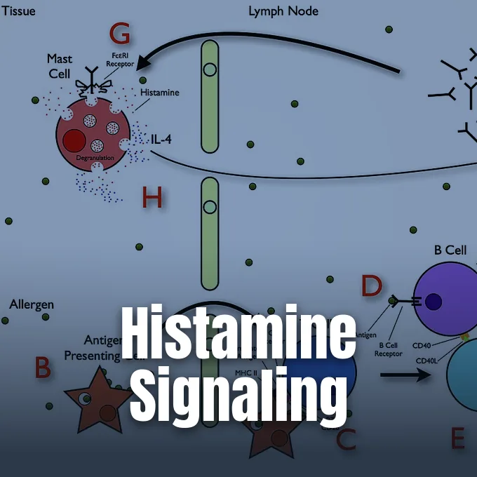
What makes this simple amino acid so powerful in the brain? In the central nervous system, it acts as the main chemical transmitter, powering an estimated 90% of excitatory synaptic connections [6]. Nerve terminals store the substance inside synaptic vesicles. When an electrical impulse arrives at the terminal, calcium rushes into the cell and triggers exocytosis, releasing the chemical transmitter directly into the narrow space of the synaptic cleft. The molecules drift across the gap to bind surface receptors on the target neuron [8].
Postsynaptic receptors split into ionotropic and metabotropic families. Ionotropic receptors open ion channels upon binding, letting sodium and calcium pass into the cell. Fast AMPA receptors create electrical impulses within a fraction of a millisecond, working alongside kainate receptors and calcium-permeable NMDA receptors to regulate baseline excitability in brain circuits [6]. In contrast, metabotropic receptors bind to G-proteins divided into Group I, Group II, and Group III. Researchers Willard and Koochekpour showed that metabotropic receptors activate downstream cascades including the PI3K/Akt, MAPK, NFkB, PLC, and Ca/CaM signaling pathways to produce enduring cellular modifications. The human mGluR1a receptor shows how these proteins fine-tune cellular responses [7].
These receptor proteins interact with several medical drugs and illicit substances. Street phencyclidine, known as PCP or Angel Dust, blocks NMDA channels without competing for the binding site, causing strong dissociative reactions and altered perception in human users. Ketamine and dextromethorphan also block this channel to alter conscious perception. On the metabotropic side, experimental drug eglumetad activates group II receptors. In bonnet macaques, or Macaca radiata, eglumetad reduced baseline cortisol levels by roughly 50%. The drug also acts on human adrenal cells, lowering aldosterone synthase and steroid production [2].
Synaptic Plasticity and Memory
Beyond fast signalling, the neurotransmitter drives lasting changes in connection strength known as long-term potentiation. This process takes place in the hippocampus and neocortex, providing the physical foundation for learning and memory. When repeated high-frequency impulses stimulate a synapse, NMDA channels shed their magnesium blocks and admit calcium ions. Calcium entry triggers biochemical cascades that insert new AMPA receptors into the synaptic membrane, making the synapse substantially more sensitive to subsequent electrical signals during cognitive tasks. Strengthening connections through this mechanism allows fleeting sensory input to become stable memories stored in brain networks across the hippocampus, neocortex, and related cognitive structures [6].
Synaptic signals also spill beyond individual connections. When neurons fire intensely, transmitter molecules escape the synaptic cleft into outer brain fluid. Extrasynaptic signaling allows adjacent synapses to talk to one another through volume transmission. The molecule also guides nerve wiring in young brains [6]. Research originally published by Mark Mattson showed that local release directs growth cone movement and synaptogenesis in developing nerves, establishing functional wiring throughout the growing nervous system [2].
In inhibitory brain circuits, the molecule serves as the direct precursor for gamma-aminobutyric acid, or GABA. The enzyme glutamate decarboxylase, called GAD, strips a carboxyl group from glutamate to produce GABA. This pathway concentrates in the cerebellum and pancreas. When patients make antibodies against GAD, GABA levels plunge, causing a rare condition called Stiff person syndrome. Because the pancreas also relies on GAD, these patients frequently suffer from autoimmune diabetes [2].
Astrocytic Clearance of Glutamate Neurotransmitter
Because loose transmitter molecules harm neurons, astrocytes keep tight control over extracellular fluid. Glial cells express excitatory amino acid transporters, or EAAT, and vesicular carriers called VGLUT to pull molecules out of the cleft [9]. In 2004, researchers crystallized a bacterial transporter homolog, uncovering how these transport proteins use sodium ions to drag transmitter molecules against steep concentration gradients into glial cells. Scientists Vandenberg and Ryan showed that this molecular pump clears the synaptic space within milliseconds [10].
Inside glial cells, imported molecules enter the glutamate-glutamine cycle. The enzyme glutamine synthetase turns glutamate into glutamine, a harmless compound that astrocytes pass back into extracellular fluid. Presynaptic neurons absorb this glutamine and use the enzyme glutaminase to rebuild glutamate for vesicles. Researchers Zhou and Danbolt noted that this cycle protects neurons by keeping active transmitter compartmentalized. Passing molecules through astrocytes prevents stray excitation while saving cellular energy, shuttling carbon backbones between brain cells without requiring de novo amino acid synthesis during ongoing neurotransmission [8].
Astrocytes do more than clean up fluid. Mahmoud and colleagues showed that extracellular transmitter stimulates calcium release inside astrocytes. That calcium surge triggers exocytosis of glutamate back toward neurons, helping pace network firing [9]. Similar non-neuronal regulation works in insects. In Drosophila fruit flies, glial transport regulates postsynaptic receptor clusters, showing that astrocyte-like control of extracellular transmitter evolved hundreds of millions of years ago in primitive ancestors [2].
Excitotoxicity in Neurological Disorders
When transport pumps fail during brain injury or stroke, accumulating extracellular transmitter turns deadly in a process called excitotoxicity [8]. Under low oxygen, cell energy drops and EAAT transporters run backward, spilling stored molecules into outer fluid. Flooding the synapse opens NMDA channels wide, driving uncontrolled calcium into neurons. Excess calcium damages mitochondria and produces toxic nitric oxide free radicals. These chemical shifts switch on pro-apoptotic transcription factors and silence survival genes, driving vulnerable nerve cells into irreversible programmed cell death during acute stroke and trauma [6].
Excitotoxic damage plays a role in stroke, amyotrophic lateral sclerosis, and Alzheimer’s disease. In epilepsy, abnormal release sparks electrical chaos. Injecting the amino acid into brain tissue produces rapid firing pulses spaced about one second apart, matching seizure patterns [6]. In psychiatry, a meta-analysis of proton magnetic resonance spectroscopy studies by Merritt and colleagues found significant glutamatergic changes across 1,686 schizophrenia patients compared to 1,451 healthy controls. The findings show how transmitter imbalance disrupts thought processing [11].
Evolutionary biologists find this signaling system across all animals with nerves. Comb jellies, or ctenophores, split off early in animal evolution and lack serotonin or acetylcholine. Yet ctenophores use distinct ionotropic glutamate receptors to trigger muscle contractions. Even primitive sea sponges use the molecule for cell-to-cell signaling without having any nervous system. Across hundreds of millions of years of evolutionary history, this amino acid has powered cellular communication from early marine life all the way to complex human cognitive networks [6].
- WEBSITE European Bioinformatics Institute. (2024). Glutamic acid (CHEBI:18237). ChEBI. [Article Link]
- WEBSITE Wikipedia. (n.d.). Glutamic acid. [Article Link]
- WEBSITE U.S. Food and Drug Administration. (2026). Questions and Answers on Monosodium glutamate (MSG). [Article Link]
- WEBSITE Wikipedia. (n.d.). Monosodium glutamate. [Article Link]
- ACADEMIC JOURNAL Walker, M. C., & Donk, W. A. v. d. (2015). The many roles of glutamate in metabolism. Journal of Industrial Microbiology & Biotechnology, 43(2-3), 419-430. [Article Link]
- WEBSITE Wikipedia. (n.d.). Glutamate (neurotransmitter). [Article Link]
- ACADEMIC JOURNAL Willard, S. S., & Koochekpour, S. (2013). Glutamate, Glutamate Receptors, and Downstream Signaling Pathways. International Journal of Biological Sciences, 9(9), 948-959. [Article Link]
- ACADEMIC JOURNAL Zhou, Y., & Danbolt, N. C. (2014). Glutamate as a neurotransmitter in the healthy brain. Journal of Neural Transmission, 121(8), 799-817. [Article Link]
- ACADEMIC JOURNAL Mahmoud, S., Gharagozloo, M., Simard, C., & Gris, D. (2019). Astrocytes Maintain Glutamate Homeostasis in the CNS by Controlling the Balance between Glutamate Uptake and Release. Cells, 8(2), 184-184. [Article Link]
- ACADEMIC JOURNAL Vandenberg, R. J., & Ryan, R. M. (2013). Mechanisms of Glutamate Transport. Physiological Reviews, 93(4), 1621-1657. [Article Link]
- ACADEMIC JOURNAL Merritt, K., Egerton, A., Kempton, M. J., Taylor, M. J., & McGuire, P. K. (2016). Nature of Glutamate Alterations in Schizophrenia. JAMA Psychiatry, 73(7), 665-665. [Article Link]
Cover image: Bin im Garten / Wikimedia Commons, CC BY-SA 3.0


