





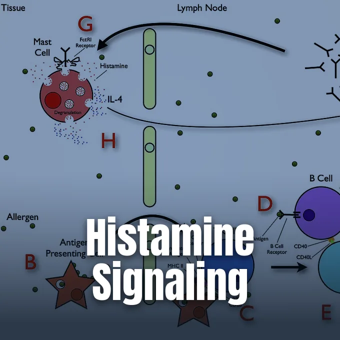
Answering what is histamine reveals an active amine that directs immune defense, stomach acid, and brain signals across human tissues. First isolated in 1910, the molecule pairs an imidazole ring with an ethylamine chain. Mast cells make it to fight off germs. When immune control slips, this amine sparks seasonal sneezing, hives, or dangerous shock. The chemical works locally in blood walls, smooth muscle, and nerve loops to preserve daily health [7].
What Is Histamine?
Histamine is an organic amine formed from histidine. Body tissues produce it without help from classic endocrine glands, prompting early biologists to call it an autacoid or local hormone. Mast cells and white blood basophils make over 90 percent of body stores, packing the amine into dense granules until danger calls [3].
The noun entered print in 1910, combining histidine with the chemical suffix amine as cataloged by Dictionary.com and Random House, Inc. During early vitamin trials in the 1920s, chemist Walter H. Eddy recorded in The Vitamine Manual that crystals often carried histamine traces from biological extracts. Beyond animal flesh, it turns up in red wine, Japanese sake, aged cheese, and ripe fruit. Microbes make it by decarboxylating food proteins with their own enzymes. High amine levels grow quickly in poorly stored fish. Because fermenting yeasts carry similar pathways, aged drinks gather small amine quantities naturally during routine storage [2].
Chemical taxonomists classify this compound as an aralkylamino base capable of binding free protons from surrounding water. The European Bioinformatics Institute lists the molecule under entry CHEBI:18295, verifying that it acts as an endogenous metabolite in both Homo sapiens and the laboratory mouse Mus musculus. The amine helps skin and mucous membranes react quickly whenever venom, cuts, or pollen breach physical barriers, alerting defense cells to danger [2].
Chemical Structure and Physical Traits
The chemical formula of histamine is C5H9N3, yielding a molecular weight of 111.1451 in reference tables. ChemSpider files the entry under ID 753 and CAS number 51-45-6, noting systematic names such as 2-(1H-imidazol-4-yl)ethanamine and 4-(2-aminoethyl)imidazole. Early pharmacologists also referred to the molecule as Ergamine, Ergotidine, β-aminoethylimidazole, and NSC 33792. Pure base melts at 84 degrees [1].
In liquid, the five-membered ring flips between two tautomers based on which ring nitrogen holds a hydrogen ion. The tele nitrogen, located farther from the ethylamine side chain and marked by the Greek letter tau, forms the preferred state known as Nτ-H-histamine. The pros nitrogen, marked by pi, remains less favored in water. The molecule carries two basic sites with distinct proton affinities. The side chain amino group shows a pKa near 9.4, whereas the ring nitrogen holds a pKa of 5.8. Because healthy human blood holds a mildly basic pH between 7.35 and 7.45, histamine travels through plasma as a single-charged cation with its positive charge resting on the side chain. It moves freely in blood [7].
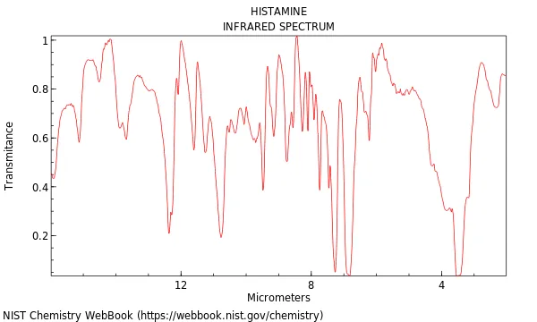
Laboratories rely on spectra to track the amine in complex samples. The Coblentz Society, Inc. logged infrared curves under reference 3858 for Wyandotte Chemicals Corp. in Wyandotte, Michigan, noting mineral oil bands near 2900, 1450, and 1380 cm-1 in the NIST Chemistry WebBook, which offers a Mol file download alongside the InChIKey NTYJJOPFIAHURM-UHFFFAOYSA-N and the average mass value 111.148. Meanwhile, analytical teams led by Liebig, Erban, and Kopka under Prof. Willmitzer at the Max Planck Institute of Molecular Plant Physiology in Golm, Germany, archived mass spectra of its 2TMS form in the Golm Metabolome Database while Kyoto University records index the clinical compound as Histamine DCF [6].
Synthesis in Mast Cells
Cells build histamine by removing a carboxyl group from the dietary amino acid L-histidine, a conversion catalyzed by L-histidine decarboxylase. This single step turns a neutral building block into a water-loving vasoactive amine. Mast cells store over 90 percent. They sit near tissue boundaries [7].
Dense clusters of mast cells patrol portals where invaders enter, guarding the nose, mouth, gut lining, skin, and outer walls of blood vessels. Inside these cells, histamine binds to heparin proteoglycans within dense storage granules. Non-mast cell pools also exist in enterochromaffin-like cells of the stomach and within neurons of the posterior hypothalamus. These cells do not hold large granule reserves like mast cells, releasing their amine directly after synthesis. Because the chemical controls diverse tasks across separate organs, local tissue management prevents unwanted leakage into general circulation so each tissue meets its own metabolic needs safely [7].
Chemical triggers can empty granules without immune help. Basic plant drugs like morphine and curare alkaloids displace histamine from heparin chains directly, prompting non-allergic skin flushing. The peptide drug polymyxin also triggers granule discharge without antibodies. In clinical medicine, the Kyoto Encyclopedia of Genes and Genomes notes under KEGG DRUG entry D08040 that doctors use histamine as a diagnostic agent to assess stomach acid output [5].
How Histamine Triggers Allergic Reactions
Classic allergy begins when the immune system mistakenly treats harmless pollen, dust, or food as a lethal threat. In sensitized hosts, plasma cells produce immunoglobulin E antibodies that lock onto high-affinity Fc epsilon RI receptors on mast cell walls. When matching allergens enter the body later, they bind across adjacent antibodies, opening calcium channels to trigger exocytosis [7].
Escaped histamine transforms local blood flow. Endothelial cells lining small venules contract, widening the tiny gaps between cells so plasma fluid, albumin, and defensive white cells leak into tissue beds to engage potential pathogens. The amine also signals smooth muscle cells to relax by driving nitric oxide release. Blood vessels swell. Leaking plasma causes red hives, heat, and puffy skin. In the nasal passages, fluid pouring through leaky capillary walls creates running noses and watery eyes to flush foreign particles away before tissue damage spreads. Airway passages narrow fast. Smooth muscle clamps down [7].
Allergic reactions also engage sensory nerves through a two-way signal loop. In a 2013 paper published in the British Journal of Pharmacology, scientists Rosa and Fantozzi explained that sensory nerve tips release substance P and calcitonin gene-related peptide during tissue irritation. These neuropeptides stimulate nearby mast cells to discharge histamine, which then acts on local nerve endings to cause persistent itching and trigger further peptide release [7].

Four Receptors Across Human Tissues
Histamine acts through four G protein-coupled receptors labeled H1, H2, H3, and H4. Each receptor sits in specific organs, joins distinct internal pathways, and triggers tailored biological actions. H1 governs smooth muscle spasms and nasal swelling, while H2 controls stomach acid and cardiac tempo across the body [7].
The H2 receptor links to adenylate cyclase through the Gs alpha subunit. Its binding spurs cyclic adenosine monophosphate production, turning on protein kinase A to modify target proteins. In oxyntic cells of the stomach lining, this signal drives gastric acid secretion, making H2 blockers like cimetidine a common therapy for peptic ulcers and reflux. Heart cells beat faster. Outside the gut, H2 receptors sit in cardiac muscle, bladder walls, and cerebellar dentate nuclei, where receptor stimulation causes smooth muscle relaxation by slowing myosin light-chain kinase. The drug betazole serves as an experimental agonist that stimulates these digestive pathways [8].
Nerve and immune cells carry the remaining receptor types. In the British Journal of Pharmacology, a research group led by Obara, Telezhkin, Alrashdi, and Chazot reported that presynaptic H3 autoreceptors control monoamine traffic, while postsynaptic H4 receptors modify neuropathic pain relief. Writing in Frontiers in Immunology, a team guided by Thangam and Church showed that H4 receptors coat mast cells and eosinophils to steer inflammatory cell movement [7].

Enzymatic Breakdown and Genetics
Free histamine must be cleared quickly to prevent blood pressure drops. Two primary enzymes break it down: diamine oxidase, encoded by the AOC1 gene, and histamine N-methyltransferase, encoded by the HNMT gene. Low activity in either enzyme lets amines linger in blood. It causes trouble [7].
Diamine oxidase acts as an export enzyme made in epithelial enterocytes lining small intestine villi. It neutralizes dietary histamine from meals before the chemical can cross into portal blood. Mutations in AOC1 weaken this digestive defense. Inherited alleles such as rs2052129, rs2268999, rs10156191, and rs1049742 reduce enzyme output, leaving carriers vulnerable to gut cramps and food sensitivities. In patients suffering from celiac damage or ulcerative colitis, worn intestinal villi fail to generate enough diamine oxidase, allowing dietary amines to seep unchecked into circulation where they trigger systemic complaints across distant organs. Intestinal lining loss multiplies amine absorption risks [7].
Histamine N-methyltransferase works inside cells across the brain, liver, and bronchial lining. Variations in HNMT alter neurotransmitter clearance. In children evaluated for attention-deficit hyperactivity disorder, the Thr105Ile and T939C alleles correlate with symptom flare-ups after drinking drinks with food dyes. In pediatric asthma groups, the HNMT-464 TT and HNMT-1639 TT genotypes appear frequently, especially among African-American children with sensitive airways [7].
Origins of Histamine Intolerance
Histamine intolerance describes a digestive issue where dietary amine intake exceeds the body’s breakdown capacity. When circulating levels surpass personal limits, people develop symptoms that resemble classic allergies without involving IgE antibodies. In Allergologia et Immunopathologia, researchers Kováčová, Buday, Gavliakova, and Plevková observed that sluggish enzymes allow foodborne histamine to spill into blood vessels and engage peripheral receptors [9].
Symptoms span multiple organs, muddying clinical diagnosis. Patients report stomach pain, bloating, loose stools, facial redness, hives, skin itch, migraines, and poor sleep. Yet histamine intolerance is not listed as a diagnosable condition in the International Classification of Diseases 11th Revision (ICD-11). Double-blind food challenge tests show erratic results, prompting medical debate over whether cases stem from pure enzyme failure or broader gut disorders. Diagnosis remains difficult. Clinical trials often produce contradictory outcomes [9].
Food poisoning differs from intolerance because massive amine doses poison anyone who eats spoiled meals. Known as scombroid poisoning, this toxic illness occurs when marine bacteria build high histamine levels in dark-meat fish of the Scombridae family, including tuna and mackerel. In contrast, healthy people break down the modest amine amounts found in Swiss Emmental cheese without trouble. When gut signs persist, doctors advise short-term low-histamine diets and diamine oxidase supplements to calm digestion [9].
Neurotransmitter Roles in the Brain
Histamine acts as a central neurotransmitter in the brain, springing almost entirely from the tuberomammillary nucleus in the posterior hypothalamus. Histaminergic axons fan out across the cortex, striatum, hippocampus, and spinal cord. Their firing tracks vigilance, firing during active daytime hours and falling quiet during non-rapid eye movement sleep [7].
Histamine heavily shapes motor control and mood within deep brain circuits. In Neuropharmacology, neuroscientists Bolam and Ellender showed that histamine adjusts signaling in the striatum, the primary gateway of the basal ganglia. Flaws in histaminergic transmission correlate with movement disorders like Tourette’s syndrome and Parkinson’s disease. Beyond receptor signaling, scientists discovered in 2012 that histamine binds directly to glutamine residues on histone proteins. This post-translational mark, called protein histaminylation, acts as a neuroepigenetic switch controlling circadian rhythm genes in brain tissue, establishing an unexpected molecular pathway that coordinates daily waking cycles [7].
The varied roles of histamine reflect its flexible molecular backbone and positive charge at blood pH. From opening blood vessels during allergy to pumping stomach acid and waking the brain, the amine remains an active chemical messenger. Insights into receptor targets and degradative enzymes continue to guide new therapies for immune and neurological diseases [7].
- WEBSITE National Institute of Standards and Technology. (2026). Histamine. NIST Chemistry WebBook, NIST Standard Reference Database Number 69. [Article Link]
- WEBSITE European Bioinformatics Institute. (2017). Histamine (CHEBI:18295). Chemical Entities of Biological Interest. [Article Link]
- DICTIONARY ENTRY Dictionary.com. (2023). Histamine. In Dictionary.com Unabridged. [Article Link]
- WEBSITE Royal Society of Chemistry. (2026). Histamine (ChemSpider ID: 753). ChemSpider. [Article Link]
- WEBSITE Kyoto Encyclopedia of Genes and Genomes. (n.d.). Histamine (KEGG DRUG: D08040). KEGG. [Article Link]
- WEBSITE Max Planck Institute of Molecular Plant Physiology. (2026). Histamine (2TMS). Golm Metabolome Database. [Article Link]
- WEBSITE Wikipedia contributors. (n.d.). Histamine. In Wikipedia, The Free Encyclopedia. [Article Link]
- WEBSITE Wikipedia contributors. (n.d.). Histamine H2 receptor. In Wikipedia, The Free Encyclopedia. [Article Link]
- WEBSITE Wikipedia contributors. (n.d.). Histamine intolerance. In Wikipedia, The Free Encyclopedia. [Article Link]
Cover image: SariSabban / Wikimedia Commons, CC BY-SA 3.0


