



Rabies virus is a lethal neurotropic pathogen that attacks the central nervous system of warm-blooded mammals, causing progressive encephalomyelitis that proves fatal in 100% of human cases once clinical signs emerge. Found on every continent except Antarctica, it transmits through the saliva of infected mammals via deep bite wounds or scratches [1],. The pathogen quietly invades peripheral nerve endings before travelling along motor pathways toward the spinal cord and brain [2]. Because early incubation produces no fever or tissue distress, exposed people feel healthy for weeks or months after an animal encounter [11]. Prompt wound care and post-exposure prophylaxis can stop the pathogen before central nerves become infected [3].
- Rabies Virus Structure
- How Rabies Virus Enters Muscle and Nerves
- Axonal Transport to the Central Nervous System
- Why Rabies Virus Triggers Hydrophobia
- Progression of the Paralytic Form
- Diagnostic Confirmation in Suspected Cases
- Post-Exposure Prophylaxis Protocols
- Global Elimination Through Mass Dog Vaccination
Rabies Virus Structure
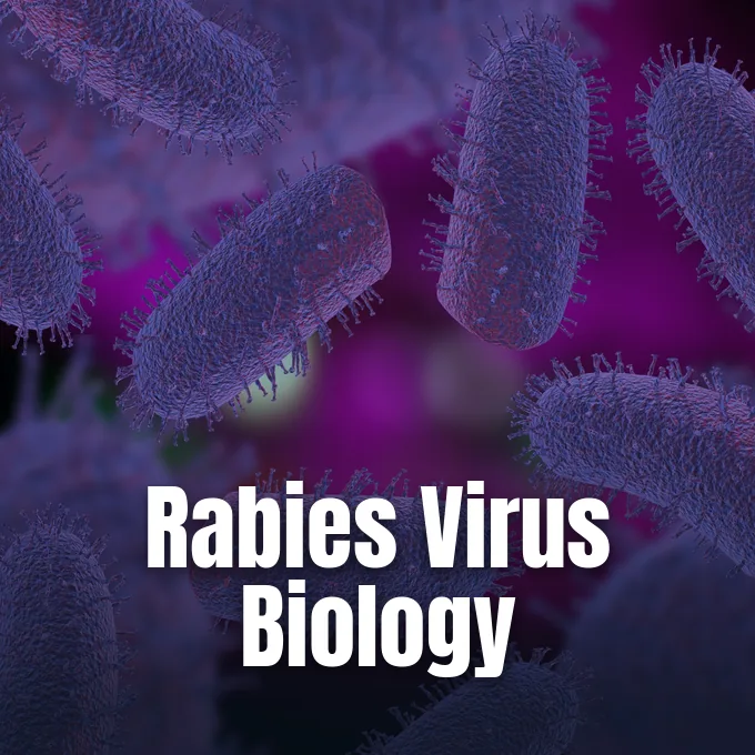
Rabies virions display an elongated bullet-like morphology that measures approximately 180 nanometers in length and 75 nanometers across their cylindrical cross-section, distinguishing the pathogen from the spherical capsids found in common human viruses [12]. The pathogen belongs to the family Rhabdoviridae and represents the type species of the genus Lyssavirus, an ancient group of negative-strand RNA viruses that infect mammals and perpetuate in reservoirs like dogs, foxes, and bats [2]. One end presents a rounded conical tip while the opposing base forms a flat planar surface [12]. A host-derived lipoprotein envelope wraps around the ribonucleoprotein core, leaving virions vulnerable to quick dissolution when exposed to lipid-dissolving household detergents, bleach, or ultraviolet sunlight [10], [12].
The single-stranded genomic RNA spans 11,615 to 11,966 nucleotides in length without segmented breaks. Five distinct genes make up this sequence: N encodes the nucleoprotein coat, P produces the phosphoprotein, M directs matrix assembly, G generates surface spikes, and L supplies the viral RNA polymerase. The nucleoprotein packages the negative-sense RNA strand tightly at a ratio of one protein unit for every nine nucleotides. This tight assembly forms an organized ribonucleoprotein core that resists host enzymes [12].
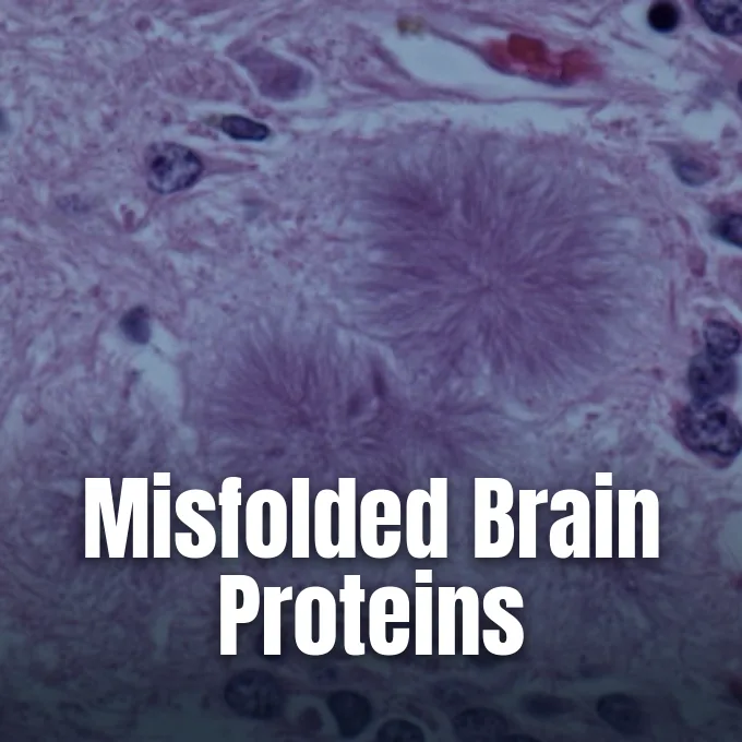
Roughly 400 trimeric spikes made of glycoprotein G protrude from the membrane to steer host cell attachment. Inside neurons, replication produces cytoplasmic clusters named Negri bodies after Italian pathologist Adelchi Negri. Negri bodies provide conclusive postmortem proof [12].
How Rabies Virus Enters Muscle and Nerves
Infection begins when a rabid carnivore or bat breaks the skin during a bite, shedding saliva into underlying muscle fibers and connective tissues without triggering prompt immune alarms [1], [2]. Intact human skin blocks viral penetration fully, making mechanical trauma an absolute requirement for successful inoculation [10]. Although virions occasionally contact nerve endings at once, rabies virus particles often replicate locally inside striated myocytes before engaging peripheral nerves [5],. The viral glycoprotein attaches to nicotinic acetylcholine receptors located at neuromuscular junctions, while neural cell adhesion molecules also facilitate cell entry. The pathogen enters quiet host cells without causing cellular death [11].
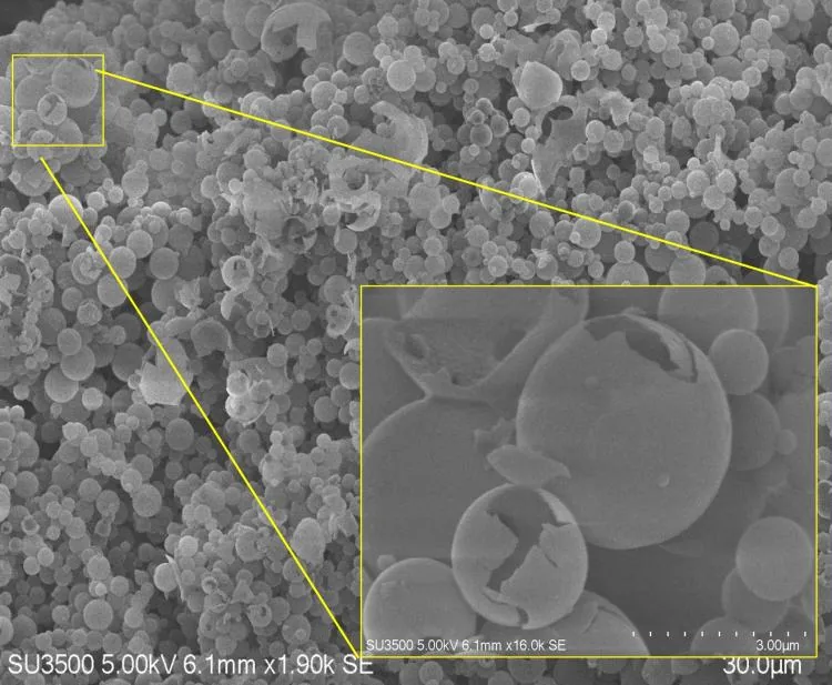
Canadian researcher K. M. Charlton and colleagues showed in 1997 that striped skunks (Mephitis mephitis) inoculated with street strains maintained detectable viral RNA in muscle fibers at the bite site for two months. Polymerase chain reaction assays showed that regional nerve trunks and spinal cord segments remained free of viral RNA during that early period. This localized pause in muscle explains why incubation periods vary widely between exposure events [5].
Once bound to surface receptors, host cell membranes pinch inward through pinocytosis, engulfing the virion inside an acidic endosome. Low pH stimulates envelope fusion, releasing the viral core into the cytoplasm [11]. Axonal transit begins along motor pathways [4].
Axonal Transport to the Central Nervous System
Inside peripheral motor axons, the viral core travels exclusively by retrograde intra-axonal transport toward spinal cord motor neurons, evading neutralizing antibodies circulating in blood vessels [4], [11]. Ultrastructural research published by virologist Frederick Murphy in 1977 established that the pathogen hijacks normal retrograde cellular pathways instead of diffusing across extracellular fluids or traveling through blood vessels [4]. The viral phosphoprotein binds to host cytoplasmic dynein light chains, linking the viral ribonucleoprotein complex to microtubule motors [11]. These motor proteins pull the pathogen to neuron cell bodies at speeds between 8 and 20 millimeters each day [2],. It travels silently [4].
This steady rate of travel explains why bite location influences the length of the asymptomatic incubation window [1],. Exposures on distant extremities like toes or fingers force traveling virions to traverse extended nerve lengths before reaching the spinal cord [2]. In contrast, deep bites to the face, neck, or head shorten this travel distance, triggering clinical symptoms within two weeks [1], [2]. Routine blood tests cannot detect the infection during this period [10].
Upon reaching spinal sensory ganglia and dorsal horns, the virus multiplies before crossing trans-synaptic gaps to neighboring neurons [2], [4]. It climbs ascending tracts into the brainstem, thalamus, and cerebellum. From there, infection spreads through the cerebral cortex [2].
Why Rabies Virus Triggers Hydrophobia
Encephalitic rabies, historically designated as furious rabies, develops in roughly 80% of human patients as invading virions spread through the limbic system, amygdala, and brainstem nuclei to disrupt normal neurological control over emotion and autonomic stability [1], [11]. Agitation and terror alternate with moments of calm lucidity [1], [11]. Autonomic storming triggers heavy sweating, dilated pupils, fever, and uncontrolled muscle twitches, while the World Health Organization reports that 40% of all rabies victims worldwide are children under 15 years old [1], [2].
Why does drinking water provoke sudden terror in furious rabies? The reaction represents a painful reflex rather than a psychological aversion [2],. When a patient attempts to swallow liquids, destruction in brainstem motor nuclei triggers violent spasms in the diaphragm and larynx [11]. The sensation of choking is so distressing that the mere sound, sight, or mention of water sparks throat spasms [1], [2]. Unable to swallow, the patient begins drooling, allowing infectious saliva to pool in the mouth [11].
Centrifugal neural transport carries rabies virus particles outward from the brain along autonomic fibers to the salivary glands and cornea [2], [4]. In the CDC Yellow Book, clinicians recorded an infected traveler who underwent cardiac catheterization because autonomic rabies mimicked a heart emergency [2].

Progression of the Paralytic Form
Roughly 20% of human rabies cases present as paralytic rabies, a quieter manifestation historically known as dumb rabies that runs an extended clinical course [1], [11]. Instead of explosive excitement and hydrophobia, patients develop progressive muscle weakness that begins at the bitten limb and ascends symmetrically or asymmetrically toward the torso [1],. This clinical picture closely resembles Guillain-Barré syndrome, leading physicians to misdiagnose the underlying viral encephalitis. Weakness slowly advances to quadriplegia and urinary incontinence [11]. Because distinctive hydrophobic spasms never emerge, public health registries often miss these cases entirely [1], [2].
The latent incubation period of rabies virus infection averages two to three months, though verified cases range from under seven days to over a year [1], [11]. Prodromal illness begins with non-specific signs including low fever, headache, tiredness, and aching muscles [1], [2]. An early hallmark is tingling, burning, or itching at the healed bite wound, reflecting viral irritation of local sensory ganglia [1], [4]. Once brain inflammation spreads, weakness progresses into stupor, coma, and fatal respiratory failure [1], [11].
Once central nervous signs emerge, rabies proves fatal in 100% of human cases, making supportive sedation the primary compassionate recourse [1]. Survival remains an unusual medical anomaly accompanied by severe neurological deficits [2]. One patient in 2004 survived bat rabies without receiving vaccination [3], [11].

Diagnostic Confirmation in Suspected Cases
Diagnosing human rabies before death poses formidable medical challenges because the earliest complaints mimic common viral prodromes like influenza, viral meningitis, or psychological distress, masking the presence of a fatal neurological infection. Unless the patient or family recalls an animal bite, attending clinicians rarely consider lyssavirus exposure during early examinations, delaying crucial risk assessments for hospital personnel [2], [3]. Surveillance data in the United States show that early clinical encounters almost always overlook the disease [2], [3]. Certainty develops only when classic hydrophobia or localized paralysis emerges in the final days of illness [1].
Antemortem testing requires multiple biological specimens collected over several days to account for intermittent viral shedding. Testing protocols evaluate saliva, serum, spinal fluid, and cutaneous biopsies taken from the nape of the neck. Cutaneous biopsies must contain hair follicles, where sensory nerves harbor viral antigen detectable by direct fluorescent antibody staining [2], [4]. Reverse transcription polymerase chain reaction assays amplify viral RNA from saliva or fluid aspirates within hours. Neutralizing antibodies in spinal fluid from an unvaccinated person confirm the disease [2].
Postmortem diagnosis in animals and humans relies on microscopic examination of brainstem and cerebellar tissue [2], [9]. In states like North Dakota, public health laws require biting wild mammals to be euthanized for direct testing [10]. Domestic dogs, cats, and ferrets that bite a human can be quarantined for ten days under veterinary observation [2],. Healthy animals after ten days were non-infectious during the bite [10].
Post-Exposure Prophylaxis Protocols
Post-exposure prophylaxis acts as an urgent biological barrier that prevents rabies virus particles from gaining access to peripheral nerve terminals after an animal bite [1]. First introduced by Louis Pasteur in 1885, immunization has advanced to modern cell-culture formulations [13]. The first line of defense is prompt wound care: washing bites and scratches with soap and running water for at least 15 minutes [1],. The viral particle possesses a delicate lipid membrane that is easily inactivated by soap, detergents, povidone-iodine, and ethanol [10],. This simple washing step flushes away viral particles before they can bind to cell receptors [11].
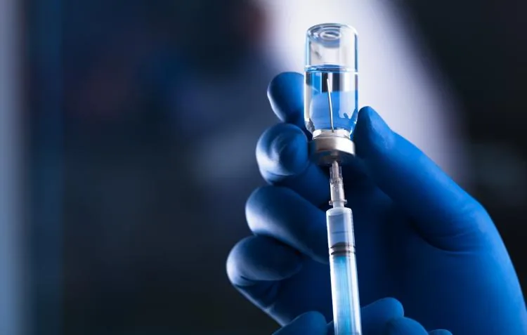
Unvaccinated bite victims require prompt passive and active immunization known collectively as PEP [1], [3]. Human rabies immune globulin (RIG) or monoclonal antibodies (RmAb) supply neutralizing coverage, and clinicians must infiltrate the full calculated dose into and around the wound margins [1], [2], [3]. Any remaining solution is injected into an anatomical site distant from the vaccine [6]. Active immunization follows with cell-culture vaccine doses given on days 0, 3, 7, and 14 [3],. Injections must enter the deltoid muscle in adults or the anterolateral thigh in infants, because gluteal fat produces inadequate antibody titers [6].

Over 29 million people receive post-exposure immunization every year around the world [1], [13]. In 2018, the average cost of post-exposure care reached US$ 108, posing a heavy economic burden in low-income regions. The World Health Organization prequalifies three vaccines globally: Verorab by Sanofi Pasteur, Rabivax-S by Serum Institute of India, and VaxiRab N by Zydus Lifesciences, while preparations like RabAvert and Imovax Rabies remain standard in North America [1], [6], [7]. Persons who received prior pre-exposure vaccination need only two booster shots on days 0 and 3, omitting immune globulin [3], [13].
Global Elimination Through Mass Dog Vaccination
Domestic dogs represent the primary reservoir for rabies across Africa and Asia, causing 99% of all human transmissions. Culling free-roaming dog packs fails to halt transmission, whereas mass dog vaccination breaks the cycle of infection. In Latin America and Caribbean nations, over 50 million dogs were vaccinated annually during peak campaigns, drastically cutting canine and human cases [1]. In the United States, canine vaccination begun after World War II reduced domestic dog rabies from 8,384 cases in 1946 to 79 cases in 2006 [3]. Wildlife species like raccoons, skunks, foxes, and bats now account for more than 90% of American animal cases, while Hawaii remains rabies free [2], [3], [9].
International health agencies collaborate under the Zero by 30 roadmap, an initiative led by the WHO, FAO, and WOAH to end dog-mediated human deaths by 2030. Gavi, the Vaccine Alliance supports vaccine access under Gavi 6.0 funding guidelines, while 17 countries have self-declared free of infection under Chapters 1.6 and 8.15 of the Terrestrial Code. As of September 2026, three countries—Bhutan, Chile, and Mexico—have earned WHO validation for eliminating dog-mediated human rabies. The World Health Organization calculates that global rabies damages exceed US$ 8.6 billion each year in economic losses and medical care. According to 2021 Global Health Estimates data, documented deaths reached 44 203 worldwide [1].
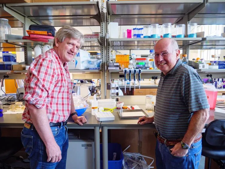
In North America, USDA Wildlife Services drops fishmeal baits containing Raboral V-RG vaccine from airplanes like the Beechcraft King Air and De Havilland Twin Otter to immunize wild raccoons against rabies virus [9]. Meanwhile, University of Colorado Boulder researchers Ted Randolph and Robert Garcea formed startup company VitriVax to create temperature-stable single-dose vaccines. These sugary beads endured heat reaching 104 degrees Fahrenheit over three full months without losing potency [8].
- WEBSITE World Health Organization. (2026). Rabies. [Article Link]
- REPORT Centers for Disease Control and Prevention. (2026). Rabies. CDC Yellow Book 2026. [Article Link]
- REPORT Manning, S. E., Rupprecht, C. E., Fishbein, D., Hanlon, C. A., Lumlertdacha, B., Guerra, M., Meltzer, M. I., Dhankhar, P., Vaidya, S. A., Jenkins, S. R., Sun, B., & Hull, H. F. (2008). Human Rabies Prevention — United States, 2008: Recommendations of the Advisory Committee on Immunization Practices. MMWR. Recommendations and Reports, 57(RR-3), 1-28. [Article Link]
- ACADEMIC JOURNAL Murphy, F. A. (1977). Rabies Pathogenesis: Brief Review. Archives of Virology, 54(4), 279-297. [Article Link]
- ACADEMIC JOURNAL Charlton, K. M., Nadin-Davis, S., Casey, G. A., & Wandeler, A. I. (1997). The Long Incubation Period in Rabies: Delayed Progression of Infection in Muscle at the Site of Exposure. Acta Neuropathologica, 94(1), 73-77. [Article Link]
- WEBSITE Sanofi Vaccines US Inc. (2026). IMOVAX RABIES (Rabies Vaccine) Prescribing Information. [Article Link]
- WEBSITE GlaxoSmithKline Biologicals SA. (2019). RabAvert Rabies Vaccine Prescribing Information. [Article Link]
- ONLINE NEWS Carlson, A. (2025). New Single-Dose, Temperature-Stable Rabies Vaccines Could Expand Global Access. CU Boulder Today. [Article Link]
- WEBSITE Animal and Plant Health Inspection Service. (2023). Rabies – Frequently Asked Questions. U.S. Department of Agriculture. [Article Link]
- WEBSITE Health and Human Services North Dakota. (2026). Rabies FAQs. [Article Link]
- WEBSITE Wikipedia contributors. (2026). Rabies. Wikipedia, The Free Encyclopedia. [Article Link]
- WEBSITE Wikipedia contributors. (2026). Rabies Virus. Wikipedia, The Free Encyclopedia. [Article Link]
- WEBSITE Wikipedia contributors. (2026). Rabies Vaccine. Wikipedia, The Free Encyclopedia. [Article Link]
- ACADEMIC JOURNAL Rao, A. K., Briggs, D., Moore, S. M., Whitehill, F., Campos-Outcalt, D., Morgan, R. L., Wallace, R. M., Romero, J. R., Bahta, L., Frey, S. E., & Blanton, J. D. (2022). Use of a Modified Preexposure Prophylaxis Vaccination Schedule to Prevent Human Rabies: Recommendations of the Advisory Committee on Immunization Practices — United States, 2022. MMWR. Morbidity and Mortality Weekly Report, 71(18), 619-627. [Article Link]


