





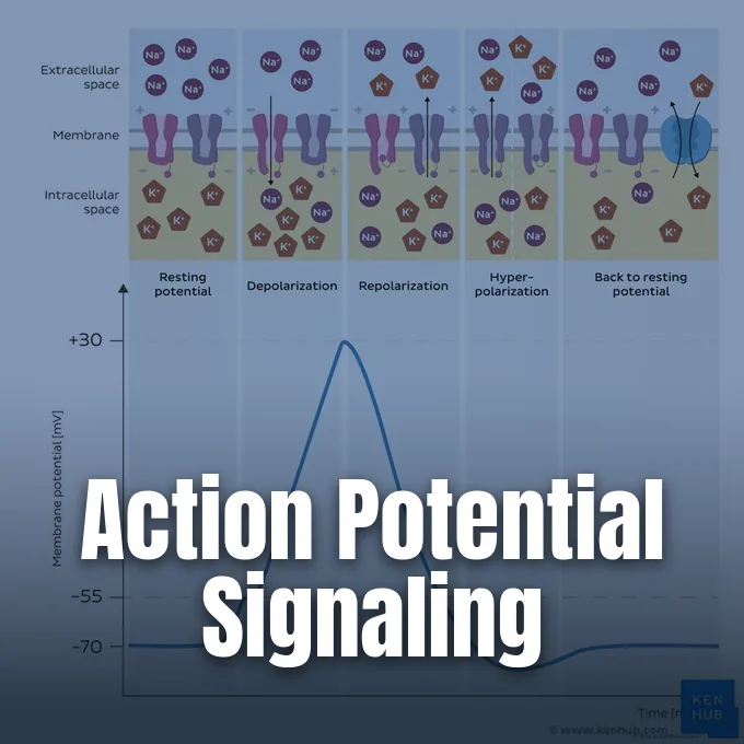
An action potential is a fast, temporary change in electrical voltage across a cell membrane that lets excitable cells send signals over long distances. While most cells keep a steady voltage difference between their interior and exterior fluids, neurons and muscle fibers can abruptly reverse this charge in response to incoming stimuli. The entire cycle takes only a few milliseconds, driven by the opening and closing of ion channels embedded in the lipid bilayer [7].
- What Is an Action Potential?
- Resting Membrane Potential and Ion Gradients
- Sodium Influx During the Depolarization Phase
- Potassium Efflux During Repolarization
- Refractory Periods in Action Potential Timing
- How Axons Propagate Electrical Signals
- Synaptic Transmission at Axon Terminals
- Diverse Roles of the Action Potential Across Tissues
What Is an Action Potential?
Neurons use these brief electrical impulses to share information throughout the nervous system, generating an action potential whenever excitation surpasses threshold [8]. When an incoming stimulus nudges a neuron’s membrane voltage past a critical point, voltage-gated ion channels open in an all-or-none sequence. If the voltage fails to reach that point, nothing happens. Once triggered, the impulse travels down the axon at full strength without decaying, meaning that a signal arriving at an axon tip meters away carries the same amplitude as it had at the axon hillock.
Early insights into this electrical signaling came from experiments on the squid giant axon, which can reach up to 1 mm in diameter and is visible to the naked eye [11]. Neuroscientist Eric H. Chudler explains that this unmyelinated axon innervates mantle muscles that propel the squid through water. Because a mammalian axon is between 100 and 1,000 times thinner, early twentieth-century recording electrodes were too large to penetrate mammalian nerve fibers without destroying them. The sheer size of the squid axon let researchers insert internal electrodes directly into the axoplasm, creating the experimental foundation for modern cellular electrophysiology and proving that electrical currents across cell membranes govern nerve transmission.
Neuroscientists describe this binary behavior as the all-or-none principle. A neuron can’t fire a half-sized spike in response to weak stimulation, nor can a stronger stimulus produce a larger wave [15]. Stimulus strength is reflected solely in firing rate rather than spike size.
Resting Membrane Potential and Ion Gradients
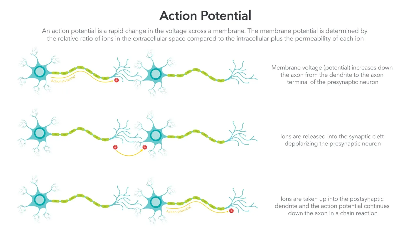
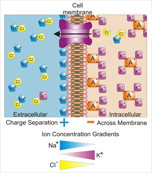
Before any action potential can occur, a cell must establish an uneven distribution of charged ions across its membrane. In human neurons, the resting membrane potential sits between -50 mV and -75 mV, with -70 mV serving as the typical benchmark [10]. This negative interior charge reflects an unequal distribution of sodium (Na⁺), potassium (K⁺), chloride (Cl⁻), and impermeable organic anions. At rest, potassium ions are concentrated inside the cytosol, while sodium and chloride ions remain predominantly in the extracellular fluid. Ion movement depends on transmembrane channel proteins that select for specific ionic radii and charges through electrochemical exclusion [12]. Leakage channels allow potassium ions to drift out of the cell down their chemical concentration gradient far more readily than sodium can enter.
Large negatively charged proteins trapped along the inner leaflet of the plasma membrane can’t follow the exiting potassium ions, leaving an excess of negative charge hugging the inner surface of the membrane. To keep these concentration gradients from dissipating over time, an active transport pump uses energy from adenosine triphosphate (ATP). Danish physiologist Jens Skou isolated this enzyme in 1957, later sharing the 1997 Nobel Prize in Chemistry [7]. For each molecule of ATP consumed, the sodium-potassium pump extrudes three Na⁺ ions while importing two K⁺ ions. This exchange maintains the concentration gradients that store potential energy for subsequent signaling events.
Sodium Influx During the Depolarization Phase
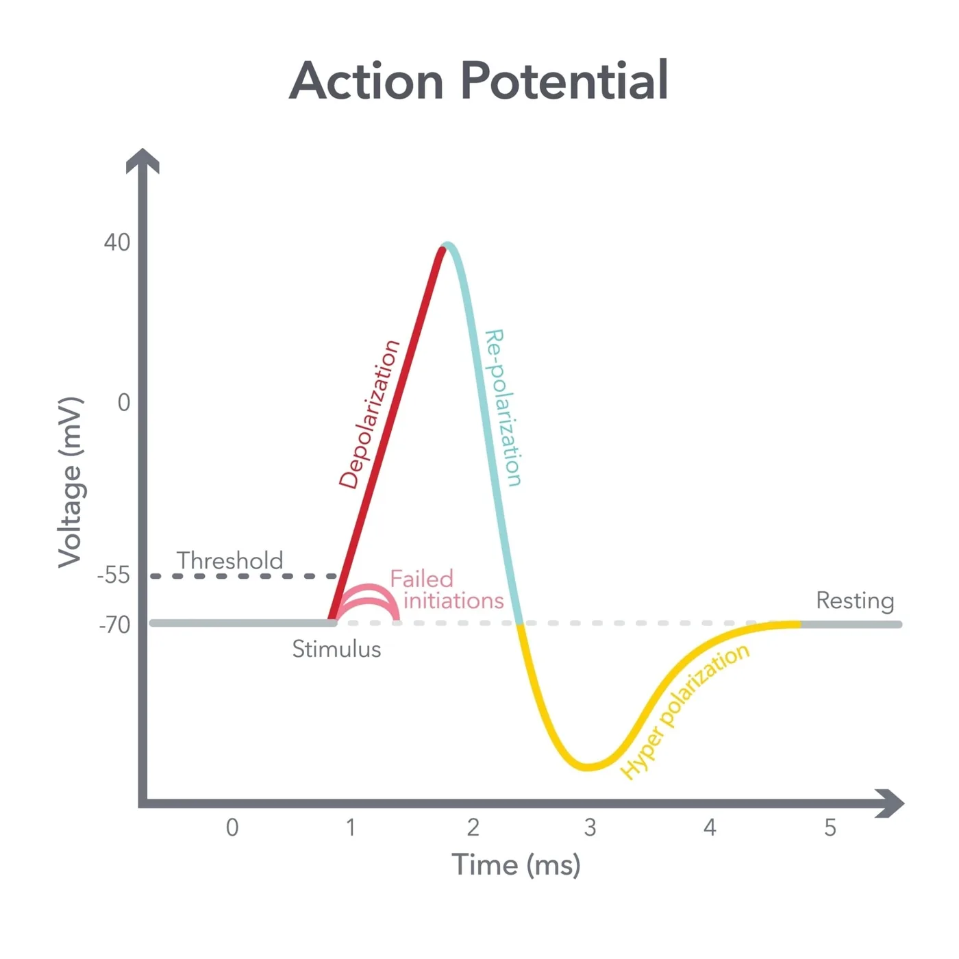
How do these resting conditions shift when an external signal arrives? When neurotransmitters bind to dendrites or sensory receptors experience pressure, small graded potentials shift the membrane voltage toward zero. If localized hypopolarization pushes the membrane potential from -70 mV up to the threshold of roughly -55 mV, voltage-gated sodium channels swing open [14]. The drop in electrical resistance allows sodium ions to surge into the cell, driven by both the steep chemical concentration gradient and the electrical attraction of the negative interior. Because sodium is roughly ten times more concentrated outside the neuron than inside, entering cations depolarize neighboring membrane patches, which in turn opens additional voltage-gated channels in a rapid cycle [9].
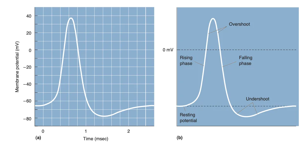
The membrane potential shoots upward past 0 mV into positive territory, an event termed the overshoot phase. In typical mammalian neurons, this spike crests between +30 mV and +40 mV, heading toward the sodium electrochemical equilibrium potential of +61 mV. Electrophysiologists study these rapid ionic shifts in the laboratory using patch-clamp techniques that record currents through single channel pores [13]. Voltage-clamp amplifiers supply opposing current to hold membrane potential at set values, measuring the millivolt shifts that occur during excitation. These measurements show that the massive voltage swing requires only a tiny fraction of available ions to cross the membrane, leaving bulk intracellular ion concentrations stable.
Potassium Efflux During Repolarization
A nerve spike can’t stay positive for long if the cell is to fire again. At the peak of depolarization, voltage-gated sodium channels automatically shut down through a process called inactivation, halting further sodium entry even though the membrane remains depolarized [10]. At roughly the same moment, voltage-gated potassium channels open their pores. Because potassium is heavily concentrated inside the cell, opening these channels prompts a swift outward rush of positive potassium ions down their electrochemical gradient.
As positive charge leaves the cytosol, the internal voltage drops sharply back toward negative levels during repolarization. However, voltage-gated potassium channels close more slowly than sodium channels inactivate [8]. This delayed closure permits potassium ions to continue leaking out, driving the membrane potential down to roughly -80 mV before leveling off.
This two-stage ionic mechanism settled a long-standing historical debate in neurophysiology. Around 1900, German physiologist Julius Bernstein suggested that excitation involved a nonselective breakdown of membrane resistance, predicting that internal voltage would collapse toward zero without ever turning positive [7]. British physiologists Alan Hodgkin and Andrew Huxley disproved that idea in 1939 by recording an actual positive reversal of roughly +40 mV inside squid giant axons. Their 1952 mathematical equations demonstrated that two distinct currents—first sodium influx, then delayed potassium efflux—fully explain the waveform, earning them the 1963 Nobel Prize in Physiology or Medicine with John Eccles.


Refractory Periods in Action Potential Timing
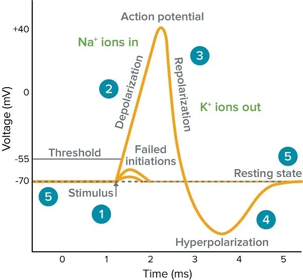
Following the initiation of an action potential, an excitable cell enters a recovery window during which it can’t generate normal impulses. This recovery period is split into two distinct stages: the absolute refractory period and the relative refractory period [14]. The absolute phase spans depolarization and the first two-thirds of repolarization, lasting about one millisecond. During this window, voltage-gated sodium channels are already open or locked in an inactive state, making it physically impossible for any stimulus to trigger another spike.
As repolarization completes its final third, the membrane enters the relative refractory period. Sodium channels begin transitioning from their inactivated state back to their resting, closed configuration [15]. A new spike can occur during this stage, but only if an incoming stimulus is substantially stronger than normal threshold requirements.
Refractory dynamics govern the maximum firing frequency of neural circuits and shape collective brain rhythms. In rat neocortical recordings, researchers found that high-frequency oscillations in the 50 to 180 Hz band correlate closely with spike timing [2]. When neurons fire rapidly during sensory processing, local field potentials register these collective bursts. Because each neuron must wait for its sodium channels to reset, refractoriness sets an upper ceiling on firing rate, preventing continuous runaway excitation across cortical networks and ensuring that signals remain organized in discrete informational packets.
How Axons Propagate Electrical Signals
Once an action potential ignites at the axonal initial segment, it must travel the entire length of the fiber without losing strength. In unmyelinated fibers, incoming sodium ions generate local currents that spread forward along the interior of the membrane, depolarizing adjacent patches to threshold [10]. Because the patch immediately behind the wavefront is locked in its refractory period, the impulse can’t double back. Conduction travels strictly forward toward the axon terminals, though continuous regeneration across every micrometer makes this process relatively slow.
Vertebrates evolved myelin sheaths to boost conduction velocity while conserving metabolic energy. Formed by oligodendrocytes in the central nervous system and Schwann cells in the periphery, myelin insulates the axon, raising membrane resistance and decreasing capacitance [10]. Gaps in this insulation, known as Nodes of Ranvier, contain dense clusters of voltage-gated sodium channels.
Electrical current flows passively through the insulated internodes and regenerates only at the nodes, an efficient process known as saltatory conduction that speeds transmission up to tens of meters per second. Axon conduction also interacts with surrounding glial cells to guide circuit development and fine-tune signal shape. Research in mouse dorsal root ganglion neurons shows that glutamate released along active axons stimulates local synthesis of myelin basic protein through Fyn kinase signaling, promoting myelination where electrical traffic is highest [4]. Furthermore, ex vivo hippocampal CA3 recordings show that astrocytic calcium signaling can broaden axonal waveforms during conduction [5]. In cortical circuits, sustained signal transmission depends on coordinated network feedback, as described in research on the primary visual cortex and its mechanisms of recurrent activity.
Synaptic Transmission at Axon Terminals
When an electrical wavefront reaches the swollen axon terminals, electrical signaling gives way to chemical communication. Depolarization of the presynaptic membrane triggers the opening of voltage-gated calcium channels [14]. Because calcium is far more concentrated outside the cell, Ca²⁺ rushes into the terminal button. This rapid calcium influx prompts synaptic vesicles to fuse with the presynaptic membrane, releasing stored neurotransmitters into the narrow synaptic cleft via exocytosis. Once released, neurotransmitter molecules diffuse across the synaptic cleft and bind to receptor proteins on the postsynaptic target cell. Depending on whether the receptor activates cation or anion channels, the incoming chemical message either excites or inhibits the receiving neuron.
Clinical pharmacology takes advantage of these mechanisms to manage pain and modify nerve excitation. Local anesthetics such as lidocaine bind to voltage-gated sodium channels from within the neuronal membrane, blocking sodium influx and preventing sensory nerves from firing pain signals [7]. Calcium signaling at the terminal also intersects with broader cellular stress pathways and metabolic maintenance. When calcium regulation falters, stress within the endoplasmic reticulum can trigger apoptotic pathways, illustrating how ion homeostasis directly preserves cell survival [3]. Neural adaptations and synaptic resilience also help preserve complex cognitive processing across decades, a dynamic observed when adults strengthen cognitive reserve by learning another language. In healthy tissue, precise ion clearance mechanisms quickly reset calcium concentrations, ensuring that subsequent electrical impulses can reliably trigger further neurotransmitter release.
Diverse Roles of the Action Potential Across Tissues
Although neurons receive the most attention, action potentials also operate in several other excitable tissues, most notably the heart. Cardiac muscle does not require nervous stimulation to beat; instead, specialized pacemaker cells in the sinoatrial node generate spontaneous electrical impulses at a rate of 60 to 100 per minute [16]. These rhythmic impulses spread through intercalated discs across the atria and ventricles, synchronizing muscular contraction to pump blood. Disruptions in this electrical sequence produce cardiac arrhythmias visible on electrocardiograms.
Ventricular cardiomyocytes exhibit a five-phase waveform that differs markedly from brief neuronal spikes. Phase 0 features rapid depolarization driven by sodium channels opening at rates of roughly 300 V/s [17]. Phase 2 introduces a prolonged plateau lasting approximately 100 milliseconds, maintained by balanced calcium influx and potassium efflux.
Action potentials also coordinate movement and hormone secretion across diverse organisms. In the nematode Caenorhabditis elegans, which lacks voltage-gated sodium channels entirely, body wall muscle contractions rely on all-or-none calcium action potentials driven by L-type calcium channels and Kv1 potassium channels [6]. Beyond muscle contraction, electrical spikes prompt pancreatic beta cells to release insulin into the bloodstream. Moreover, resting membrane potentials in non-excitable tissues regulate cell proliferation, cell volume control, and wound healing, demonstrating that bioelectric dynamics extend far beyond the classical nervous system [1].
- ACADEMIC JOURNAL Kadir, L. A., Stacey, M. W., & Barrett‐Jolley, R. (2018). Emerging Roles of the Membrane Potential: Action Beyond the Action Potential. Frontiers in Physiology, 9, 1661-1661. [Article Link]
- ACADEMIC JOURNAL Watson, B. O., Ding, M., & Buzsáki, G. (2017). Temporal coupling of field potentials and action potentials in the neocortex. European Journal of Neuroscience, 48(7), 2482-2497. [Article Link]
- ACADEMIC JOURNAL Bahar, E., Kim, H., & Yoon, H. (2016). ER Stress-Mediated Signaling: Action Potential and Ca2+ as Key Players. International Journal of Molecular Sciences, 17(9), 1558-1558. [Article Link]
- ACADEMIC JOURNAL Wake, H., Lee, P. R., & Fields, R. D. (2011). Control of Local Protein Synthesis and Initial Events in Myelination by Action Potentials. Science, 333(6049), 1647-1651. [Article Link]
- ACADEMIC JOURNAL Sasaki, T., Matsuki, N., & Ikegaya, Y. (2011). Action-Potential Modulation During Axonal Conduction. Science, 331(6017), 599-601. [Article Link]
- ACADEMIC JOURNAL Gao, S., & Zhen, M. (2011). Action potentials drive body wall muscle contractions in Caenorhabditis elegans. Proceedings of the National Academy of Sciences, 108(6), 2557-2562. [Article Link]
- WEBSITE Guy-Evans, O. (2025). Action Potential: Definition, Steps, and Examples. Simply Psychology. [Article Link]
- WEBSITE Pan, S. (2024). Action Potential – Definition, Properties, Generation, Mechanism, Conduction. Biology Notes Online. [Article Link]
- WEBSITE Mukherjee, S. (2023). Action Potential – Definition, Phases, Examples, and Graph. Science Facts. [Article Link]
- WEBSITE Khanna, A. (2023). The Action Potential. TeachMePhysiology. [Article Link]
- WEBSITE Chudler, E. H. (2020). Action Potential. Neuroscience for Kids, University of Washington. [Article Link]
- WEBSITE Halpern, J. (2020). The Action Potential. Biology LibreTexts. [Article Link]
- WEBSITE Molecular Devices. (n.d.). What is an Action Potential? Membrane Potential & Graph. Molecular Devices. [Article Link]
- WEBSITE Kenhub. (2019). Action Potential: Key Facts, Steps and Phases. Kenhub. [Article Link]
- WEBSITE Wikipedia contributors. (n.d.). Action potential. In Wikipedia, The Free Encyclopedia. [Article Link]
- WEBSITE Wikipedia contributors. (n.d.). Cardiac action potential. In Wikipedia, The Free Encyclopedia. [Article Link]
- WEBSITE Wikipedia contributors. (n.d.). Ventricular action potential. In Wikipedia, The Free Encyclopedia. [Article Link]


