






Understanding what is cerebrospinal fluid begins with the clear, water-like liquid that bathes the brain and spinal cord [12]. Secreted inside the deep ventricles of the skull, the fluid wraps around delicate CNS tissue to provide protection [3]. It keeps the heavy brain buoyant, preserves intracranial stability, and clears cellular waste [12].
- What Is Cerebrospinal Fluid?
- Early Medical Observations of Brain Fluid
- How the Choroid Plexus Produces Fluid
- Circulation Through the Ventricular Cavities
- Why Cerebrospinal Fluid Is Important for Neural Health
- Normal Chemical Composition and Baseline Values
- How Cranial and Spinal CSF Leaks Differ
- Diagnostic Lumbar Punctures for Clinical Evaluation
What Is Cerebrospinal Fluid?
Medical science defines cerebrospinal fluid as an ultrafiltrate of blood plasma that fills the ventricles and subarachnoid spaces of the CNS. In healthy adults, the total fluid volume ranges between 125 and 150 milliliters at any single moment, though measurements can extend from 140 to 270 milliliters. The body renews this supply several times daily. Capillary beds produce fresh fluid at 0.2 to 0.7 milliliters every minute, yielding between 500 and 700 milliliters over twenty-four hours [3].
Fluid distribution changes as humans grow from birth to maturity, influencing how doctors administer spinal anesthetics and interpret intracranial pressure. Newborn infants carry approximately 4 milliliters per kilogram of body weight, whereas growing children average 3 milliliters per kilogram and mature adults hold between 1.5 and 2 milliliters per kilogram. Because young infants possess a much larger fluid volume relative to their body size, pediatric anesthesiologists at major hospitals must calculate higher weight-adjusted drug doses to achieve effective spinal blocks during surgery. Young age protects patients. Children experience lower rates of post-puncture headaches following lumbar punctures, as higher relative fluid capacity buffers against sudden volume drops [12].
Inside the cranium, fluid volume divides between internal cavities and external surfaces. The four brain ventricles hold only about 25 milliliters of fluid at any given time, meaning that over eighty percent surrounds the cerebral surface and spinal cord [3]. Fluid circulates without pause.
Early Medical Observations of Brain Fluid
Medical awareness of intracranial fluid began in antiquity, yet accurate understanding took more than two thousand years to develop. Ancient Greek physician Hippocrates (460–375 BC) noted watery fluid surrounding the brain when recording early descriptions of congenital hydrocephalus. Centuries later, Roman anatomist Galen (130–200 AD) described what he termed an excremental liquid inside the cerebral ventricles, mistakenly asserting that the brain purged this liquid outward through the nasal passages. Old autopsies missed the liquid. Dissectors severed the head from the neck, draining all fluid and blood before examination [4].
Emanuel Swedenborg made a key discovery between 1741 and 1744 after attending medical dissections in France, Germany, and Italy. He found clear liquid. In an unpublished manuscript discovered in Stockholm a century later, Swedenborg described this liquid as spirituous lymph and showed that it flows from the fourth ventricle into the spinal cord [4].
During the eighteenth and nineteenth centuries, European investigators charted fluid pathways through careful anatomical dissections. Swiss physiologist Albrecht von Haller showed in 1747 that ventricular water is secreted inside brain cavities and absorbed by veins, warning that excess fluid causes hydrocephalus. Italian professor Domenico Cotugno documented fluid in the inner ear labyrinth, while French physician Francois Magendie discovered the median aperture of the fourth ventricle. Heinrich Quincke popularized diagnostic lumbar punctures in 1891. In 1912, French neurologist William Mestrezat published the first accurate chemical analysis of the fluid, and American neurosurgeon Harvey Cushing proved in 1914 that the choroid plexus secretes it [4].
How the Choroid Plexus Produces Fluid
Most fluid production takes place within vascular tissue folds known as the choroid plexuses [3]. These structures reside inside the two lateral ventricles as well as the third and fourth ventricles, but they are absent from the cerebral aqueduct [12]. Histologically, each choroid plexus consists of capillary loops lined by thin fenestrated endothelial cells. Modified cuboidal ependymal cells cover these vessels, presenting microvilli that expand contact with the ventricular lumen [3].
Fluid generation operates through a two-step transport mechanism that builds the blood-cerebrospinal fluid barrier [3]. First, blood pressure drives a plasma filtrate out through leaky capillary pores into the surrounding connective tissue space. Next, choroid epithelial cells actively transport ions across their tightly joined membranes to establish an osmotic gradient [12]. Apical membranes contain high concentrations of Aquaporin 1 (AQP1) water channels and sodium-potassium ATPase pumps, while carbonic anhydrase converts dissolved carbon dioxide and water into bicarbonate and hydrogen ions [3]. This enzymatic exchange drives sodium, chloride, and bicarbonate into the ventricle, pulling water along to create fresh, isotonic fluid [12]. Tight junctions block larger blood proteins [3].
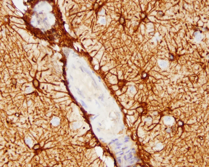
How do extra-choroidal tissues contribute to this daily output? Ependymal cells lining the ventricular walls lack tight junctions, allowing fluid to seep between brain tissue and the cavities. Water filtered from parenchymal microvessels into the brain interstitial fluid drains continuously into the ventricles, regulated by astrocytic Aquaporin 4 channels [3].
Circulation Through the Ventricular Cavities
Circulation follows a clear anatomical route through the interconnected chambers of the CNS. Secreted within the lateral ventricles, fluid passes through the paired interventricular foramina into the midline third ventricle. From there, it flows downward through the narrow cerebral aqueduct of Sylvius into the fourth ventricle, located anterior to the cerebellum [12]. The fluid then exits the ventricular system through three separate openings: a single median aperture known as the foramen of Magendie and two bilateral openings called the foramina of Luschka [4]. A small volume also enters the central canal of the spinal cord [12].
Once outside the fourth ventricle, fluid bathes the expansive subarachnoid space between the arachnoid membrane and the deeper pia mater [3]. This fluid envelope extends upward over the cerebral hemispheres and downward around the spinal cord to the second sacral vertebra. A direct link connects this space to the inner ear. Through the narrow perilymphatic duct, cerebrospinal fluid remains continuous with inner ear perilymph in approximately 93 percent of individuals, forming a physiological bridge between cranial and auditory fluid spaces [12]. Pressure shifts cause ear symptoms. Sudden fluid loss or intracranial hypotension frequently triggers tinnitus or muffled hearing, as pressure drops alter delicate sensory mechanics within the inner ear labyrinth [8].
Arachnoid villi protrude through the dura mater into the superior sagittal sinus to return fluid to venous blood, functioning as one-way valves that allow outflow while preventing backflow into the subarachnoid space [3]. Reabsorption balances daily production.


Why Cerebrospinal Fluid Is Important for Neural Health
Buoyancy is the primary physical defense provided by cerebrospinal fluid. An adult human brain has an actual physical mass of about 1400 to 1500 grams, yet when completely suspended in fluid, its effective net weight drops to between 25 and 50 grams. Without this neutral buoyancy, the massive weight of the cerebral cortex would crush delicate blood vessels against the rigid cranial floor and starve lower neurons of blood supply [12]. Suspension neutralizes gravity.
Mechanical buffering cushions the central nervous system against physical impacts. During rapid head movements or accidental collisions, the surrounding fluid acts as a shock absorber, dissipating kinetic energy before brain tissue strikes the skull. Fluid also shifts into the spinal canal to prevent dangerous spikes in intracranial pressure [12].
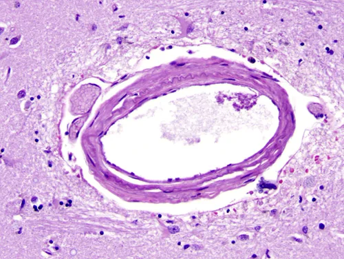
Beyond structural protection, cerebrospinal fluid drives the brain’s waste clearance network, known as the glymphatic system. Cerebral arteries penetrating into brain tissue are surrounded by narrow channels called perivascular or Virchow-Robin spaces, which are bordered externally by astrocytic end-feet forming the glia limitans. Pulsations pump the fluid. Arterial wall movements drive subarachnoid fluid into these periarterial channels, forcing it to mix with interstitial fluid and wash through extracellular matrix spaces to clear toxic beta-amyloid and tau proteins. Impairment of this clearance pathway may contribute to neurodegenerative conditions such as Alzheimer’s disease [3].

Normal Chemical Composition and Baseline Values
Healthy cerebrospinal fluid appears perfectly clear and colorless, resembling water. Laboratory analysis measures a specific gravity of 1.006 to 1.008, with an osmolarity of approximately 281 milliosmoles per liter. The fluid maintains an acid-base balance slightly more acidic than arterial blood, presenting a normal pH of 7.28 to 7.32 [5]. Opening pressure measured during lumbar puncture with the patient lying flat on their side normally registers between 100 and 180 millimeters of water (8 to 15 millimeters of mercury). Pressure rises to 200 to 300 millimeters when the patient sits upright [3].
Total protein concentration in healthy cerebrospinal fluid remains low. Lumbar fluid contains only 15 to 45 milligrams per deciliter, which is less than one percent of plasma concentration [3]. Albumin makes up 56 to 76 percent of this fraction, while larger immunoglobulins remain excluded by the blood-brain barrier [5].
Cellular content, glucose, and electrolyte balances reflect the fluid’s protective, non-inflammatory state. Normal adult fluid contains zero red blood cells and no more than zero to five mononuclear white blood cells per microliter [5]. Glucose averages between 50 and 75 milligrams per deciliter, maintaining roughly two-thirds of circulating blood glucose via GLUT1 transporter proteins [3]. Sodium levels hover between 135 and 150 millimoles per liter, while chloride concentrations reach 700 to 750 milligrams per deciliter, exceeding plasma chloride to balance lower protein anion counts. Glutamine concentrations range between 6 and 15 milligrams per deciliter. Because the brain converts toxic metabolic ammonia into glutamine, measuring elevated glutamine levels provides reliable diagnostic evidence when evaluating patients with hepatic encephalopathy or valproate toxicity [5].
How Cranial and Spinal CSF Leaks Differ
When structural breaches develop in the tough dura mater, fluid escapes into surrounding anatomical spaces [1]. Clinicians point out that cranial leaks and spinal leaks represent two completely distinct medical disorders with different causes, symptoms, and risks [6]. Cranial leaks occur when defects form in the anterior skull base, particularly across the cribriform plate, sphenoid bone, or ethmoid sinus following head trauma or sinus surgery [14]. Clear fluid drips from the nose or ear [1]. This opening creates a direct passage between the outside air and the brain, exposing patients to dangerous bacterial meningitis [6].
In contrast, spinal fluid leaks occur along the spinal canal, where escaping fluid drains into soft paraspinal tissues without communicating through the skin. Because there is no open passage to air, spinal leaks do not carry an elevated risk of meningitis [6]. Instead, spinal fluid loss leads to spontaneous intracranial hypotension (SIH), characterized by a marked reduction in internal cranial volume [11]. The hallmark symptom is a severe orthostatic headache that worsens when standing or sitting upright and eases when lying flat on the back [8]. Gravity pulls remaining fluid downward, causing the unsupported brain to sag inside the skull [2]. Head MRI scans often reveal diffuse pachymeningeal enhancement, while brain sagging may cause the cerebellar tonsils to descend like an acquired Chiari I malformation [9].

Spontaneous spinal leaks affect at least one in 20,000 individuals annually [13]. Women develop spinal leaks twice as often as men, with symptoms most commonly appearing between ages 33 and 52 [2]. Causes include ruptured meningeal diverticula, calcified spurs, and CSF-venous fistulas, especially in patients with Marfan syndrome [13].
Diagnostic Lumbar Punctures for Clinical Evaluation
Diagnostic sampling of cerebrospinal fluid relies on lumbar puncture, commonly called a spinal tap. Clinicians insert a needle between lower lumbar vertebrae into the subarachnoid space to measure opening pressure and withdraw fluid samples [7]. Laboratory tests distinguish traumatic needle punctures from true subarachnoid hemorrhage [3]. When an advancing needle tears a small vein, red blood cells appear in the first collection tube and clear progressively across subsequent tubes. In contrast, true hemorrhage produces uniform blood across all tubes and generates xanthochromia, showing bilirubin absorbance peaks at 415 and 476 nanometers [5].
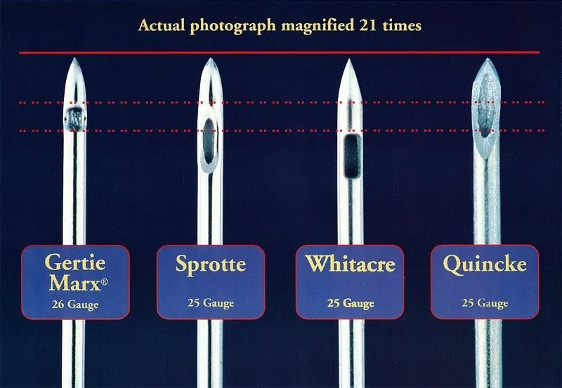
Lumbar punctures also carry a risk of persistent dural leakage, producing post-dural puncture headaches (PDPH) in susceptible individuals. Traditional sharp cutting needles slice through dural fibers, leaving a defect that leaks fluid for days or weeks. Modern clinical guidelines recommend atraumatic pencil-point needles, such as Gertie Marx, Sprotte, or Whitacre designs, which spread fibers apart rather than cutting them. At Cedars-Sinai Medical Center in Los Angeles, neurosurgeon Wouter Schievink and neuroradiologist Marcel Maya routinely employ atraumatic needles to minimize procedural fluid leaks during dynamic CT myelography and digital subtraction myelography (DSM). Interventional neuroradiologist Linda Gray Leithe at Duke University also uses small-gauge pencil-point needles for CT myelography, and radioisotope cisternography can further help identify elusive leakage sites [7].
Treatment for persistent leaks follows a stepwise medical approach [1]. Conservative care begins with horizontal bed rest, oral hydration, and caffeine intake to stimulate choroid production [8]. When headaches persist, doctors perform an epidural blood patch, injecting the patient’s own blood outside the dural tear to form a sealing clot [1]. Canadian Consensus Guidelines advise remaining flat for twenty-four hours after patching, while clinicians monitor for rebound intracranial hypertension [11].
- WEBSITE MedlinePlus Medical Encyclopedia. (2026). CSF leak. [Article Link]
- WEBSITE Cleveland Clinic. (2023). What Are the Symptoms of a CSF Leak? [Article Link]
- WEBSITE Agamanolis, D. P. (2023). Neuropathology: The Normal CSF and Abnormalities of CSF. [Article Link]
- ACADEMIC JOURNAL Hajdu, S. I. (2003). Discovery of the Cerebrospinal Fluid. Annals of Clinical & Laboratory Science, 33(3), 334-336. [Article Link]
- WEBSITE Medscape. (2026). Cerebrospinal Fluid Analysis: Reference Range, Interpretation, Collection and Panels. [Article Link]
- WEBSITE Spinal CSF Leak Foundation. (2018). Are all CSF leaks similar? [Article Link]
- WEBSITE Spinal CSF Leak Foundation. (2015). Iatrogenic CSF Leaks from Diagnostic Lumbar Punctures – a commentary. [Article Link]
- WEBSITE Northwestern Medicine. (2026). Cerebrospinal Fluid Leak (CSF Leak) FAQ. [Article Link]
- ACADEMIC JOURNAL Maher, C. O., Meyer, F. B., & Mokri, B. (2000). Surgical Treatment of Spontaneous Spinal Cerebrospinal Fluid Leaks. Neurosurgical Focus, 9(1), e2. [Article Link]
- WEBSITE CSF Leak Association. (2018). Raising Awareness and Supporting Recovery for Cerebrospinal Fluid (CSF) Leaks. [Article Link]
- WEBSITE Spinal CSF Leak Canada. (2026). Canadian Consensus Guidelines for the Diagnosis and Management of Spontaneous Intracranial Hypotension. [Article Link]
- WEBSITE Wikipedia. Cerebrospinal fluid. [Article Link]
- WEBSITE Wikipedia. Cerebrospinal fluid leak. [Article Link]
- WEBSITE Wikipedia. Cerebrospinal fluid rhinorrhoea. [Article Link]
Cover image: James Heilman, MD / Wikimedia Commons, CC BY 3.0


