




For families learning about clinical genetics, what is phenylketonuria is one of the most studied inherited conditions in medicine. Phenylketonuria, widely known as PKU, is an autosomal recessive disorder caused by a lack of the liver enzyme phenylalanine hydroxylase. Without this working protein, the human body cannot turn the dietary amino acid phenylalanine into tyrosine. Unprocessed Phe builds to toxic levels in the blood, crossing into nervous tissue where it harms normal infant brain growth [1].
What Is Phenylketonuria?
Norwegian physician Asbjörn Fölling first described the condition in Norway in 1934 after examining two young siblings whose urine turned dark green when mixed with ferric chloride. Fölling isolated phenylpyruvic acid in their samples, revealing an unrecognized error of amino acid breakdown [6]. In 1953, German pediatrician Horst Bickel showed that a low-protein diet prevents brain harm [3].
Public health screening programs report that phenylketonuria affects about one in 12,000 infants worldwide, but local rates diverge widely among different human groups. Why does prevalence differ so noticeably among human populations? In Turkey, clinical registries document the highest frequency in the world at roughly one case per 2,600 births, largely driven by parental consanguinity. Medical surveys in Ireland and Scotland record frequencies near one in 5,300 newborns, while northern European and East Asian groups average one in 10,000 cases. By comparison, national registries in Finland identify fewer than one case per 100,000 births, and Japan records one in 125,000 infants [7]. These geographic contrasts illustrate how ancestral lineages and founder variants shape rare genetic patterns across continents [12].
Current clinical consensus groups like the American College of Medical Genetics and Genomics (ACMG) view phenylalanine hydroxylase deficiency as an unbroken metabolic spectrum. Classic PKU occurs when untreated blood Phe levels rise above 1,200 µmol/L, far exceeding typical physiological levels of 35 to 120 µmol/L [7]. Milder forms, known as variant PKU or mild hyperphenylalaninemia, involve partial residual enzyme activity that lets children safely process modest amounts of dietary protein without cognitive harm [1].

How PAH Deficiency Disrupts Brain Chemistry
The molecular basis of the disorder involves pathogenic variants in the PAH gene located on chromosome 12q23.2, which spans 13 exons and encodes the phenylalanine hydroxylase enzyme. This liver enzyme functions as a tetramer that breaks down dietary Phe into tyrosine [7]. When mutations destroy its activity, blood Phe levels surge steeply [12].
Because Phe is a large neutral amino acid, it crosses into cerebral tissue by binding to the large neutral amino acid transporter known as LNAAT. When blood Phe levels reach abnormally high numbers, the excess molecules flood and saturate these specific transport channels at the blood-brain barrier. This saturation blocks other amino acids, particularly tyrosine and tryptophan, from entering brain cells in adequate amounts [12]. Deprived of these raw building blocks, brain tissue cannot make essential neurotransmitters like dopamine, norepinephrine, and serotonin [7]. Without sufficient neurotransmitter output to guide synaptic communication, neural networks cannot form properly during early childhood, leading to progressive cognitive decline [12].
High blood Phe also harms the cellular wiring of the central nervous system by slowing myelin sheath growth around nerve fibers. Magnetic resonance imaging scans of untreated patients reveal white matter injury alongside changes within the thalamus, hippocampus, and cerebral cortex. Laboratory studies further suggest that excess free Phe forms toxic amyloid-like clumps that trigger neural cellular stress [12]. Left unmanaged, these biochemical disruptions cause lasting injury to cerebral networks [7].
Autosomal Recessive Inheritance
Inheritance of the condition follows a classic autosomal recessive pattern, meaning a child must inherit two defective PAH alleles to develop symptoms. When both parents carry one mutated gene, each pregnancy carries a 25% chance of an affected baby, a 50% chance of an asymptomatic carrier, and a 25% chance of normal genes [4]. Carriers show no health complications [1].
Genetic researchers cataloged in the BioPKU database have identified more than 900 distinct mutations within all 13 exons of the gene, establishing broad allelic diversity among affected populations. Missense mutations make up roughly 62% of identified disease alleles, while small or large deletions make up 13% and splicing defects represent 11% [7]. Because individuals commonly inherit different pathogenic variants from each parent, about 76% of affected patients are compound heterozygotes rather than homozygous for a single mutation. The precise pairing of these two mutated alleles dictates how much residual enzyme function remains, heavily shaping clinical severity and patient response to cofactor therapies [1]. Even among siblings carrying identical genetic variants, environmental factors and secondary metabolic traits can lead to subtle variations in cognitive outcomes [7].

The survival of pathogenic alleles in human history led scientists to explore the heterozygote advantage hypothesis. British researcher Louis Woolf suggested that carrying a single mutated allele gave protective biological resistance against ochratoxin A, a toxic fungal compound that contaminated stored grain and caused pregnancy loss. This reproductive advantage allowed carrier frequencies to stay high in ancestral European farming communities despite the fatal toll of untreated classic PKU [12].
Clinical Symptoms of Untreated PKU
How does a newborn show signs of illness after birth? Newborns with phenylalanine hydroxylase deficiency look completely healthy during their first days because maternal liver enzymes clear excess amino acids across the placenta before birth. Symptoms only emerge gradually as routine milk feeding introduces natural protein into the infant’s digestive system [5]. Without dietary management, blood Phe levels rise steadily, causing affected babies to miss milestones, lose interest in surroundings, and develop acquired microcephaly as head and brain growth falters [4].
Unchecked metabolic buildup harms cognitive function and motor coordination over the first year of life. Untreated infants often display brisk deep tendon reflexes, purposeless arm jerking, and recurring seizures [4]. Many children suffer from attention deficit disorders, mood disturbances, and profound, irreversible intellectual disability [7].
The disease also causes tell-tale outward signs that aid pediatricians during physical exams. Because phenylalanine hydroxylase deficiency prevents normal synthesis of tyrosine, the body produces diminished amounts of melanin pigment, leading to unusually fair skin, light blonde hair, and blue eyes compared to unaffected family members [4]. As excess Phe accumulates, alternative breakdown pathways convert the surplus into phenylacetic acid. This specific carboxylic acid is excreted through sweat, breath, and urine, imparting a characteristic musty or mousy odor [5]. Many untreated children also develop dry skin and chronic eczema that resist standard topical treatments until Phe intake is brought under careful metabolic control [1].


Newborn Blood Screening
Universal newborn screening transformed phenylketonuria from a devastating cause of lifelong institutionalization into a manageable metabolic condition. American microbiologist Robert Guthrie pioneered a simple bacterial inhibition assay using dried blood spots collected on filter paper from an infant’s heel prick. Known as the Guthrie test, this reliable method allowed public health systems to detect high blood Phe before irreversible brain damage occurred. Today, newborn screening is standard practice in North America, Europe, Australia, and parts of Asia [12].
Public health laboratories in the United States and Europe now analyze dried blood spots using automated tandem mass spectrometry systems rather than bacterial growth plates. These instruments measure Phe levels and calculate the ratio of Phe to tyrosine within a single minute [12]. A positive screening result triggers prompt confirmatory quantitative plasma testing and complete genetic sequencing [4].
A critical phase of newborn diagnosis involves ruling out rare defects in tetrahydrobiopterin metabolism, which account for roughly 1% to 2% of hyperphenylalaninemia cases. Tetrahydrobiopterin, abbreviated as BH4, functions as an indispensable nonprotein cofactor required by phenylalanine hydroxylase, tyrosine hydroxylase, and tryptophan hydroxylase. Biallelic mutations in genes such as GCH1, PTS, or QDPR cause tetrahydrobiopterin deficiency, leaving infants with profound neurological deficits even if dietary Phe is restricted. To detect these cofactor disorders, medical teams evaluate urinary pterin excretion and measure dihydropteridine reductase activity in red blood cells. Infants diagnosed with BH4 synthesis defects need replacement therapies using synthetic cofactor formulations and neurotransmitter precursors like L-DOPA and 5-hydroxytryptophan to support neurological development [7].
Dietary Management and Protein Limits
The cornerstone of medical treatment is a strict, lifelong diet that eliminates high-protein foods to prevent Phe accumulation. Patients must avoid animal proteins like beef, poultry, pork, fish, eggs, milk, and cheese, as well as plant sources such as soybeans, beans, nuts, and peas [2]. Furthermore, people with PKU must strictly avoid the artificial sweetener aspartame, sold under brand names like NutraSweet, because it digests directly into free phenylalanine [4].
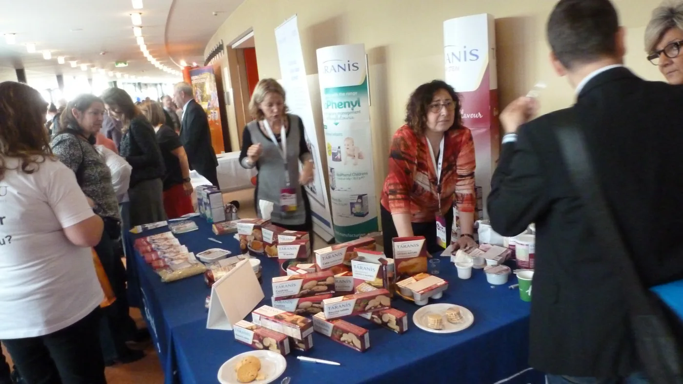
Because strict protein restriction deprives the body of essential building blocks, patients must consume tailored medical amino acid formulas every day. Infants drink synthetic formulas devoid of Phe, occasionally supplemented with calculated amounts of breast milk or standard formula to provide enough amino acids for normal growth [2]. Leading medical nutrition manufacturers such as Nutricia, Vitaflo, and Lactalis supply specialized amino acid formulas, low-protein breads, and modified pastas [9]. Clinical nutritionists continuously monitor food intake to ensure patients do not suffer deficiencies in calcium, iron, vitamin D, and vitamin B12 [7].
In past decades, pediatricians believed adolescents could safely discontinue dietary restrictions once major brain growth finished around age six or eight. Subsequent clinical tracking revealed that adults who discontinued the diet suffered executive function decline, memory lapses, depression, and noticeable white matter alterations on neuroimaging [7]. European PKU guidelines published in the Orphanet Journal of Rare Diseases by researchers Francine J. van Spronsen and Anita MacDonald now mandate a lifelong dietary approach, setting target blood Phe ranges between 120 and 360 µmol/L for children up to age 12 and capping adult levels below 600 µmol/L [12].
Risks During Pregnancy
Maternal phenylketonuria poses serious health risks when women with the disorder enter pregnancy without tight dietary control. Phenylalanine moves across the placenta, concentrating in fetal circulation at levels roughly 50% higher than in maternal blood. High intrauterine Phe concentrations act as a potent teratogen, increasing the likelihood of congenital heart disease, microcephaly, developmental delay, and low birth weight under 2,500 grams [11].
To prevent maternal PKU syndrome, guidelines from the American College of Obstetricians and Gynecologists (ACOG) emphasize preconceptional counseling and rigorous metabolic care. Women planning a pregnancy must follow a prescribed low-Phe diet for at least three months prior to conception, keeping blood levels between 120 and 360 µmol/L [7]. Blood Phe levels must be monitored frequently through home blood spot collection during all three trimesters. If maternal blood Phe stays high during early gestation, the risk of serious fetal cardiovascular malformations and spontaneous miscarriage rises steeply. Conversely, when women achieve and sustain target blood levels before conceiving and throughout pregnancy, their babies develop normally without intellectual impairment or anatomical birth defects [11].

Living with maternal PKU demands extraordinary personal discipline, social accommodation, and meticulous meal preparation. Describing her pregnancy experience in the United Kingdom, adult patient Eleanor Williamson wrote: “I’ve carried scales with me to restaurants, sent dietary essays to friends who have hosted me, and done blood tests at home three days a week.” While maintaining such rigorous routines creates daily challenges, this commitment shields the developing child from irreversible harm and supports a healthy pregnancy outcome [11].
Medical Therapies and Support
In recent years, pharmacological agents developed with support from the U.S. Food and Drug Administration (FDA) have broadened treatment strategies beyond strict dietary restriction alone. The agency approved sapropterin dihydrochloride, marketed as Kuvan, for individuals with BH4-responsive phenylketonuria. Sapropterin acts as a synthetic cofactor that stabilizes mutant phenylalanine hydroxylase enzymes, stimulating residual catalytic activity and helping responsive patients lower blood Phe or consume more natural dietary protein [2].
Researchers backed by the National Institute of Child Health and Human Development (NICHD) continue developing novel medical options to lessen the heavy daily burdens of metabolic care. Large neutral amino acid supplements compete with Phe at the blood-brain barrier to reduce cerebral entry, while bovine whey glycomacropeptide formulations provide palatable, naturally low-Phe protein alternatives. Biomedical investigators are also testing enzyme substitution therapies and liver-directed gene therapies to degrade Phe independently of defective hepatic enzymes [2].
Patient organizations offer practical educational resources, advocacy networks, and emotional solidarity for families navigating metabolic disorders. The European Society for Phenylketonuria coordinates with national groups, including the German society DIG PKU and the French association Les Feux Follets, to standardize patient care in European countries [8]. Reflecting on collective advocacy efforts, ESPKU President Eric Lange said: “The publication of this consensus paper is a significant milestone in the collective effort to improve the treatment and management of PKU in Europe.” Global advocates celebrate International PKU Day every June 28, honoring the birthdays of screening pioneer Robert Guthrie and dietary therapy founder Horst Bickel while promoting universal newborn testing worldwide [10].
- WEBSITE National Library of Medicine. Phenylketonuria: MedlinePlus Genetics. [Article Link]
- WEBSITE National Institute of Child Health and Human Development. What are common treatments for phenylketonuria (PKU)? [Article Link]
- ACADEMIC JOURNAL Bickel, H., Bachmann, C., Beckers, R., et al. (1981). Neonatal mass screening for metabolic disorders. European Journal of Pediatrics, 137(2), 133–139. [Article Link]
- WEBSITE MedlinePlus Medical Encyclopedia. Phenylketonuria. [Article Link]
- WEBSITE Madriella Doula Network. Phenylketonuria (PKU). [Article Link]
- WEBSITE MarkerDB. Phenylketonuria Condition Record. [Article Link]
- WEBSITE Medscape. Phenylketonuria (PKU): Background, Pathophysiology, Etiology. [Article Link]
- WEBSITE European Society for Phenylketonuria and Allied Disorders Treated as Phenylketonuria. Consensus Paper. [Article Link]
- WEBSITE Domenighetti, L., & Berthe-Dequin, V. (2015). Congrès de l’ESPKU : témoignage de Véronique Berthe-Dequin. Les Feux Follets. [Article Link]
- WEBSITE Excemed. Eric Lange talks about the changing role of patient organisations. [Article Link]
- ONLINE NEWS Williamson, E. (2017). Maternal PKU – Why I’m Having A Burrito Delivered To The Labour Ward. HuffPost UK. [Article Link]
- WEBSITE Wikipedia. Phenylketonuria. [Article Link]


