




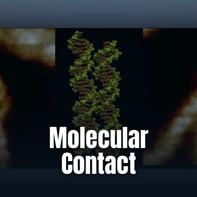
DNA pairing brings together molecules that carry the same negative charge and should repel one another. Researchers at the Universities of Sheffield and York have now imaged two double helices (the familiar twisted structures of DNA) aligned groove for groove. Their study combines those images with computer simulations to investigate DNA–DNA recognition. The clearest experimental result is a precise side-by-side arrangement consistent with a proposed electrostatic zipper. [1]
DNA Pairing Between Double Helices
The Sheffield and York team examined contacts between two already formed double helices. Each molecule retained its double-helical structure as it approached another molecule, making the observation different from two single DNA strands joining to form one double helix. The distinction matters for interpreting the widely reported description of DNA ‘zipping together’: the researchers investigated how neighboring helices recognize and contact one another along their outer surfaces. [1, 3]
Cells bring DNA regions together during genetic recombination, chromosome organization and gene regulation, even though the molecules’ negatively charged backbones create an electrical obstacle to close contact. Proteins contribute to these cellular processes. The new study asks how DNA’s own structure and surrounding ions might also contribute, using purified molecules to examine a physical interaction separately from the many other components of a cell. [3, 4]
Thomas E. Catley, Victor Velasco-Berrelleza, Daniel E. Rollins, Alice L. B. Pyne and Agnes Noy report the work in Nucleic Acids Research. Their paper, Imaging and mechanism of DNA–DNA recognition mediated by divalent ions, links structural observations to a possible molecular explanation. The study offers evidence for a mechanism under laboratory conditions; it does not establish how frequently living chromosomes use it. [1]
Why Would Charged Molecules Meet?
Alexei Kornyshev and Sergey Leikin proposed an explanation in 2001 in Physical Review Letters. Their theory considered how ions around DNA could change the electrical environment of neighboring helices, creating patterns that favor contact when the molecules adopt a suitable alignment. DNA’s helical geometry matters here. The relevant charges occupy a structured surface, rather than a smooth rod with identical electrical properties everywhere along its length. [2, 3]
The electrostatic DNA zipper model describes neighboring helices fitting together through complementary patterns of charge. Salt ions can help create those patterns. Because the molecules twist, the proposed arrangement resembles two interlocking spiral staircases, with their grooves facing one another in a repeating pattern that can support contact along the helices. Kornyshev, identified by the University of York account as an Imperial College London professor, helped develop the theoretical basis. [2, 4]
Earlier experiments had already suggested that DNA pairing could involve recognition without proteins. ZME Science describes observations from 2009 involving long, similar DNA molecules, followed by work in the fungus Neurospora crassa linking regularly spaced matching sequences to alignment. More recent nanoscale measurements also suggested stronger attraction between matching molecules in the presence of certain positive ions. The Sheffield–York images now add a structural view to that experimental history. [3]

What Did the Microscope Show?
Catley and colleagues used atomic force microscopy (a method for mapping surface topography) to examine DNA in liquid containing nickel, calcium or magnesium ions. The team imaged purified DNA on mica. In the clearest images, neighboring molecules aligned major groove with major groove and minor groove with minor groove, matching the geometry predicted by the zipper model. The images resolve the arrangement of the helices; the explanation involving individual ion bridges comes from the accompanying simulations. [1, 3]
Across larger sets of DNA fragments containing 339 base pairs, approximately 10–14 percent appeared paired. Most fragments remained unpaired. Contacts typically extended for about 14–16 nanometers, equivalent to roughly three or four turns of the helix. The overall pairing rates did not differ significantly among nickel, calcium and magnesium, even though the simulations suggested differences in how those ions supported individual contacts. Those percentages describe DNA pairing in the experimental samples, rather than a rate for chromosomes inside living cells. Nickel helped the researchers obtain their highest-resolution images, a practical detail that also matters when considering how closely the experiment resembles conditions in a cell. [3]


How Could Ions Hold DNA Together?
Velasco-Berrelleza performed simulations that followed atoms and ions around the DNA molecules. The calculations suggested that divalent ions (ions carrying two positive charges) could interact with both helices at once, partly offsetting the repulsion between their backbones. Each ion could effectively provide a small bridge. The simulations supply a candidate mechanism for the close contacts visible in the microscope, while allowing the researchers to compare the behavior of different ions. [3, 4]
“Microscopy shows us what happens, but the simulations allow us to uncover the molecular mechanism behind it,” Velasco-Berrelleza said in the University of York account reproduced by ScienceDaily. [4]
Nickel favored particularly stable contact at GTAC. The simulations repeatedly identified that short DNA sequence as a meeting point, suggesting that local sequence can influence how firmly two molecules associate when nickel ions are present. Calcium and magnesium also supported contacts. Their arrangements were more varied. The findings indicate that similar overall pairing frequencies can coexist with differences in the molecular details of how individual helices interact. [3]

Must the Sequences Match Exactly?
The researchers observed many contacts between stretches that did not match perfectly. Initial contact did not require an exact sequence match. Their results suggest a possible two-stage process in which ions first help nearby helices approach, after which compatible sequences and shapes allow the association to become more stable and extend. The proposed sequence of events explains how nonspecific encounters could precede more selective DNA pairing, although the study does not establish that sequence inside a living cell. [3]
Noy, from York’s School of Physics, Engineering and Technology, suggests the results could help researchers identify genomic regions especially involved in pairing. Cancer is a possible research connection. Mutations that disrupt normal cellular processes could make such regions relevant to disease, but this study reports neither a cancer treatment nor a demonstrated diagnostic method. The potential medical relevance concerns understanding DNA interactions before translating that understanding into any application. [4]
DNA maintenance also includes processes such as light-driven repair by photolyase, which PerEXP Teamworks has covered separately. The present experiment concerns physical contacts between DNA molecules; it does not test photolyase activity or show that ion-mediated pairing repairs damaged DNA. [1, 3]
What Remains Unresolved Inside Cells?
The Sheffield–York team worked with purified DNA on a supporting surface, while living chromosomes encounter proteins, molecular crowding and a much more complex environment. Nickel was especially useful for imaging. In simulations with more cell-like mixtures of potassium and magnesium or calcium, parallel pairing succeeded in only a minority of runs. DNA pairing inside a living nucleus remains an open experimental question for this mechanism, even with the new structural evidence. [3]
Researchers could eventually use sequence-dependent contacts to design customized DNA structures for biotechnology, according to the University of York account. That possibility connects with the broader subject of polymer–DNA origami nanostructures in living systems. The current paper establishes no ready-to-use design system. It identifies interactions that might become useful if scientists can understand and control their dependence on sequence, ions and surroundings. [4]
Can cells exploit the same groove alignment under their own chemical conditions? Future experiments would need to connect the laboratory geometry to DNA behavior in a cellular environment. Noy’s suggested search for regions with stronger pairing tendencies offers one direction, while the differing behavior of nickel, calcium and magnesium leaves another: determining which contacts persist when the surrounding ions more closely resemble those around living chromosomes. [3, 4]
- ACADEMIC JOURNAL Catley, T. E., Velasco-Berrelleza, V., Rollins, D. E., Pyne, A. L. B., & Noy, A. (2026). Imaging and mechanism of DNA–DNA recognition mediated by divalent ions. Nucleic Acids Research, 54(16). [Article Link]
- ACADEMIC JOURNAL Kornyshev, A. A., & Leikin, S. (2001). Sequence recognition in the pairing of DNA duplexes. Physical Review Letters, 86(16), 3666–3669. [Article Link]
- ONLINE NEWS Puiu, T. (2026, September 11). Scientists watched two DNA double helices ‘zip’ together for the first time. ZME Science. [Article Link]
- ONLINE NEWS University of York. (2026, September 10). Scientists capture two DNA strands zipping together for the first time. ScienceDaily. [Article Link]
APA 7: PerEXP Teamworks. (2026, September 12). Two double helices reveal how DNA pairing can work.


