





Every living cell relies on an orderly waste disposal network to survive starvation, clear debris, and renew worn parts. Cellular cleanup takes place through autophagy, a conserved biological pathway that delivers damaged organelles, protein clumps, and invading microbes to acidic compartments for breakdown. By breaking down these captured structures into basic building blocks like amino acids and lipids, the cell keeps tissues nourished during metabolic hardship. Healthy cells maintain a steady baseline turnover of these components, safeguarding tissue integrity long before acute starvation occurs [2].
What Is Autophagy?
Autophagy means self-eating in Greek. Through this catabolic route, eukaryotic cells break down their own parts inside lysosomes to stay alive during metabolic hardship. Belgian biochemist Christian de Duve discovered lysosomes at the University of Louvain in 1949, and in 1955 he collaborated with microscopist Alex Novikoff from the University of Vermont to record the first electron micrographs of these digestive sacs. De Duve soon recognized that cells deliver parts of their cytoplasm to these organelles. In 1963, de Duve and his student Russell Deter coined the term autophagy to describe glucagon-induced liver cleanup [2].
In 1954, Werner Straus tracked labeled proteins into digestive droplets, demonstrating that enzymes actively break down foreign molecules inside animal cells. At the Rockefeller Institute in 1962, Keith R. Porter and Thomas Ashford observed lysosomes moving toward the center of liver cells after glucagon exposure, while related research on the lysosome cellular recycling center explains how these acidic compartments isolate hydrolases to degrade internal macromolecules. Hruban and Spargo confirmed in 1963 that this focal degradation occurs during normal differentiation rather than injury [6].
The lysosome handles large debris that other breakdown systems cannot manage. While the proteasome degrades individual unfolded proteins one by one—a mechanism recognized with the 2004 Nobel Prize in Chemistry awarded to Aaron Ciechanover, Avram Hershko, and Irwin Rose—it cannot digest whole organelles. Autophagy provides the necessary machinery for bulk organelle removal [1].

How Autophagosomes Capture Cellular Cargo
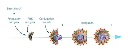
Autophagosome assembly begins when a flat membrane cistern called a phagophore forms in the cytoplasm. Marilyn Farquhar and her colleagues at the University of California, San Francisco, first identified these cup-shaped precursors in 1966 while studying pituitary secretory cells. The curving rim expands around nearby cellular cargo until its opposing edges meet and fuse into a sealed double-membrane vesicle, a process whose lipid source remained debated until 2010 when Jennifer Lippincott-Schwartz used fluorescent tags to prove that the outer mitochondrial membrane serves as the main lipid donor, supported by lipid transfers from the endoplasmic reticulum [2].
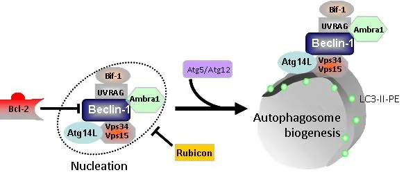
Membrane growth relies on two sequential ubiquitin-like conjugation cascades, beginning when Atg12 binds covalently to Atg5 to build a multimeric protein scaffold that shapes vesicle curvature. The second cascade cleaves MAP1LC3 into cytosolic LC3-I, which transfer enzymes then couple to phosphatidylethanolamine along both boundaries of the growing phagophore. This lipidated form is designated LC3-II. Because LC3-II stays attached to the autophagosomal membrane throughout vesicle maturation, laboratory researchers monitor it as a dependable biochemical marker of autophagic activity [5].
After the vesicle closes, it travels along microtubules to merge with lysosomes. Small Rab GTPases, such as Rab7 and Rab24, guide this membrane fusion to form an autolysosome. Lysosomal enzymes then break down the inner vesicle and its cargo, releasing free amino acids for protein synthesis [5].
Four Pathways of Cellular Autophagy
Eukaryotic cells rely on four distinct routes of lysosomal breakdown: macroautophagy, microautophagy, chaperone-mediated autophagy, and crinophagy. Macroautophagy is the main pathway for enclosing large cytoplasmic regions and whole organelles inside newly synthesized double-membrane vesicles. Unlike macroautophagy, microautophagy bypasses intermediate transport vesicles completely by folding the lysosomal surface inward or projecting outward to engulf surrounding cytoplasm directly for rapid enzymatic digestion. Crinophagy disposes of surplus secretory granules by fusing them with lysosomes. Originally called autophagocytosis in early scientific literature, these distinct pathways allow cells to adjust their degradative power according to the exact size, location, and chemical nature of cellular waste [6].
Chaperone-mediated autophagy operates with singular selectivity by targeting individual soluble proteins rather than whole organelles. Client proteins display a specific pentapeptide amino acid motif recognized by cytosolic hsc70 chaperone complexes. After binding this motif, the chaperone guides the unfolded client to lysosomal-associated membrane protein 2A on the lysosomal surface, allowing the substrate protein to thread through a membrane pore into the lumen for rapid enzymatic breakdown. This single-protein translocation spares neighboring organelles [6].
Selective macroautophagy targets specific organelles using dedicated receptor proteins. Mitophagy removes damaged mitochondria through the coordinated action of PINK1 and parkin enzymes, supported by receptors like NIX and BNIP3. Similarly, lipophagy breaks down lipid droplets to release fatty acids during energy deficits, a pathway discovered in mice in 2009 [6].
The Discovery of Yeast ATG Genes
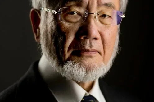
Genetic control of cellular recycling remained an unsolved biological mystery for decades. Born in Fukuoka in 1945, Japanese cell biologist Yoshinori Ohsumi established his independent laboratory at the University of Tokyo in 1988 after working at Rockefeller University in New York. Ohsumi selected baker’s yeast, Saccharomyces cerevisiae, to study vacuolar protein turnover. In yeast, a large compartment called the vacuole corresponds to the lysosome in mammalian cells. Because yeast cells are small, scientists questioned whether fungal cells even performed autophagy until Ohsumi reasoned that blocking vacuolar proteases while starving cells of nitrogen would force autophagosomes to accumulate visibly inside the vacuole. In 1992, Ohsumi published landmark light microscope images in the Journal of Cell Biology showing swollen vacuoles packed with undegraded autophagic bodies, establishing yeast as an ideal experimental model [1].
Ohsumi and Miki Tsukada exposed yeast cells to chemical mutagens to isolate mutants that failed to accumulate vacuolar vesicles during nutrient starvation. In 1993, they reported the first essential autophagy genes, designated APG. Michael Thumm at the University of Stuttgart and Daniel J. Klionsky at the University of Michigan independently identified matching genes in Saccharomyces cerevisiae while studying the cytoplasm-to-vacuole targeting pathway. Fungal researchers unified this terminology under the standardized ATG notation in 2003. Klionsky launched the scientific journal Autophagy two years later [6].
Beth Levine and Daniel Klionsky organized the first Gordon Research Conference on autophagy in Waterville in 2003. Ohsumi’s discoveries reshaped modern cell biology, earning him the 2016 Nobel Prize in Physiology or Medicine from the Nobel Assembly at Karolinska Institutet [8].

Why Does Autophagy Decline With Age?
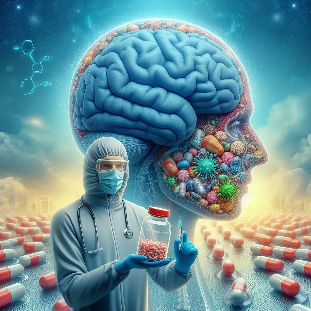
Autophagy slows as tissues grow older because lysosomes gradually lose their interior acidity, transport along microtubules slows down, and nutrient-sensing kinase cascades lose their sensitivity to changing metabolic cues. In aging post-mitotic tissues like the brain, long-lived neurons cannot dilute accumulated waste through cell division. Genetic and clinical studies led by David C. Rubinsztein at the University of Cambridge show that slowing clearance allows aggregate-prone toxic proteins to accumulate unchecked, including mutant huntingtin in Huntington’s disease, alpha-synuclein in Parkinson’s disease, and tau tangles in Alzheimer’s disease [3].
Stimulating autophagic degradation has emerged as a promising strategy to protect vulnerable brain circuits from progressive neurodegeneration. In experimental cell cultures and animal models, upregulating clearance reduces the concentration of toxic protein aggregates and delays motor symptoms. Emerging experimental strategies for drugging toxic proteins in neurodegenerative illness show how small-molecule modulators can clear insoluble aggregates before permanent neuronal cell death occurs. Restoring the natural clearance of toxic oligomers helps preserve synaptic communication in the brain [3].
Therapeutic activation of autophagic cleanup requires caution because if downstream lysosomes lack functional enzymes or cannot fuse with incoming autophagosomes, artificially boosting vesicle production creates damaging cellular congestion. Accumulated vesicles leak hydrolytic enzymes that accelerate cell death rather than promoting recovery. Clinical treatments must verify that the entire digestive route is fully operational before attempting pharmacological induction [3].
Selective Cleanup Through Chaperone Systems
To survive repeated mechanical force and continuous physical strain, contractile tissues like skeletal muscle, heart tissue, and lungs depend on chaperone-assisted selective autophagy to identify and clear damaged proteins. The system relies on molecular chaperones HSPA8 and HSPB8, which cooperate with the cochaperone BAG3 and the ubiquitin ligase STUB1 to recognize unfolding nonnative client proteins. When muscle contraction unfolds the actin-crosslinking protein filamin, this chaperone assembly binds the damaged region, allowing STUB1 to attach polyubiquitin tags that recruit the autophagic adaptor p62/SQSTM1 to guide membrane precursors around the client for lysosomal destruction [7].
Membrane closure during this chaperone-directed process depends on BAG3 binding the actin-associated protein SYNPO2. This interaction recruits a VPS18-containing protein complex that drives the rapid fusion of precursor membranes around the tagged client, enclosing it inside an autophagosome for subsequent lysosomal digestion. The sealed vesicle then merges with a lysosome for rapid enzymatic breakdown. Similar organelle breakdowns occur in motor neurons, where mitochondrial membrane disruption in ALS impairs cellular transport. Defective membranes and stalled clearance pathways combine to undermine neuromuscular health [7].
Unfolded proteins aggregate when this chaperone pathway fails under prolonged stress. Human genetic studies demonstrate that mutations in BAG3 and related chaperone genes cause severe muscular dystrophy and cardiomyopathy. Continuous chaperone-assisted clearance is needed to preserve contractile strength and protect heart muscle against mechanical wear [7].
Roles in Cancer and Viral Defense
The relationship between cellular recycling and cancer progression changes dynamically as tumors develop. In healthy tissues, baseline clearance functions as a tumor suppressor by destroying damaged mitochondria, which limits the reactive oxygen species that cause chromosome breaks and dangerous genomic mutations. At Columbia University in 1999, Beth Levine proved that deleting one copy of the Beclin1 gene caused spontaneous tumors in mice, an observation matching human pathology where 40 to 75 percent of sporadic ovarian and breast cancers lack one copy. Eileen White later verified this link in vivo [2].
Once tumors form, cancer cells exploit the recycling pathway to survive metabolic stress. The center of a solid tumor often lacks adequate blood vessels, subjecting malignant cells to low oxygen and nutrient starvation. Cancer cells ramp up self-digestion to harvest recycled amino acids and maintain energy production. Research by Iman Tavassoly at Mount Sinai and Virginia Tech shows how the balance between autophagic survival and apoptotic cell death dictates whether tumor cells withstand chemotherapy [9].
Autophagy also coordinates host defense against intracellular microbes, an immune response called xenophagy. Cellular receptors including CALCOCO2/NDP52 and LC3 direct invading bacteria and viruses into autophagosomes for destruction. However, certain viruses have evolved molecular strategies to evade capture or hijack autophagic membranes to assist their own replication [2].
Endurance Exercise and Organelle Turnover
Physical exercise triggers extensive metabolic remodeling across skeletal muscle tissues. Research led by Anthony M. J. Sanchez, Henri Bernardi, Guillaume Py, and Robin B. Candau at the University of Montpellier demonstrates that sustained endurance activity activates autophagic flux in skeletal muscle. This response is coordinated by Forkhead box class O transcription factors, designated FOXO, and the nutrient-sensitive MTORC1 complex. Endurance exercise suppresses MTORC1 while activating FOXO, initiating lysosomal cleanup of worn mitochondria to supply working muscle fibers with recycled nutrients [4].
Nutritional timing strongly governs exercise-induced cleanup, as Anthony M. J. Sanchez documented that prolonged endurance running or cycling in a fasted state produces the greatest rise in autophagic flux markers and mitophagy messenger RNA. Conversely, resistance training and heavy weightlifting preferentially stimulate the ubiquitin-proteasome system and activate lipid kinases like DAGK that drive muscle protein synthesis instead of bulk catabolism. Fasted endurance work stimulates cellular cleanup, while resistance training builds new muscle mass [4].
Tracking autophagic flux in living tissue requires sensitive biochemical assays. Research consortiums including the Autophagy, Inflammation, and Metabolism Center study how modifications across the Atg8 protein family remodel membranes during cellular stress. These physiological discoveries provide practical, science-based guidelines for designing exercise and fasting regimens that sustain cellular health over a lifetime [10].
- PRESS RELEASE Nobel Media AB. (2016). The 2016 Nobel Prize in Physiology or Medicine to Yoshinori Ohsumi. [Article Link]
- ONLINE NEWS Scitable by Nature Education. (2007). Lysosomes and Autophagy. [Article Link]
- ACADEMIC JOURNAL Djajadikerta, A., Keshri, S., Pavel, M., Prestil, R., Ryan, L., & Rubinsztein, D. C. (2020). Autophagy Induction as a Therapeutic Strategy for Neurodegenerative Diseases. Journal of Molecular Biology, 432(8), 2799-2821. [Article Link]
- ACADEMIC JOURNAL Sanchez, A. M. J., Bernardi, H., Py, G., & Candau, R. B. (2014). Autophagy is essential to support skeletal muscle plasticity in response to endurance exercise. American Journal of Physiology-Regulatory, Integrative and Comparative Physiology, 307(8), R956-R969. [Article Link]
- WEBSITE HADb Consortium. (2025). Human Autophagy Database (HADb). [Article Link]
- WEBSITE Wikipedia contributors. (2026). Autophagy. In Wikipedia, The Free Encyclopedia. [Article Link]
- WEBSITE Wikipedia contributors. (2026). Chaperone-assisted selective autophagy. In Wikipedia, The Free Encyclopedia. [Article Link]
- CONFERENCE PAPER Gordon Research Conferences. (2003). Autophagy in Stress, Development and Disease Conference Program. [Article Link]
- BOOK Tavassoly, I. (2015). Dynamics of Cell Fate Decision Mediated by the Interplay of Autophagy and Apoptosis in Cancer Cells. Springer Cham. [Article Link]
- ACADEMIC JOURNAL Autophagy, Inflammation, and Metabolism Center. (2026). Ubiquitylation & Atg8ylation Journal Overview. [Article Link]


