





A standard low-carbohydrate regimen forces the body to burn fats for fuel instead of glucose, driving cells into an altered metabolic state called ketosis. While popular culture views high-fat meal plans as a uniform therapy, laboratory evidence shows that biological outcomes depend on the specific fats consumed. A study published in Science Advances shows that ketogenic diets containing high amounts of long-chain saturated fatty acids cause mitochondrial strain and prompt inflammatory macrophages to accumulate in rodent organs [1]. Researcher David Gius and his team at the University of Cincinnati Cancer Center discovered this response while studying how different lipid mixtures shape immune responses in living tissues.
Lipid Types in Ketogenic Diets
Dietary fats differ by carbon chain length and saturation, which alters how metabolic enzymes process them. Physician-scientist David Gius, who serves as a professor in the Department of Radiation Oncology at the University of Cincinnati College of Medicine and treats patients at UC Health, led the investigation. In cell cultures and animal models, medium-chain fats and unsaturated fatty acids preserved anti-inflammatory properties, but long-chain saturated fats provoked immune activity [4].
When cells process long-chain saturated fatty acids (LC-SFAs), their inner machinery faces metabolic strain. In both murine and human macrophage tissue cultures, these long-chain molecules promoted M1-like polarization, pushing white blood cells toward an active inflammatory defense state. Primary macrophages taken from mice fed this high-fat diet showed high levels of proinflammatory ceramides and hexosylceramides. By contrast, medium-chain triglycerides and unsaturated oils did not cause this macrophage shift, showing that ketone production itself does not harm immune cells [1].
While earlier clinical reports praised carbohydrate restriction for lowering baseline inflammation, parallel trials documented heightened inflammatory activity in animal tissues, leaving researchers puzzled until lipid chemistry resolved the contradiction. “Our previous studies have found that all ketogenic diets are not the same, with different fats causing different metabolic changes,” Gius said [4].

Crisco Versus Coconut Oil in Mice
To test how fat types act inside a living body, researchers fed mice three separate diets for 21 days. One group ate regular rodent chow, a second received a keto diet based on coconut oil, and a third ate a keto diet formulated with Crisco shortening. The Crisco formulation contained about 26 percent long-chain saturated fatty acids derived from palm oil, whereas the coconut oil diet contained only 12 percent long-chain saturated fatty acids despite having similar total fat density [3].
Tissue checks at three weeks showed marked biological differences. Animals fed the Crisco diet accumulated high numbers of proinflammatory macrophages in the heart, kidney, liver, brain, and white fat tissue, whereas mice on the coconut oil plan kept inflammatory markers close to regular chow controls. Shorter-chain fats prevented this immune surge. White blood cells did not flood rodent tissues, showing that ketogenic diets do not cause inflammation by default [3].
“Our findings suggest that the proinflammatory effects of long-chain saturated fatty acids may emerge only beyond a critical threshold of exposure,” the authors wrote in Science Advances. ScienceAlert editor Carly Cassella said shortening contains fewer long-chain fats than lard, yet it still triggered macrophage build-up [3].

Cellular Pathways Behind Immune Signaling
When macrophages break down excess long-chain saturated fats, strained mitochondria release metabolic distress cues that trigger defense networks, including inflammasome activation and the cGAS-STING pathway. Cyclic guanosine 5′-monophosphate-adenosine 5′-monophosphate synthase detects misplaced nucleic acids, and stimulator of interferon genes activates downstream defensive transcription programs [1].
When the investigators tested knockout mice engineered without Sting in their myeloid lineage, the inflammatory consequences of the high-fat diet vanished completely, showing that the cGAS-STING system is necessary for macrophage buildup. Sung-Jen Wei led these genetic validation experiments [1]. Mitochondrial stress releases the signals that activate this innate sensor, prompting the authors to conclude that “the proinflammatory consequences of ketogenic diets are not intrinsic to ketone metabolism” [3].
Metabolic tests revealed that long-chain fats disrupt lipid balance inside immune cells. Levels of ceramides and hexosylceramides climbed in primary macrophages, producing stress signals that caused white blood cells to release cytokines. When lipid overload strains mitochondrial membranes, the resulting leak of organelle contents acts as an internal alarm that recruits inflammatory cells into healthy organ tissues [1].

Why Ketogenic Diets Were Created for Epilepsy
Long before athletes used low-carbohydrate eating to cut weight, clinicians in the 1920s developed ketogenic diets as a targeted medical treatment for pediatric patients with hard-to-control epilepsy who did not respond to standard anticonvulsant therapies. Seizure suppression guided this clinical innovation. For more than 70 years, medical teams have used carbohydrate restriction to alter brain fuel supply and prevent recurrent seizures [2]. In brain tissue, ketone bodies offer an alternative energy source that helps stabilize nerve cell signals [3].
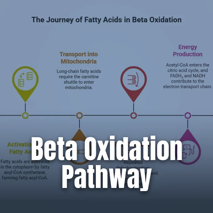
When dietary carbohydrates fall, the liver converts fatty acids through beta-oxidation into three ketone molecules: acetoacetate (AcAc), acetone, and beta-hydroxybutyrate (β-OHB) for circulation through body tissues. In healthy adult humans, circulating ketone levels fluctuate between 100 and 250 micromolar during normal daily rhythms, but concentrations of β-OHB rise between 1 and 8 millimolar under structured ketogenic feeding, prolonged physical exercise, or deep fasting. Diabetic ketoacidosis pushes total ketone concentrations up to 25 millimolar in acute patients [2].
Although beta-hydroxybutyrate supplies cellular fuel when carbohydrate intake drops, sustained ketone elevation alters gene expression over time, yet pediatric neurologists still prescribe ketogenic diets because seizure control outweighs potential metabolic hazards [2]. Findings from David Gius and Nathaniel Bowman reporting for BIOENGINEER.ORG suggest that replacing heavy saturated fats with medium-chain triglycerides could spare pediatric patients unwanted inflammatory strain during prolonged medical therapy [5].
Heart Function and Long-Term Tissue Fibrosis
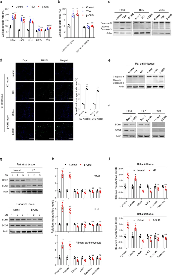
The cardiovascular effects of prolonged ketosis remain an active subject of study in cell biology. A laboratory investigation published in Signal Transduction and Targeted Therapy examined how extended high-fat feeding affected rat heart tissue over a period of 16 weeks. Researchers found that prolonged high levels of β-OHB reduced mitochondrial creation, lowered cell respiration, and increased cardiomyocyte apoptosis in cardiac muscle. These cellular injuries led to cardiac fibrosis, where stiff scar tissue replaced working muscle cells [2].
Echocardiograms showed weakened cardiac pumping capacity in the high-fat group, along with thicker left ventricular walls (LVPWd) and thicker right ventricular anterior walls (RVAW). In mechanistic tests tracking HCM human cardiomyocytes, H9C2 rat myoblasts, and HL-1 heart cells, elevated β-OHB acted as a histone deacetylase 2 (HDAC2) inhibitor, promoting histone acetylation at the promoter of the Sirt7 gene to suppress mitochondrial ribosome-encoding genes. Cellular assays in mouse embryonic fibroblasts (MEFs) and 3T3 cells showed no such damage, but doctors detected similar elevations of β-OHB and SIRT7 in human atrial fibrillation heart tissues [2].
Caloric restriction (CR) controls confirmed that low energy was not causing the tissue scars. In rat feeding trials, caloric restriction produced only mild ketone increases and caused zero cardiac fibrosis or functional impairment, showing that excessive β-OHB accumulation drove the cardiac damage. Direct intraperitoneal injections of β-OHB into rats caused atrial fibrosis and raised levels of type I collagen, type III collagen, and alpha-smooth muscle actin (α-SMA) [2].

Clinical Trials and Human Cancer Studies
In cancer biology, researchers have tested whether low-carbohydrate diets can starve tumors by exploiting the Warburg effect. Preclinical trials showed promise: a meta-analysis by Li, Zhang, and Dai in Frontiers in Nutrition reviewed 38 animal studies from PubMed, Embase, and Web of Science, finding that high-fat feeding reduced tumor volume and prolonged survival in rodents. In addition, work by Otto in BMC Cancer showed that ketogenic diets delayed gastric adenocarcinoma growth in NMRI nude mice [7]. Yet human trials tell a different story. A review by Römer, Dörfler, and Huebner in Clinical and Experimental Medicine analyzed 39 studies across Cochrane, EMBASE, and Medline covering 770 cancer patients, finding no evidence of improved survival or direct tumor shrinkage. Adherence was low across patient cohorts [6].
Oncologists still investigate whether ketogenic diets can reshape the tumor microenvironment to support immunotherapy. Gius said specific dietary fats might alter immune infiltration to enhance cancer drug efficacy [4]. A clinical trial has begun at the University of Texas Health Science Center in San Antonio, where Gius launched the project [3]. A second trial will open at the University of Cincinnati Cancer Center to evaluate how tailored fatty acids guide patient immune responses [4].
Translating these metabolic findings from rodent models to human health requires careful clinical observation. Laboratory mice eat feed with fixed fat percentages, but humans eat varied meals containing diverse fats and carbohydrates [3]. “We followed the research data while studying a different aspect of ketogenic diets and found something unexpected,” Gius said regarding the Cincinnati project. Future dietary advice in medical care will focus on fat quality rather than treating all low-carb plans as identical [4].
- ACADEMIC JOURNAL Wei, S., Schell, J., Sezaki, M., Baseviciene, A., Qian, W., Chen, W. H., Trevino, R., Sreenivas, P., Chocron, E. S., Jiang, H., Dong, F. F., Chang, J., Nazarullah, A., Huang, G., Munkácsy, E., Horikoshi, N., & Gius, D. (2026). Ketogenic diets induce proinflammatory macrophage accumulation through long-chain saturated fatty acids driving cGAS-STING signaling. Science Advances, 12(46). [Article Link]
- ACADEMIC JOURNAL Xu, S., Tao, H., Cao, W., Cao, L., Lin, Y., Zhao, S., Xu, W., Cao, J., & Zhao, J. (2021). Ketogenic diets inhibit mitochondrial biogenesis and induce cardiac fibrosis. Signal Transduction and Targeted Therapy, 6(1). [Article Link]
- ONLINE NEWS Cassella, C. (2026). Some Ketogenic Diets May Increase Inflammation And Mitochondrial Stress. ScienceAlert. [Article Link]
- ONLINE NEWS University of Cincinnati Cancer Center. (2026). Some ketogenic diets found to cause mitochondrial stress and lead to inflammation. Medical Xpress. [Article Link]
- ONLINE NEWS Bowman, N. (2026). Ketogenic Diets Rich in Long-Chain Saturated Fats Increase Inflammation, Study Finds. BIOENGINEER.ORG. [Article Link]
- ACADEMIC JOURNAL Römer, M., Dörfler, J., & Huebner, J. (2021). The use of ketogenic diets in cancer patients: a systematic review. Clinical and Experimental Medicine, 21(4), 501-536. [Article Link]
- ACADEMIC JOURNAL Li, J., Zhang, H., & Dai, Z. (2021). Cancer Treatment With the Ketogenic Diet: A Systematic Review and Meta-analysis of Animal Studies. Frontiers in Nutrition, 8. [Article Link]
APA 7: PerEXP Teamworks. (2026, November 14). Saturated Fatty Acids Drive Inflammation on Ketogenic Diets. PerEXP Teamworks. https://perexpteamworks.com/en/ketogenic-diets-inflammation-saturated-fats/


