




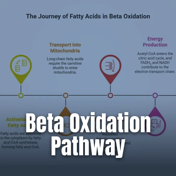
Cells rely on fat reserves for long-term survival, and beta oxidation serves as the central metabolic route that unlocks this fuel whenever circulating glucose concentrations fall below baseline levels [3]. When energy demands rise during fasting or hard muscular work, tissues dismantle fatty acid chains two carbons at a time inside the mitochondrial matrix [8]. Fat holds far more energy than glycogen [3]. The repeating enzyme cycle clips off acetyl-CoA units while loading high-energy electrons onto FAD and NAD+ carriers that feed respiratory complexes [1]. Human adults store roughly 100,000 kcal as triglycerides, whereas glycogen stores provide barely 2,000 kcal [3]. Through this iterative breakdown, living cells convert dense carbon tails into steady ATP to support continuous work without depleting blood sugar [5].
- What Is Beta Oxidation?
- Fatty Acid Activation for Mitochondrial Entry
- Four Reactions of the Beta Oxidation Cycle
- Net ATP Yield from Palmitate
- Odd Chains and Double Bonds
- Peroxisomal Breakdown of Very Long Chains
- Hormonal Signals Regulating Beta Oxidation Rates
- Clinical Disorders Linked to Defective Enzymes
What Is Beta Oxidation?
Beta oxidation breaks down fatty acid chains into two-carbon acetyl-CoA units inside mitochondria [8]. The name comes from the beta carbon [3]. As Sagar Aryal and Nidhi Dewangan explain in Microbe Notes, specific enzymes repeatedly oxidize this second carbon position to a carbonyl group before cutting the carbon-carbon bond [1]. In eukaryotic cells, the cycle runs within the mitochondrial matrix of the heart, liver, and skeletal muscle, whereas bacteria carry out the entire breakdown in their cytosol [6].
Fatty acids supply a dense fuel source because their carbon tails carry many reduced hydrogen bonds [2]. One gram of fat delivers roughly nine kilocalories of usable energy, whereas one gram of carbohydrate or protein yields barely four kilocalories [3]. In their 2008 textbook Biochemistry, Reginald Garrett and Charles Grisham at Twayne Publishers note that glucose yields 6.3 moles of ATP per carbon, while saturated fatty acids produce 8.1 moles. Darik Benson at University of California Davis confirms these ratios under Chemistry LibreTexts Page ID 452 [2]. Red blood cells cannot burn fats because they lack mitochondria. Brain neurons also depend on glucose and ketone bodies [8].
When food intake ceases, adipose tissue mobilizes stored fat through lipolysis run by adipose triglyceride lipase and hormone-sensitive lipase [1]. The released fatty acids travel through the blood to muscle and liver tissue to provide ATP when glycogen runs out [4]. In Current Protocols in Toxicology, G. W. Rogers and colleagues describe high-throughput tests that measure fatty acid-driven oxygen consumption in isolated rat liver mitochondria to evaluate metabolic drugs [14]. Japanese chemist S. Uchinomiya and coworkers in Chemical Communications developed fluorescent probes that illuminate fatty acid breakdown inside living cells [15]. Researchers use these tools to track fuel shifts [14].
Fatty Acid Activation for Mitochondrial Entry
Free fatty acids carry a negative charge at physiological pH, which prevents them from diffusing freely across cell membranes. Transport proteins from the SLC27 family move these molecules into the cytoplasm so metabolism can begin [8]. In Biology Notes Online, Sourav Pan explains that fatty acids must undergo priming before catabolic enzymes can process them. The enzyme fatty acyl-CoA synthetase, abbreviated FACS, links the fatty acid to coenzyme A, consuming one molecule of ATP and making fatty acyl-CoA, AMP, and inorganic pyrophosphate, or PPi. Pyrophosphatase promptly cleaves this PPi into two phosphate molecules [6].
Priming costs the cell two high-energy phosphate bonds because ATP splits into AMP [3]. The upfront investment prepares the fuel, yet the activated acyl chain remains trapped outside the matrix [6]. Short-chain fatty acids with fewer than twelve carbons cross membranes freely, but common long-chain varieties cannot penetrate the inner mitochondrial membrane on their own [4]. David L. Nelson and Michael M. Cox note in Lehninger Principles of Biochemistry that this barrier separates cytosolic pools from matrix enzymes. The cell needs a carrier [5].
To overcome this barrier, cells use the carnitine shuttle [1]. On the outer membrane, carnitine palmitoyltransferase I, or CPT-I, swaps coenzyme A for carnitine to form acylcarnitine [8]. A carrier called carnitine translocase, known as CAT, moves this acylcarnitine across the inner membrane while returning free carnitine back into the cytosol [1]. Inside the matrix, carnitine palmitoyltransferase II, or CPT-II, transfers the acyl group back onto coenzyme A to regenerate fatty acyl-CoA [8]. CPT-I acts as the gatekeeper. Malonyl-CoA, the first compound in fat synthesis, blocks CPT-I whenever cells store fuel. The block stops cells from burning fat while making it [3].
Four Reactions of the Beta Oxidation Cycle
Inside the mitochondrial matrix, fatty acyl-CoA enters the core beta oxidation cycle, which runs through four sequential enzymatic reactions [1]. The first reaction is dehydrogenation catalyzed by acyl-CoA dehydrogenase, an enzyme that extracts two hydrogen atoms from the alpha and beta carbons of the fatty chain [7]. Removal of these hydrogens creates a trans double bond and reduces flavin adenine dinucleotide to FADH2 [8]. Cells produce distinct forms of this dehydrogenase tailored to chain lengths, including short, medium, long, and very-long varieties [3]. The reaction yields trans-delta-2-enoyl-CoA [5].
In the second step, the enzyme enoyl-CoA hydratase adds a water molecule across that newly formed double bond [5]. Stereospecific hydration yields L-3-hydroxyacyl-CoA, placing a hydroxyl group specifically on the beta carbon while preparing the intermediate for the subsequent oxidative step [6]. The reaction requires no energy input from nucleotide cofactors [5].
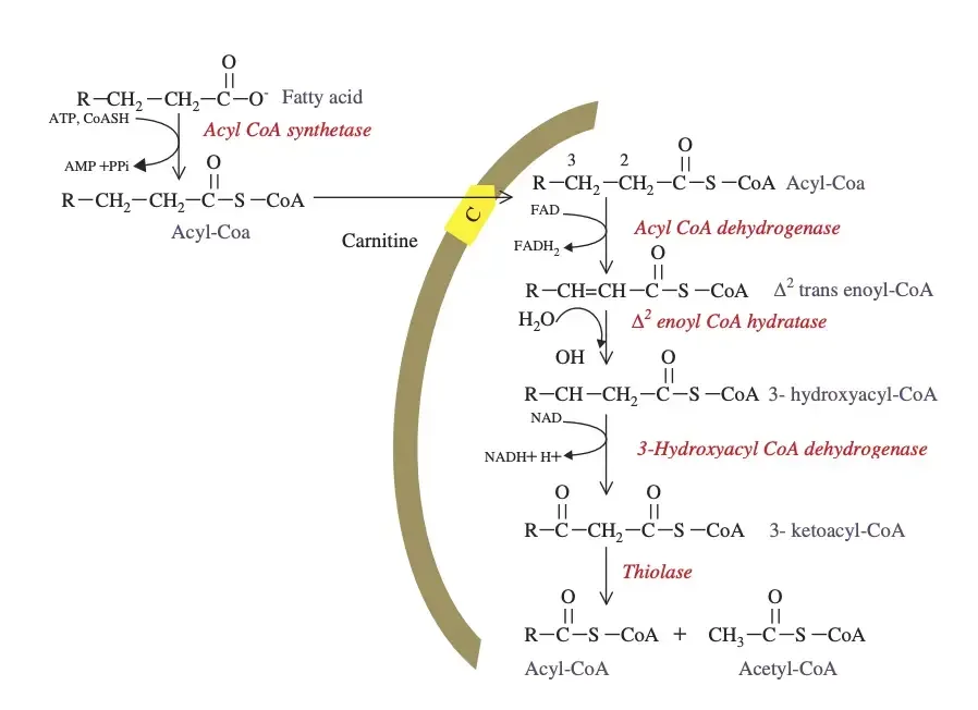
The third step is another dehydrogenation, carried out by 3-hydroxyacyl-CoA dehydrogenase, which oxidizes the beta-hydroxyl group into a keto group while reducing NAD+ to NADH and a proton [8]. The resulting product is 3-ketoacyl-CoA. The fourth and final reaction of the turn is thiolytic cleavage, which is catalyzed by beta-ketothiolase [6]. A fresh molecule of coenzyme A attacks the bond between the alpha and beta carbons, splitting the molecule apart [8]. Thiolytic cleavage releases one acetyl-CoA unit along with a shortened acyl chain designated Cn-2-acyl-CoA [6]. The shortened acyl chain, which already carries its coenzyme A attachment, loops back to step one to repeat the four-reaction sequence until the tail is fully consumed [3].
Net ATP Yield from Palmitate
Palmitate, a sixteen-carbon saturated fat abundant in human tissue, illustrates the energy yield of this pathway [3]. Dismantling palmitate takes seven complete cycles through the four enzymatic reactions, generating eight acetyl-CoA molecules, seven FADH2 carriers, and seven NADH reducing equivalents [4]. Each cycle removes two carbons, but the seventh turn splits a four-carbon butyryl-CoA into two separate acetyl-CoA units [3]. In Science Facts, Santanu Mukherjee reports that burning one mole of palmitic acid yields roughly 2,340 kcal of energy [7].
Current biochemical consensus values use P/O ratios of roughly 2.5 ATP per NADH and 1.5 ATP per FADH2, while each acetyl-CoA yields ten ATP in the citric acid cycle [3]. With these factors, the seven NADH generate 17.5 ATP, the seven FADH2 supply 10.5 ATP, and the eight acetyl-CoA units yield eighty ATP [4]. Subtracting the initial priming cost of two ATP equivalents results in a net yield of 106 ATP per palmitate molecule [3].
Older textbook models that assumed whole ratios of three ATP per NADH and two per FADH2 calculated a higher total of 129 ATP for the same palmitate chain [7]. Peter Raven notes in his 2005 text Biology that fats also produce large amounts of water, or H2O, through respiratory oxygen reduction [2]. A general formula describes the yield for any even-numbered saturated fatty acid with n carbon atoms: 0.5 times n minus one cycles occur, producing n divided by two acetyl-CoA molecules. In algebraic notation based on modern P/O ratios, net ATP equals seven times n minus six [8]. Camels and killer whales rely on this internal H2O production when liquid drinking water is unavailable. Fat thus provides hydration as well as fuel [2].


Odd Chains and Double Bonds
Natural diets contain fats with odd numbers of carbons and varying degrees of unsaturation, requiring cells to deploy accessory enzymes [3]. Odd-chain fatty acids appear in plant lipids, marine organisms, and ruminant fats [8]. These chains proceed through the standard four reactions until the final round cleaves a five-carbon intermediate into one molecule of acetyl-CoA and one three-carbon propionyl-CoA [5].
The conversion of this three-carbon propionyl fragment into a citric acid cycle intermediate takes three specialized enzymatic reactions [6]. In the first step of this conversion, propionyl-CoA carboxylase attaches a bicarbonate ion to propionyl-CoA using biotin and one molecule of ATP to form D-methylmalonyl-CoA. Next, methylmalonyl-CoA epimerase converts this molecule into L-methylmalonyl-CoA [8]. Then methylmalonyl-CoA mutase, which requires vitamin B12 as a coenzyme, rearranges the carbon skeleton into succinyl-CoA [6].
Succinyl-CoA enters the citric acid cycle directly. When metabolic needs change, the liver turns these cycle intermediates into blood glucose. Natural unsaturated fats present a different geometric problem because their cis double bonds block enoyl-CoA hydratase. An auxiliary enzyme named cis-delta-3-enoyl-CoA isomerase shifts the double bond into a trans-delta-2 position, which allows standard matrix enzymes to continue the cycle without interruption. Conjugated polyunsaturated fats need 2,4-dienoyl-CoA reductase, which uses NADPH to clear double bonds before beta oxidation proceeds [8]. The pathway readily burns common dietary oils like oleic acid and linoleic acid. Helper enzymes handle both double bonds [6].

Peroxisomal Breakdown of Very Long Chains
Mitochondria cannot process very-long-chain fatty acids containing more than twenty-two carbons, nor can their standard matrix enzymes handle bulky, heavily branched dietary fats that arrive from food [8]. Cells route these bulky lipids to peroxisomes, which possess the enzymatic machinery to shorten them. In the International Journal of Molecular Sciences, M. Tahri-Joutey and coauthors explain that peroxisomal oxidation degrades very long chains until they reach approximately eight carbons, forming octanoyl-CoA [13].
Once trimmed to this manageable length, the shortened acyl chains travel to mitochondria for complete degradation into acetyl-CoA. Although peroxisomal oxidation uses four steps similar to the mitochondrial cycle, its energy handling is distinct. In the initial step, acyl-CoA oxidase transfers high-energy electrons directly to molecular oxygen rather than passing them into the respiratory chain, producing hydrogen peroxide, or H2O2, and heat. Catalase degrades this H2O2 into water and oxygen to prevent oxidative damage [8].
In addition, reducing equivalents from NADH cannot be oxidized inside peroxisomes, so they are exported to the cytosol [8]. Peroxisomes also process branched fats such as phytanic acid, which carries a methyl group on its beta carbon that stops normal cleavage. Cells bypass this block through alpha oxidation, an auxiliary pathway that removes a single carbon from the carboxyl end using phytanoyl-CoA dioxygenase and the lyase HACL1. This reaction yields pristanic acid, which can then proceed through standard beta oxidation. Defects in this alpha-oxidation pathway cause Refsum disease, a disorder leading to neurological damage from phytanic acid accumulation [9]. Peroxisomal transport defects also cause adrenoleukodystrophy [3].
Hormonal Signals Regulating Beta Oxidation Rates
Hormonal signaling mechanisms ensure that beta oxidation accelerates during energy deficits and slows down when circulating nutrients are plentiful after feeding [1]. After a meal, high insulin levels promote glucose utilization and fatty acid synthesis [4]. Insulin stimulates the formation of malonyl-CoA, which acts as an allosteric inhibitor of carnitine palmitoyltransferase 1 [3]. The inhibition stops fatty acyl entry into mitochondria [1].
When malonyl-CoA binds to CPT-I, it shuts down the carnitine shuttle, preventing long-chain fatty acyl groups from entering the mitochondrial matrix while lipogenesis is active. The feedback loop prevents a wasteful cycle where the cell makes and degrades fatty acids at the same time. During fasting or heavy exercise, glucagon and adrenaline stimulate fuel mobilization [1]. These hormones lower malonyl-CoA levels and stimulate hormone-sensitive lipase in adipose tissue. In working muscle, activation of AMP-activated protein kinase further accelerates fatty acid oxidation by phosphorylating acetyl-CoA carboxylase, which reduces malonyl-CoA levels during sustained physical exercise [4].

With CPT-I disinhibited, fatty acids enter mitochondria to meet energy needs [3]. Thyroid hormones and cortisol also influence metabolic rate and support lipolysis during prolonged fasting [4]. When fasting continues for several days, the liver oxidizes so much fat that acetyl-CoA accumulates far faster than the citric acid cycle can process it. Liver mitochondria divert this excess acetyl-CoA into water-soluble ketone bodies, including acetoacetate and beta-hydroxybutyrate [3]. These ketones enter the bloodstream to fuel the brain and heart during starvation. In uncontrolled type 1 diabetes, uninhibited fatty acid breakdown generates dangerous ketone levels, resulting in diabetic ketoacidosis. The resulting condition causes metabolic acidosis and severe dehydration [4].
Clinical Disorders Linked to Defective Enzymes
In the Annals of Neurosciences, Dr. V. A. Vishwanath explains that genetic mutations in beta oxidation enzymes produce inborn errors of metabolism that impair cellular energy production. The most common disorder is medium-chain acyl-CoA dehydrogenase deficiency, known as MCAD deficiency [12]. In roughly one in 15,000 live births, MCAD deficiency prevents infants from tapping their medium-chain fat reserves whenever an illness or overnight fast depletes glucose. Fasting triggers hypoketotic hypoglycemia, lethargy, and coma [3].
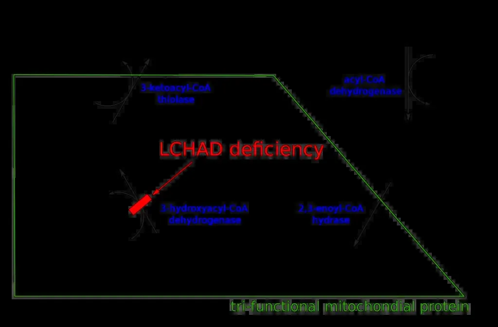
Routine newborn screening detects this condition early, and the main therapy is avoiding prolonged fasting [3]. S. M. Houten and R. J. A. Wanders review these defects in the Journal of Inherited Metabolic Disease, noting that clinical severity varies with diet [5]. Mutations in CPT-II impair long-chain fat entry into muscle mitochondria, triggering muscle pain and rhabdomyolysis after prolonged exercise or cold exposure [3]. Work by Dr. J. W. Miklas and coauthors showed that mutations in HADHA, a subunit of the mitochondrial trifunctional protein, disrupt cardiolipin remodeling and impair fatty acid oxidation in human cardiomyocytes. The defect leads to arrhythmias and sudden infant death syndrome [11].
In peroxisomal disease, the genetic condition adrenoleukodystrophy inspired the movie Lorenzo’s Oil, based on the real-life struggle of Lorenzo Odone [3]. Metabolic reprogramming toward fatty acid oxidation also occurs in cancer. Research by Dr. Y. Tan and colleagues published in Nature Communications showed that platinum-resistant ovarian cancer cells shift from glycolysis to fatty acid uptake and beta oxidation to survive chemotherapy-induced oxidative stress. Small molecule inhibitors of beta oxidation restored drug sensitivity in experimental tumor models [10]. In cardiovascular medicine, experimental drugs like etomoxir inhibit CPT-I to force failing heart tissue to burn glucose instead of fats [3]. These findings show that beta oxidation remains central to modern drug discovery and therapeutic interventions [14].
- WEBSITE Aryal, S., & Dewangan, N. (2023). Beta oxidation of fatty acid: Steps, uses, diagram. Microbe Notes. [Article Link]
- WEBSITE Benson, D. (2013). Beta-oxidation. Chemistry LibreTexts. [Article Link]
- WEBSITE Unseel. (2026). Beta-oxidation: Burning fat two carbons at a time. [Article Link]
- WEBSITE Bio-chem_hub. (2024). Understanding beta oxidation: Energy yield explained. [Article Link]
- WEBSITE BD Editors. (2017). Beta oxidation: Definition, steps and quiz. Biology Dictionary. [Article Link]
- WEBSITE Pan, S. (2024). Beta-oxidation of fatty acid: Definition, steps, importance. Biology Notes Online. [Article Link]
- WEBSITE Mukherjee, S. (2023). Beta-oxidation of fatty acid: Definition, pathway, and diagram. Science Facts. [Article Link]
- WEBSITE Wikipedia. (2026). Beta oxidation. [Article Link]
- WEBSITE Wikipedia. (2026). Alpha oxidation. [Article Link]
- ACADEMIC JOURNAL Tan, Y., Li, J., Zhao, G., Huang, K., Cárdenas, H., Wang, Y., Matei, D. E., & Cheng, J. (2022). Metabolic reprogramming from glycolysis to fatty acid uptake and beta-oxidation in platinum-resistant cancer cells. Nature Communications, 13(1), 4554. [Article Link]
- ACADEMIC JOURNAL Miklas, J. W., Clark, E. C., Levy, S., Detraux, D., Leonard, A., Beussman, K. M., Showalter, M. R., Smith, A. S., Hofsteen, P., Yang, X., Macadangdang, J. R., Manninen, T., Raftery, D., Madan, A., Suomalainen, A., Kim, D., Murry, C. E., Fiehn, O., Sniadecki, N. J., & Ruohola‐Baker, H. (2019). TFPa/HADHA is required for fatty acid beta-oxidation and cardiolipin re-modeling in human cardiomyocytes. Nature Communications, 10(1), 4671. [Article Link]
- ACADEMIC JOURNAL Vishwanath, V. A. (2016). Fatty acid beta-oxidation disorders: A brief review. Annals of Neurosciences, 23(1), 51-55. [Article Link]
- ACADEMIC JOURNAL Tahri-Joutey, M., Andreoletti, P., Surapureddi, S., Nasser, B., Cherkaoui‐Malki, M., & Latruffe, N. (2021). Mechanisms mediating the regulation of peroxisomal fatty acid beta-oxidation by PPARalpha. International Journal of Molecular Sciences, 22(16), 8969. [Article Link]
- ACADEMIC JOURNAL Rogers, G. W., Nadanaciva, S., Swiss, R. L., Divakaruni, A. S., & Will, Y. (2014). Assessment of fatty acid beta oxidation in cells and isolated mitochondria. Current Protocols in Toxicology, 60(1), 25.3.1-19. [Article Link]
- ACADEMIC JOURNAL Uchinomiya, S., Matsunaga, N., Kamoda, K., Kawagoe, R., Tsuruta, A., Ohdo, S., & Ojida, A. (2020). Fluorescence detection of metabolic activity of the fatty acid beta oxidation pathway in living cells. Chemical Communications, 56(20), 3023-3026. [Article Link]


