






Understanding what is pellagra reveals how a profound lack of niacin or tryptophan harms human biology. When cells run short of these building blocks, tissues in the brain and gut lose their energy supply. In clinical reports from the United States and Europe, doctors note that the condition damages skin, bowels, and nerves. Left untreated, the disease causes lasting disability. Simple oral vitamin therapy clears symptoms within days [1].
- What Is Pellagra and How Does It Develop?
- The Four Cardinal Clinical Manifestations
- How Cellular Energy Fails in Vitamin B3 Deficiency
- Primary Versus Secondary Niacin Deficiency
- Why Untreated Maize Triggered Historical Outbreaks
- Diagnostic Evaluation and Laboratory Findings
- Therapeutic Management Protocols
- Food Fortification in Global Populations
What Is Pellagra and How Does It Develop?
The human body obtains niacin from food or makes it from dietary tryptophan. After gut uptake, cells convert niacin into nicotinamide adenine dinucleotide, known as NAD, and its partner NADP. More than 400 enzymes rely on these coenzymes to turn food calories into ATP, powering living cells throughout human tissues [1].
When cellular pools of NAD fall below normal thresholds, organs that demand high amounts of energy and undergo fast cell turnover suffer first. Intestinal lining cells cannot regenerate quickly enough to maintain their mucosal barrier, leading to irritation, poor uptake, and swelling from the mouth down to the bowels. The skin also loses its defense mechanisms against solar radiation, while neurons in the brain face energetic failure that impairs basic memory, mood regulation, and mental focus. In clinical reviews from Harvard and the Cleveland Clinic, researchers point out that these cellular deficits explain why symptoms span three separate organ systems across patients in Asia and Africa [7].
Medical historians trace the name pellagra to the northern Italian phrase pelle agra, which translates to rough skin in Lombard vernacular. Physician Francesco Frapolli introduced the term in Milan after observing the condition among poor farming families whose daily meals depended on plain corn. Clinicians today recognize that pellagra is not an infection, but a body-wide failure stemming from a cellular shortfall of nicotinamide cofactors across vulnerable populations in southern Europe [13].
The Four Cardinal Clinical Manifestations
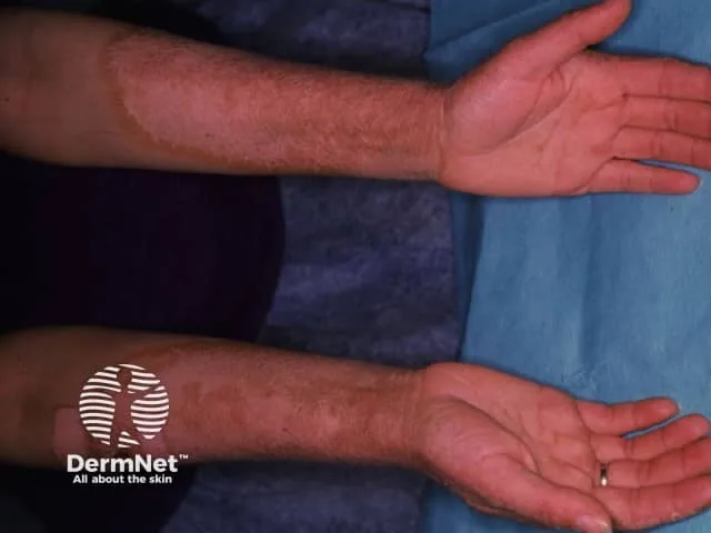
Medical students across Europe and America memorize the hallmark signs of pellagra through the classic clinical mnemonic of the 4 Ds: dermatitis, diarrhea, dementia, and death. Skin signs usually develop first, often appearing during spring and summer when solar rays trigger an inflammatory reaction on uncovered body surfaces. What starts as a red patch resembling a common sunburn soon thickens into dry, brown, scaly plaques that peel under friction [15].
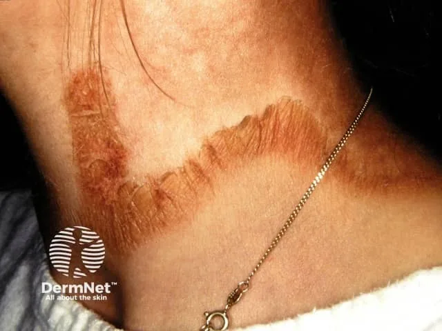
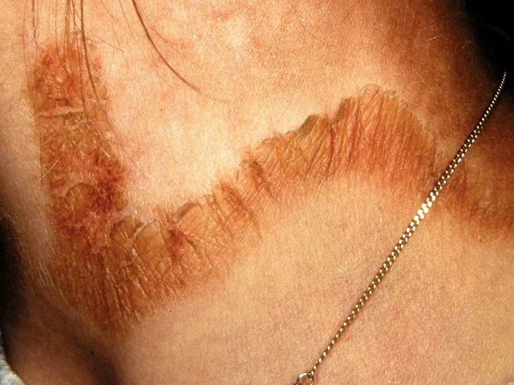
The characteristic rash shows bilateral symmetry across the face, forearms, backs of the hands, and lower legs. A telltale sign is Casal necklace, a broad, dark collar of hyperpigmented skin that encircles the lower neck and upper chest. Spanish court physician Don Gaspar Casal documented this pattern in 1735 among impoverished peasants in Asturias, where locals called it mal de la rosa. His friend Juan Sevillano published these observations in Madrid years later. In modern clinical reports from DermNet in New Zealand, dermatologists explain that patients also develop oral sores, swollen lips, and a bright red tongue called glossitis that makes chewing difficult and painful [5].
Intestinal swelling produces chronic watery diarrhea, which causes fluid loss, dangerous electrolyte drops, and rapid weight loss. Neurological signs begin quietly with fatigue, insomnia, headaches, and apathy before advancing to confusion, paranoia, hallucinations, and marked loss of motor coordination. As clinical researchers Tom Spies and J. Frostig documented in classic medical studies, these emotional and sensory disruptions reflect deep cellular starvation in the central nervous system [13].
How Cellular Energy Fails in Vitamin B3 Deficiency
At the molecular scale, nicotinamide adenine dinucleotide plays a key role as an electron carrier in cellular respiration. When a patient lacks vitamin B3, cells cannot make enough ATP through mitochondrial pathways. In a biochemical analysis from the United Kingdom, researcher A. A. Badawy notes that energetic failure quickly triggers cell injury across vital tissues [14].
NAD also serves as the sole substrate for poly(ADP-ribose) polymerase, an enzyme family known as PARP that repairs damaged DNA within cell nuclei. When sunlight penetrates unprotected skin, ultraviolet photons break DNA strands in epidermal keratinocytes, requiring fast enzymatic repair to prevent cell death. In a person lacking niacin, depleted NAD pools stall PARP activity, allowing DNA damage to accumulate until skin cells undergo apoptosis. In a medical review published in Poland, researcher P. Hałubiec and colleagues in Kraków showed that this repair failure directly drives cutaneous sun sensitivity in deficient patients [16].
The human liver can produce one milligram of niacin from roughly 60 milligrams of dietary tryptophan, but this process needs riboflavin and vitamin B6 as active cofactors. If metabolic disease, poor diet, or drug interference disrupts this pathway, the brain faces a dual shortage of both niacin and serotonin. Medical experts at Patient.info in London emphasize that this biochemical mechanism explains why depression, irritability, and anxiety frequently emerge before cutaneous sores become visible [12].
Primary Versus Secondary Niacin Deficiency
Doctors divide the causes of pellagra into two clinical categories: primary and secondary. Primary deficiency arises directly from an inadequate dietary intake of bioavailable niacin and tryptophan. It occurs mainly in poor farming regions where families eat monotonous diets dominated by untreated corn or sorghum. In field studies across the Deccan Plateau of India and rural communities in Africa, researchers found that populations relying on sorghum suffer from high dietary leucine, an amino acid that blocks niacin synthesis [9].
Secondary pellagra occurs when a person eats enough nutrients but their body cannot absorb or use them properly. In developed nations, chronic alcohol use disorder is the most common cause, because heavy ethanol intake irritates the gut lining and impairs liver function. Medical teams at Medscape report that digestive illnesses like Crohn’s disease, chronic diarrhea, and Roux-en-Y gastric bypass surgery can also disrupt normal gut absorption and precipitate deficiency [11].
Certain genetic traits and pharmaceutical drugs also provoke secondary illness by altering normal biochemistry. In Hartnup disease, an inherited defect in the SLC6A19 gene prevents normal absorption of tryptophan from the small intestine and kidneys. Neuroendocrine carcinoid tumors divert up to 70% of circulating tryptophan into serotonin production, leaving little for niacin synthesis. Medications like isoniazid, ethionamide, 6-mercaptopurine, and 5-fluorouracil also interfere with B-vitamin pathways, producing identical deficiency states in clinical patients [13].



Why Untreated Maize Triggered Historical Outbreaks
The global spread of maize after European contact created a puzzling medical mystery that baffled doctors for more than a century. Native American populations in Mexico and Mesoamerica had consumed corn as their main staple for thousands of years without developing deficiency diseases, yet European peasants fell ill as soon as they adopted the crop. The secret was nixtamalization, an ancient culinary process where dried corn is soaked and cooked in an alkaline limewater solution before milling. The alkaline bath breaks the chemical bonds that trap niacin in an indigestible complex called niacytin, releasing free nicotinic acid that human intestines can easily absorb [13].
European farmers adopted the high-yielding crop but discarded the lime-soaking step, paving the way for sweeping health crises. Endemic pellagra spread across Spain, northern Italy, France, and Egypt throughout the eighteenth and nineteenth centuries, sickening thousands of tenant farmers who survived on unfortified cornmeal. In the early twentieth century, the American South faced an even larger catastrophe as agricultural reliance on cotton left poor sharecroppers dependent on a cheap diet of fatback, molasses, and untreated corn. Between 1906 and 1940, pellagra caused more than 100,000 deaths across southern states like South Carolina and Georgia [13].
In 1914, the United States Public Health Service appointed Dr. Joseph Goldberger to investigate outbreaks in South Carolina orphanages, prisons, and mental hospitals. Goldberger observed that while institutionalized children and inmates regularly developed symptoms, doctors and nurses remained healthy despite breathing the same air and handling sick patients. By adding fresh milk, eggs, and lean meat to institutional meals, he cured the sick at the Spartanburg Pellagra Hospital and proved the illness was dietary [11].
Diagnostic Evaluation and Laboratory Findings
Recognizing pellagra requires careful physical examination and a thorough dietary history. Early symptoms often mimic other dermatological conditions. In clinical guidelines from Medicover Hospitals in India, physicians inspect sun-exposed skin for symmetrical rashes, examine the mouth for glossitis, and ask patients about unusual diets or alcohol use to spot deficiency early [10].
When laboratory confirmation is necessary, doctors measure the excretion of niacin breakdown products in a patient’s urine. Healthy kidneys excrete measurable quantities of N-methylnicotinamide and 2-pyridone, but these metabolites drop to very low levels during clinical deficiency. A urinary ratio between these two metabolites of less than two indicates severe tissue depletion, while measuring red blood cell concentrations of NAD and NADP provides objective confirmation of cellular energy failure [12].
Physicians must distinguish pellagra from other disorders that cause photosensitive rashes, including lupus erythematosus, porphyria cutanea tarda, and drug phototoxicity. Dermatologists at DermNet New Zealand, including Casey P. Schukow of Michigan State University, point out that skin biopsy reveals epidermal necrosis and hyperkeratosis. In clinical practice, the most definitive test is a therapeutic trial: when patients take oral niacin supplements, digestive distress and skin inflammation improve within days [5].
Therapeutic Management Protocols
The standard medical treatment for pellagra is oral administration of nicotinamide, a water-soluble form of vitamin B3. The World Health Organization in Geneva recommends giving 300 mg daily in divided doses for three to four weeks. Clinicians prefer nicotinamide over nicotinic acid because it does not trigger flushing, headache, or sharp drops in blood pressure [5].
Clinical recovery begins quickly once therapy starts, offering prompt relief from debilitating symptoms. Intestinal inflammation and diarrhea generally subside within 24 to 48 hours, allowing the gut lining to heal and absorb nutrients properly. In clinical summaries from Isymptom and the Merck Manual in the United States, authors note that confusion and apathy clear within days, while skin plaques heal over two to four weeks when protected from sunlight [8].

Because pellagra rarely occurs in isolation, comprehensive medical care also requires addressing general malnutrition and concurrent micronutrient deficits. Patients routinely receive a complete B-complex supplement containing riboflavin, thiamine, and vitamin B6 to support cellular pathways and ensure proper tryptophan metabolism. Long-term care involves eating a diverse, protein-rich diet with milk, eggs, poultry, and fish, while managing underlying bowel disease or alcohol use disorder [7].
Food Fortification in Global Populations
The chemical identity of the anti-pellagra factor was solved in 1937 when agricultural chemist Conrad A. Elvehjem, working at the University of Wisconsin, identified nicotinic acid as the active compound that cured black tongue in dogs. Human trials quickly confirmed that the vitamin cured pellagra, opening the door to prevention programs. During the late 1930s and 1940s, the United States introduced mandatory grain enrichment programs, adding niacin, thiamine, and iron to flour across North America [11].
Despite these successes, pellagra still resurfaces in humanitarian emergencies, refugee settlements, and disaster zones where normal food supply chains break down completely. In 2019, an epidemiological field study documented an outbreak of roughly 2300 cases in the Nhamatanda District of Mozambique after Cyclone Idai devastated local crop harvests and left families subsisting on unfortified maize. Similar outbreaks have struck displaced populations in post-war Angola, Malawi, Zimbabwe, and Nepal. These health crises prove that communities relying on narrow food rations face severe risks unless relief groups like the United Nations World Food Program distribute fortified grains, legumes, and supplemental B vitamins [11].
In modern affluent nations, pellagra persists as a rare diagnosis that can easily elude physicians who consider the disease extinct. As clinical researchers in Poland, the Netherlands, and Denmark have reported, sporadic cases continue to appear in individuals with severe anorexia nervosa, older adults living in isolation, and cancer patients. Keeping clinical awareness alive helps doctors recognize the warning signs of niacin deficiency promptly, sparing patients from long-term neurological injury through inexpensive vitamin therapy [16].
- WEBSITE Cleveland Clinic. (2023). Pellagra: Definition, Symptoms & Treatment. [Article Link]
- WEBSITE MedlinePlus. (2024). Pellagra: MedlinePlus Medical Encyclopedia. [Article Link]
- WEBSITE Dix, M. (2017). Pellagra: Pictures, Symptoms, Causes, and Treatment. Healthline Media. [Article Link]
- ONLINE NEWS Zambon, V. (2020). Pellagra: Definition, symptoms, pictures, and more. Medical News Today. [Article Link]
- WEBSITE Ngan, V., Schukow, C. P., & Keefe, M. (2021). Pellagra (vitamin B3 or niacin deficiency). DermNet. [Article Link]
- WEBSITE Kinton-Vieira, J. (2026). Pellagra: Symptoms, Causes & Treatment. Tua Saúde. [Article Link]
- WEBSITE Deshmukh, A. (2026). Pellagra: Causes, Symptoms, Treatment & Prevention. AskDocDoc. [Article Link]
- WEBSITE Isymptom Editorial Team. (2026). Pellagra (Niacin Deficiency): The 4 Ds, Causes, Treatment. Isymptom. [Article Link]
- WEBSITE HarvestHeal. (2026). Pellagra — Foods & Natural Support. HarvestHeal. [Article Link]
- WEBSITE Medicover Hospitals. (2026). Pellagra: Causes, Symptoms and Treatment Options. [Article Link]
- WEBSITE Medscape. (2026). Pediatric Pellagra: Background, Epidemiology, Etiology. [Article Link]
- WEBSITE Vakharia, K., & Tidy, C. (2023). Pellagra. Patient.info. [Article Link]
- WEBSITE Wikipedia contributors. (2026). Pellagra. Wikipedia, The Free Encyclopedia. [Article Link]
- ACADEMIC JOURNAL Badawy, A. A. (2014). Pellagra and Alcoholism: A Biochemical Perspective. Alcohol and Alcoholism, 49(3), 238-250. [Article Link]
- ACADEMIC JOURNAL Hegyi, J., Schwartz, R. A., & Hegyi, V. (2004). Pellagra: dermatitis, dementia, and diarrhea. International Journal of Dermatology, 43(1), 1-5. [Article Link]
- ACADEMIC JOURNAL Hałubiec, P., Leończyk, M., Staszewski, F., Łazarczyk, M., Jaworek, A. K., & Wojas-Pelc, A. (2022). Pathophysiology and clinical management of pellagra — a review. Folia Medica Cracoviensia, 61(3), 125-137. [Article Link]


