



To understand what is apoptosis, biologists examine how multicellular bodies quietly dismantle damaged or surplus cells without injuring neighboring tissue. Often called programmed cell death, this regulated biological routine lets organisms shape growing organs and eliminate damaged cells that could cause illness. Unlike sudden cell damage from physical injury, the process follows an orderly biochemical path that avoids inflammatory swelling. Every day, tens of billions of cells quietly collapse and vanish, preserving bodily health through balanced cellular turnover [8].
What Is Apoptosis?
Apoptosis represents an orderly form of cellular self-destruction that occurs naturally across multicellular organisms and in certain single-celled eukaryotes such as yeast [8]. According to The American Heritage Dictionary of the English Language, degradative enzymes dismantle cells that carry genetic programming for a fixed lifespan or suffer acute stress from toxic compounds and irradiation [6]. The body uses this mechanism to maintain homeostatic balance. Without controlled death, accumulating cells would disrupt organ function.
The scale of this daily clearance remains enormous in human biology. An average adult human loses between 50 and 70 billion cells each day through apoptosis, while children between 8 and 14 years old shed roughly 20 to 30 billion cells daily. These rates match the continuous production of new cells through division, keeping overall tissue volume stable throughout adult life. When this delicate equilibrium breaks down, pathology follows swiftly. Insufficient death allows damaged cells to survive and multiply, whereas excessive destruction can cause progressive tissue atrophy across vital organs [8].
Unlike accidental traumatic injury, this self-destruction cannot stop once executioner enzymes begin degrading cellular structures [8]. The commitment step seals cellular fate through controlled proteolytic digestion. Phagocytic white blood cells quickly identify and consume the shrinking cell pieces before toxic chemicals leak into extracellular fluid [3]. In single-celled fungi like Saccharomyces cerevisiae, ancient homologues such as Bir1p protect against premature death, showing that regulated suicide systems evolved early in evolutionary history [7]. This swift clean-up prevents swelling in nearby tissue.

Historical Discovery of Cell Clearance
Early observations of programmed cell loss date back more than a century before researchers understood its molecular basis. German scientist Carl Vogt was first to describe the general principle of developmental cell death in 1842 while studying toad development. Later, in 1885, anatomist Walther Flemming delivered a detailed anatomical description of regressing tissue, noting how dying cells shrank and vanished from healthy beds [8].
Modern investigation of the process revived in 1965 when Australian pathologist John Kerr used electron microscopy at the University of Queensland to distinguish controlled cellular death from traumatic necrosis. Kerr soon joined Alastair Currie and Andrew Wyllie at the University of Aberdeen, where the three investigators collaborated on a 1972 paper published in the British Journal of Cancer. Seeking an appropriate name for this biological phenomenon, Kerr, Wyllie, and Currie consulted James Cormack, a professor of Greek language at Aberdeen, who suggested reviving the classical term apóptōsis, which describes the natural dropping of petals from flowers or leaves falling from autumn trees [8].
Cormack noted that ancient medical writers Hippocrates and Galen had used variations of the word to describe falling bone fragments or scabs shedding from wounds. Decades later, Kerr shared the Paul Ehrlich and Ludwig Darmstaedter Prize with Boston biologist H. Robert Horvitz on March 14, 2000. In 2002, Horvitz, Sydney Brenner, and John Sulston received the Nobel Prize in Physiology or Medicine for identifying genetic pathways controlling cell death in Caenorhabditis elegans [8].
Intrinsic Mitochondrial Pathways
The intrinsic pathway, often called the mitochondrial pathway, responds to internal signals triggered when a cell experiences extreme intracellular stress. Cellular damage caused by ultraviolet radiation, hypoxia, heat shock, nutrient starvation, or high free fatty acid concentrations prompts internal sensors to initiate self-destruction [8]. At the center of this response sits the Bcl-2 protein family, which contains both pro-survival guardians like Bcl-2, Bcl-xL, and Bcl-w, and pro-death executioners like BAX and BAK [9], [10].
In healthy mammalian cells, BAX remains mostly in the liquid cytosol, but upon stress signals it changes shape and inserts into the mitochondrial outer membrane. Structural studies reveal that BAX contains nine α-helices, where a hydrophobic groove formed along α2 to α5 binds activating partners before forming an oligomeric pore known as MAC alongside the voltage-dependent anion channel (VDAC) [10]. This pore formation leads to mitochondrial outer membrane permeabilization, causing cytochrome c and the protein SMAC, also called DIABLO, to pour into the cytosol [2], [10].
Once in the cytosol, cytochrome c binds to Apaf-1 and ATP to assemble a wheel-like protein complex called the apoptosome. The apoptosome recruits pro-caspase-9, cleaving it into active caspase-9, which then unleashes executioner caspase-3 to digest structural proteins [8]. In a theoretical study released as an unreviewed preprint, biophysicist Subhadip Raychaudhuri modeled how strong stimuli trigger rapid deterministic responses, whereas weaker stress yields stochastic, cell-to-cell variability in apoptotic activation [1]. Researchers also explore how external physical forces alter behavior in living tissue, studying how light-induced rapid cell responses alter intracellular dynamics.
Extrinsic Death Receptor Signaling
While internal distress drives the mitochondrial route, external signals from neighboring cells can command a cell to die through the extrinsic pathway. This mechanism relies on specialized death receptors on the cell surface that belong to the tumor necrosis factor receptor superfamily. When extracellular signaling ligands bind to these receptors, they cluster together and recruit adaptor proteins to assemble the death-inducing signaling complex beneath the plasma membrane [8].
The best-known extrinsic route involves tumor necrosis factor-alpha (TNF-α) binding to the receptor TNFR1. Receptor binding brings in adaptor proteins known as TRADD and FADD, which recruit and activate initiator caspase-8. Another extrinsic route operates through Fas ligand binding to the Fas receptor (Apo-1 or CD95), while TRAIL proteins bind to death receptors DR4 and DR5. Synthetic molecules like luminescent iridium complex-peptide hybrids have even been designed to mimic TRAIL and stimulate death receptors on resistant targets. Active caspase-8 directly cleaves downstream executioner caspases 3 and 7 to complete cellular demolition [8], [9].
Extrinsic death signals also face viral and cellular barricades that can stop the chain reaction. Cowpox virus produces a serpin called crmA, or cytokine response modifier A, which forms an irreversible complex with caspase-8 and caspase-1, preventing inflammatory signaling and blocking extrinsic death [9]. By freezing caspase-8 in an inactive state, the virus shields infected host cells from death receptor signals, allowing viral replication to continue inside living cytoplasm [7], [9].


Cellular Inhibitors and Caspase Control
Because caspase activation causes irreversible death, cells express endogenous guardians known as inhibitor of apoptosis (IAP) proteins to restrain rogue proteases. All members of the IAP family share a conserved structural motif called the baculovirus IAP repeat (BIR) domain, which spans approximately 70 amino acids. The first identified IAP members came from insect baculoviruses, designated Cp-IAP and Op-IAP, which evolved to block host caspases during infection. The human genome encodes eight distinct IAP proteins: XIAP, cIAP1, cIAP2, NAIP, Survivin, Bruce, Livin, and ILP-2 [9].
Among these family members, XIAP acts as the most potent caspase blocker by directly binding active caspase-3, caspase-7, and caspase-9 [7], [9]. Cellular IAP proteins also receive instructions from inflammatory pathways. In 1997, researchers showed that the transcription factor NF-κB directs the expression of human c-IAP2 to suppress TNF-induced cytotoxicity, creating a positive feedback loop that helps cells withstand cytokine attacks. Similarly, type I and type II interferons delay neutrophil apoptosis through STAT3 activation and cIAP2 upregulation [7].
To overcome IAP barriers when death becomes necessary, the mitochondria release natural inhibitor antagonists during stress. Proteins known as SMAC, or DIABLO, along with Omi, also known as HtrA2, exit permeable mitochondria to bind the BIR3 domain of XIAP [2], [7]. This physical binding dislodges caspases from inhibition, enabling executioner enzymes to finish dismantling the cell. Without SMAC release, elevated IAP levels can halt apoptosis even after upstream stress signals fire [2].
Why Does Apoptosis Avoid Inflammation?
Why does apoptosis proceed quietly while traumatic tissue injuries provoke painful swelling? The answer lies in the orderly structural demolition of the dying cell. When executioner caspases become active, the cell shrinks, separates from adjacent cells, and displays rhythmic bubbling movements called membrane blebbing [8]. Inside, degradative enzymes condense chromatin and break nuclear DNA into small fragments, neatly packing cellular contents rather than allowing them to rupture [6], [8].
As the dying cell collapses, it breaks into small, membrane-enclosed packages known as apoptotic bodies. These packages retain intact outer membranes, preventing intracellular digestive enzymes and damage-associated molecular patterns from spilling into surrounding tissue [8]. Phagocytic cells, primarily tissue macrophages, recognize exposed surface signals on the apoptotic bodies and engulf them swiftly through receptor-mediated phagocytosis [3], [8]. Immunologist Ilya Metchnikoff shared the 1908 Nobel Prize for discovering phagocytic clearance, a concept expanded in modern pathology volumes such as Phagocytosis of Dying Cells by Dmitri V. Krysko and Peter Vandenabeele that describe how defective clearance promotes autoimmune illnesses [3].
This rapid phagocytic disposal distinguishes apoptosis from accidental necrosis. In necrosis, physical injury or severe infection causes the cell membrane to burst, releasing intracellular contents and attracting immune cells that trigger swelling and inflammation [3], [8]. If macrophages fail to clear apoptotic bodies in time, secondary necrosis can occur, potentially exposing nuclear debris that sparks chronic lupus-like autoimmune disorders [3].
Developmental Roles in Embryonic Modeling
Programmed cell death plays a fundamental role in shaping the physical architecture of multicellular organisms during embryonic growth. In developing human embryos, hands and feet initially form as solid paddle-like structures. The clean separation of individual fingers and toes occurs only when the webbing tissue between the digits undergoes genetically scheduled apoptosis, carving out distinct digits before birth [8].
Similar developmental sculpting takes place throughout the growing nervous system and immune system. Developing brains generate far more neurons than survive into adulthood, with surplus cells undergoing apoptosis if they fail to form functional synaptic connections. Likewise, developing immune cells that display autoreactive receptors against the body’s own tissues are eliminated through apoptosis in the thymus, preventing dangerous autoimmune reactions before immune cells enter general circulation [8].
Throughout adult life, controlled cell death maintains structural equilibrium in rapidly renewing tissues. Epithelial cells lining the digestive tract and skin continually shed through apoptosis as fresh progenitor cells replace them from basal layers [6], [8]. Erythrocytes and neutrophils similarly face programmed clearance when their functional lifespan concludes [6], [7]. Through this unending renewal, multicellular life preserves organ function and prevents aged cells from accumulating detrimental genetic damage [8].
Targeting Programmed Cell Death in Cancer Therapy
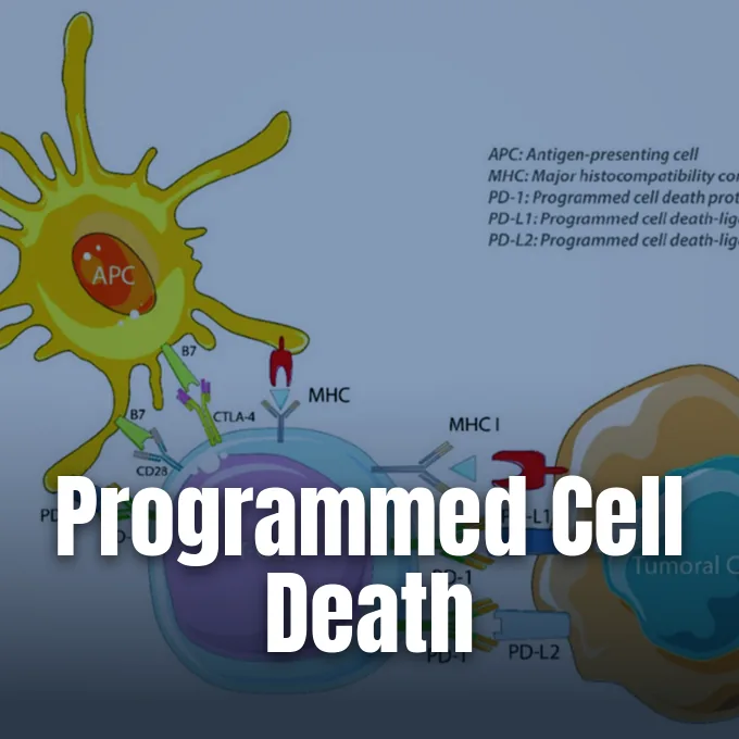
The evasion of programmed cell death represents one of the defining hallmarks of cancer described in seminal reviews in Cell. In healthy tissue, severe DNA damage prompts the tumor suppressor protein p53 to activate BAX and initiate apoptosis, eliminating potential malignancies [4], [10]. In malignant tumors, however, mutations in p53 or somatic frameshift mutations in the BAX gene disable this safety switch. Pioneering oncologist Dr. Stan Korsmeyer characterized how chromosomal translocations in follicular lymphoma cause BCL2 overexpression, shielding malignant cells from death signals [4].
To overcome cancer cell survival, modern pharmaceutical research designs therapies that reactivate latent apoptotic pathways. Small molecules called SMAC mimetics, such as xevinapant (also known as AT 406 or Debio-1143, developed through collaborations involving Ascenta Therapeutics, Debiopharm, EMD Serono, and Merck & Co), target IAP proteins to trigger autocrine TNF-α cell killing [2], [5]. Other experimental compounds include the BH3 mimetic ABT-737, which frees BAX to form mitochondrial pores, along with dietary flavonoids investigated for restoring apoptotic sensitivity in resistant tumor lines [4], [10]. When apoptotic mechanisms stall, therapies may target alternative pathways such as leveraging cellular cannibalism in cancer therapy to destroy malignant populations.
Yet manipulating cell death in tumors reveals biological complexities. High-grade cancers often exhibit an apoptosis paradox, where ongoing cell death within the tumor microenvironment conditions nearby immune cells to support cancer survival and drug resistance. Understanding these intricate molecular balances helps clinicians determine when promoting apoptosis eradicates malignant clones and when surviving tumor niches evade treatment [4].
- PREPRINT Raychaudhuri, S. (2010). A minimal model of signaling network elucidates cell-to-cell stochastic variability in apoptosis. arXiv. [Article Link]
- ACADEMIC JOURNAL McCarthy, N. (2008). Smac it to them! Nature Reviews Cancer, 8(3), 165. [Article Link]
- BOOK Krysko, D. V., & Vandenabeele, P. (Eds.). (2009). Phagocytosis of dying cells: From molecular mechanisms to human diseases. Springer Dordrecht. [Article Link]
- ACADEMIC JOURNAL Spurgers, K. B., Chari, N. S., Bohnenstiehl, N. L., & McDonnell, T. J. (2006). Molecular mediators of cell death in multistep carcinogenesis: A path to targeted therapy. Cell Death & Differentiation, 13(8), 1360–1369. [Article Link]
- REPORT Springer Nature. (2026). Xevinapant – Merck & Co: Drug evaluation and development status. AdisInsight. [Article Link]
- DICTIONARY ENTRY HarperCollins Publishers. (2022). Apoptosis. In The American Heritage Dictionary of the English Language. [Article Link]
- WEBSITE Google Scholar. (2016). Recent literature on inhibitors of apoptosis proteins. [Article Link]
- WEBSITE Wikipedia contributors. (2026). Apoptosis. In Wikipedia, The Free Encyclopedia. [Article Link]
- WEBSITE Wikipedia contributors. (2026). Inhibitor of apoptosis. In Wikipedia, The Free Encyclopedia. [Article Link]
- WEBSITE Wikipedia contributors. (2026). Apoptosis regulator BAX. In Wikipedia, The Free Encyclopedia. [Article Link]
Cover image: Alexandre Perrier, Audrey Didelot, Pierre Laurent-Puig, Hélè / Wikimedia Commons, CC BY 4.0


