




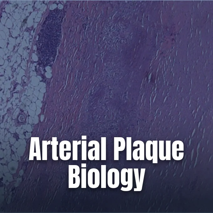
What is atherosclerosis? It is a chronic inflammatory disease where cholesterol, fatty substances, and immune cells collect inside the vessel wall to create thick deposits called plaques [12]. Healthy arteries stay flexible and permit steady blood circulation, but retained lipids gradually narrow the lumen and stiffen the vascular wall over decades of quiet accumulation [6]. Plaque growth restricts oxygen delivery to tissues and causes most heart attacks and strokes worldwide [15].
- What Is Atherosclerosis?
- Endothelial Damage and Lipid Retention
- How White Blood Cells Form Foam Cells
- Vascular Smooth Muscle Cells in Plaque Progression
- Why Plaque Buildup Often Starts in Childhood
- Major Systemic and Genetic Risk Factors
- Clinical Symptoms Across Different Arteries
- Preventing Hardening of the Arteries
What Is Atherosclerosis?
The medical term refers specifically to atheromatous plaque formation inside the tunica intima, the inner layer of large and medium arteries [12]. People commonly call it hardening of the arteries, though arteriosclerosis is an umbrella category that includes other forms of arterial stiffening [6]. Because plaques accumulate slowly beneath the endothelial lining, the arterial lumen often stays open for years as the vessel expands outward to preserve blood flow [12]. Flow adaptation conceals early stages of disease from everyday physical sensation.
Autopsy records and clinical registries show how silently plaques can grow before causing symptoms. In 2004, United States medical data indicated that in about 66% of men and roughly 47% of women, the very first sign of atherosclerotic disease was an acute heart attack or sudden cardiac death [12]. Many individuals never receive an early warning because resting blood flow remains adequate until a plaque obstructs more than seventy percent of the arterial passage. The disease mainly targets vessels under hemodynamic stress, including coronary arteries that feed heart muscle and carotid arteries that supply the brain [9].
Vascular lesions appear in specific locations rather than scattering evenly along the circulatory tree. Atheromas tend to cluster at vessel branch points and curves where blood flow becomes turbulent [9]. When blood swirls unpredictably, low shear stress disturbs the protective endothelial surface and allows circulating lipids to enter the subendothelial space [7]. Local flow patterns explain why two adjacent segments of the same artery can display completely different plaque burdens.

Endothelial Damage and Lipid Retention
The vascular lining acts as a barrier that regulates vessel tone and prevents unwanted cellular adhesion. When friction from high blood pressure, cigarette smoke toxins, or elevated blood glucose damages endothelial cells, that protective barrier weakens [3]. Damaged endothelial cells begin displaying adhesion molecules that bind circulating white blood cells and let lipid particles leak into the vessel wall [9]. The European Society of Cardiology describes this initiating sequence as the response-to-retention concept, where trapping cholesterol-rich particles within the intima drives subsequent stages of lesion development [7].
Particle size determines which circulating lipoproteins can penetrate the arterial lining. All apolipoprotein-B (apoB) containing particles with a diameter under 70 nanometers can cross the endothelium and enter the intima. Low-density lipoprotein (LDL) serves as the most abundant atherogenic carrier in plasma, transporting cholesteryl esters into vulnerable subendothelial pockets. Other apoB particles, such as very low-density lipoprotein remnants and lipoprotein(a), also squeeze through the endothelial junction. Once inside, negatively charged proteoglycans bind these particles tightly, preventing them from returning to the bloodstream [7].
Trapped lipoproteins do not stay inert inside the vessel wall. Local free radicals and enzymes alter the trapped LDL particles through oxidation, creating oxidized LDL that irritates neighboring vascular cells [12]. Oxidized LDL impairs the normal release of nitric oxide from endothelial cells, which prevents arteries from dilating properly and causes local vasoconstriction during periods of exertion [7]. The modified particles also stimulate endothelial cells to secrete chemokines, transforming a simple lipid depot into a nidus of chronic inflammation [15].
How White Blood Cells Form Foam Cells
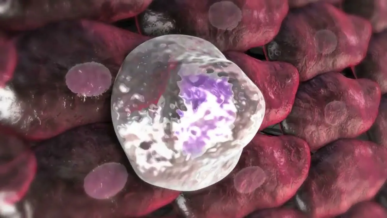
Inflammatory signals draw circulating monocytes toward the damaged arterial surface. Endothelial cells express vascular cell adhesion molecule-1 (VCAM-1) and intercellular adhesion molecules that snag passing monocytes, allowing them to roll along the lining and squeeze between cells into the intima. Macrophage colony-stimulating factor (M-CSF) prompts these monocytes to differentiate into tissue macrophages [12]. These scavenger cells internalize oxidized LDL through scavenger receptors, but unlike normal LDL receptors, scavenger pathways lack feedback inhibition and continue packing lipids until macrophages swell into fat-stuffed foam cells [9].
Accumulated foam cells group together to form the earliest gross manifestation of the disease, known as fatty streaks [9]. Under light microscopy, fatty streaks appear as flat yellow patches that do not obstruct arterial blood flow [12]. Peter Libby from Harvard Medical School pointed out in Arteriosclerosis, Thrombosis, and Vascular Biology that cytokines released by macrophages act as messengers that sustain this localized immune reaction [15]. As foam cells age, metabolic stress triggers cell apoptosis, causing them to burst and spill crystalline cholesterol, cellular debris, and digestive enzymes into the surrounding extracellular matrix [9].
Dying foam cells leave behind a lipid-rich necrotic core in the center of the plaque. The necrotic core attracts further waves of inflammatory cells, including T-lymphocytes that release interferon-gamma and accelerate tissue breakdown [12]. Macrophages also release matrix metalloproteinases, enzymes that dissolve collagen fibers and weaken the protective framework of the vessel [7]. Over time, persistent inflammation shifts the lesion from a harmless fatty streak into an unstable atheroma [15].
Vascular Smooth Muscle Cells in Plaque Progression
Vascular smooth muscle cells (VSMCs) normally reside in the media, keeping vessel tone through contraction. In atheromas, smooth muscle cells migrate into the intima and produce collagen to build a fibrous cap over the lipid core. Pathologists once viewed these cells as simple cap builders that prevented plaque rupture. Genetic lineage tracing reviewed by Martin Bennett and colleagues in Circulation Research shows that smooth muscle cells undergo phenotypic switching, adopting macrophage-like or bone-forming traits [14].
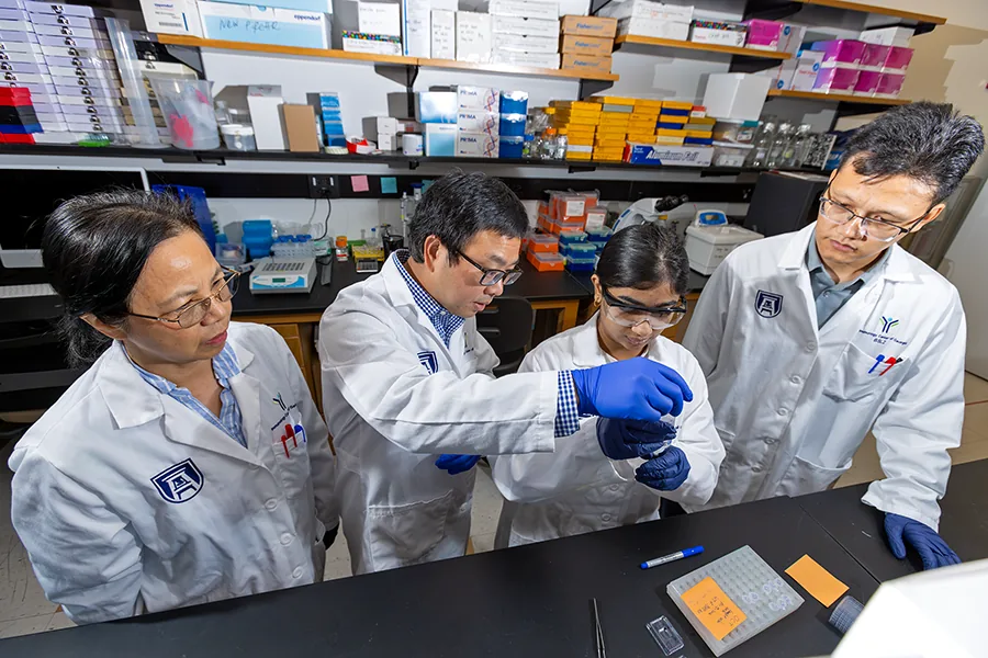
At the Immunology Center of Georgia, researcher Kunzhe Dong received a five-year, $3.8 million NIH grant in 2026 to study how long noncoding RNA controls this smooth muscle shift. Dong studies PRDM16-DT, a molecule abundant in healthy smooth muscle cells. “With this grant, we will determine how PRDM16-DT – which is highly enriched in healthy blood vessels and smooth muscle cells, and contains genetic variants associated with atherosclerosis – regulates muscle cell function in disease,” Dong said. Co-director Klaus Ley added that smooth muscle cells drive plaque growth [1].
When Dong’s team removed the mouse equivalent of PRDM16-DT, the animals developed heavier plaque buildup and increased arterial inflammation [1]. Separately, a 2015 preprint on arXiv by Jicun Wang-Michelitsch and Thomas Michelitsch proposed that plaque growth follows a Misrepair mechanism. This preprint has not undergone peer review, but it suggests that repeated repairs cluster smooth muscle cells unevenly and accelerate lipid trapping [4]. Cellular shifts explain why plaques vary in size along the same artery.


Why Plaque Buildup Often Starts in Childhood
Atherosclerosis is widely perceived as an affliction of middle age, yet pathology records reveal that vascular damage begins decades earlier. A 1953 autopsy study published in the Journal of the American Medical Association examined 300 American battle casualties from the Korean War whose average age was 22 years. The investigators observed gross evidence of coronary atherosclerosis in 77% of the soldiers, with some showing vessels clogged by 90% or more [11]. The findings startled the medical community by proving that arterial disease takes root long before clinical chest pain or heart attacks occur.
Later pediatric studies confirmed that silent vascular changes emerge during grade school. In the Bogalusa Heart Study, researchers examined young individuals who died from accidents between the ages of 3 and 26, discovering that fatty streaks were present in the arteries of 100% of children by age ten. Blood cholesterol levels stood out as the leading predictor of how extensively streaks covered the arterial wall [11]. Intracoronary ultrasonography in the United States similarly found that one in six teenagers already displays abnormal intimal thickening in major arteries [9].
When high circulating cholesterol persists through adolescence, benign fatty streaks slowly transition into raised fibromuscular atheromas. Autopsy scoring systems developed on thousands of young accident victims show that high non-HDL cholesterol raises the risk of advanced coronary lesions eightfold compared to smoking alone. By age thirty, an individual with unmanaged childhood hypercholesterolemia faces a one-in-three chance of carrying advanced arterial lesions [11]. Early nutritional education helps protect lifelong vascular integrity.

Major Systemic and Genetic Risk Factors
Clinical research identifies several modifiable habits and chronic conditions that accelerate arterial injury. Cigarette smoking damages endothelial function and accounts for nearly one-third of deaths caused by coronary heart disease in the United States. High blood pressure exerts chronic mechanical shear stress against arterial linings, accounting for more atherosclerosis deaths nationwide than any other changeable risk factor [3]. Metabolic disorders such as type 2 diabetes further damage vessels because elevated blood glucose glycosylates lipoproteins and impairs the body’s natural antioxidant defense systems [9].
Endocrine and genetic imbalances also influence how quickly plaques accumulate in multiple vessel beds. In American Family Physician, Richard Sadovsky reported that overt hypothyroidism increases LDL levels, induces diastolic hypertension, and alters vascular smooth muscle function. Starting low-dose levothyroxine replacement therapy at 12.5 to 25 micrograms per day helps lower total cholesterol and improves myocardial contractility and efficiency in hypothyroid patients [8]. Furthermore, inherited disorders like familial hypercholesterolemia affect about 1 in 300 people, driving severe lifelong cholesterol elevation and premature arterial stiffening [3].
Inherited variants in the apolipoprotein E (APOE) gene show associations with arterial disease spread. In a 2024 study of 1,074 patients published in the International Journal of General Medicine, Youni Lin and colleagues found that the APOE E3/E4 genotype frequency was lower in patients with multi-bed atherosclerosis than in those with single-bed disease (11.4% versus 17.8%) [10]. Meanwhile, experimental research in Atherosclerosis by J. F. Berbée and colleagues showed that deuterium-reinforced polyunsaturated fatty acids reduced aortic root lesion area by 26% in mice fed a 0.15% cholesterol diet by limiting lipid peroxidation [5]. Genetic background thus shapes both lipid handling and arterial vulnerability.
Clinical Symptoms Across Different Arteries
Symptoms emerge only after arterial narrowing slows blood delivery or when an unstable plaque ruptures to spawn an obstructive thrombus. In coronary heart disease, diminished myocardial perfusion causes angina pectoris, marked by chest tightness, shortness of breath, cold sweats, and nausea during physical exertion [2]. If an atheromatous fibrous cap ruptures, exposed thrombogenic material triggers blood clotting that can block a coronary artery, causing acute myocardial infarction [9]. The severity of symptoms reflects the speed of arterial occlusion and the metabolic demands of downstream tissue.
Plaques in arteries supplying the head and neck present grave neurological risks. Carotid artery disease often develops without early symptoms, though physicians can sometimes detect turbulent flow by listening for an abnormal whooshing sound called a bruit with a stethoscope. Severe narrowing or dislodged emboli can produce transient ischemic attacks or full strokes, causing facial numbness, one-sided weakness, and speech difficulties [2]. In intracranial vessels, cerebral atherosclerosis disrupts blood supply to deep brain structures, causing localized tissue ischemia and microbleeds [13]. Emerging studies investigate whether statins for healthy people to prevent heart disease might also confer long-term cognitive protection against vascular dementia.

Extracranial and systemic arteries also suffer when atherosclerosis advances unchecked. Peripheral artery disease (PAD) leads to intermittent claudication, a condition where patients experience aching leg pain, muscle cramping, and heaviness while walking that consistently resolve after resting. Renal artery stenosis impairs blood delivery to the kidneys, prompting fluid retention, fatigue, and resistant hypertension. Mesenteric artery narrowing causes severe post-meal abdominal pain and involuntary weight loss as digestion demands blood flow the stiffened vessels can’t deliver [2]. Recognizing these regional patterns helps clinicians identify systemic vascular disease before catastrophic organ damage occurs.
Preventing Hardening of the Arteries
Halting the progression of vascular disease begins with addressing root metabolic drivers before plaques calcify and ulcerate. Quitting tobacco use is the single most effective personal lifestyle change, quickly improving endothelial function and reducing heart disease risk. Adopting a balanced dietary pattern that minimizes saturated fat and eliminates industrial trans fats helps lower circulating atherogenic particles [6]. The American Heart Association’s Life’s Essential 8 guidelines advise engaging in at least 150 minutes of moderate aerobic exercise weekly, which improves insulin sensitivity and stimulates endothelial nitric oxide production [3].
Pharmacological therapy plays a central role when lifestyle adjustments can’t sufficiently lower circulating lipid levels. Clinical practice guidelines emphasize evaluating statin therapy for primary prevention of cardiovascular disease in individuals who exhibit elevated baseline risk [6]. Statins inhibit HMG-CoA reductase in hepatocytes, upregulating LDL receptors to clear atherogenic particles from the circulation. The European Society of Cardiology supports “the lower, the better” approach for LDL cholesterol, because lower circulating apoB concentrations decrease subendothelial retention and allow plaque stabilization [7].
When arterial blockages become critical, revascularization procedures restore vital organ perfusion. Interventional cardiologists use percutaneous coronary intervention with drug-eluting stents to prop open narrowed coronary passages and prevent acute vessel re-occlusion. In multivessel coronary disease or severe carotid stenosis, surgeons perform coronary artery bypass grafting or carotid endarterectomy to bypass or surgically clear obstructive atheromas [12]. Although mechanical interventions clear flow channels, ongoing lifestyle vigilance and lipid control remain necessary to prevent new lesions from taking root elsewhere in the vascular tree [6].
- ONLINE NEWS Austin, M. (2026). IMMCG researcher receives $3.8 million NIH grant to study atherosclerosis. Jagwire. [Article Link]
- WEBSITE National Heart, Lung, and Blood Institute. (2024). Atherosclerosis symptoms. [Article Link]
- WEBSITE National Heart, Lung, and Blood Institute. (2024). Atherosclerosis causes and risk factors. [Article Link]
- PREPRINT Wang-Michelitsch, J., & Michelitsch, T. M. (2015). Misrepair mechanism in the development of atherosclerotic plaques. arXiv. [Article Link]
- ACADEMIC JOURNAL Berbée, J. F., Mol, I. M., Milne, G. L., Pollock, E., Hoeke, G., Lütjohann, D., Monaco, C., Rensen, P. C., van der Ploeg, L. H., & Shchepinov, M. S. (2017). Deuterium-reinforced polyunsaturated fatty acids protect against atherosclerosis by lowering lipid peroxidation and hypercholesterolemia. Atherosclerosis, 264, 100-107. [Article Link]
- WEBSITE MedlinePlus. (2026). Atherosclerosis. [Article Link]
- WEBSITE European Society of Cardiology. (2020). What is the role of lipids in atherosclerosis and how low should we decrease lipid levels? E-Journal of Cardiology Practice, 19. [Article Link]
- ACADEMIC JOURNAL Sadovsky, R. (2004). Treating hypothyroidism reduces atherosclerosis risk. American Family Physician, 69(3), 656. [Article Link]
- WEBSITE Lecturio. (2025). Atherosclerosis: Concise medical knowledge. [Article Link]
- ACADEMIC JOURNAL Lin, Y., Yang, M., Liu, Q., Cai, Y., Zhang, Z., Xu, C., & Luo, M. (2024). Apolipoprotein E gene epsilon4 allele is associated with atherosclerosis in multiple vascular beds. International Journal of General Medicine, 17, 5039-5048. [Article Link]
- WEBSITE Greger, M. (2014). Stopping heart disease in childhood. NutritionFacts.org. [Article Link]
- WEBSITE Wikipedia. (2026). Atherosclerosis. [Article Link]
- WEBSITE Wikipedia. (2026). Cerebral atherosclerosis. [Article Link]
- ACADEMIC JOURNAL Bennett, M. R., Sinha, S., & Owens, G. K. (2016). Vascular smooth muscle cells in atherosclerosis. Circulation Research, 118(4). [Article Link]
- ACADEMIC JOURNAL Libby, P. (2012). Inflammation in atherosclerosis. Arteriosclerosis, Thrombosis, and Vascular Biology, 32(9), 2045-2051. [Article Link]
- ACADEMIC JOURNAL Björkegren, J., & Lusis, A. J. (2022). Atherosclerosis: Recent developments. Cell, 185(10), 1630-1645. [Article Link]
- ACADEMIC JOURNAL Schaftenaar, F. H., Frodermann, V., Kuiper, J., & Lutgens, E. (2016). Atherosclerosis. Current Opinion in Lipidology, 27(3), 209-215. [Article Link]


