




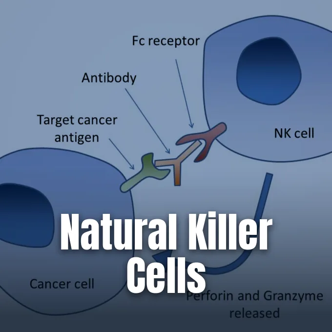
Natural killer cells are cytotoxic white blood cells of the innate immune system that destroy virus-infected and cancer targets without prior antigen priming. Unlike adaptive T and B cells that depend on gene rearrangement, these sentinels rely on germline-encoded receptors that distinguish abnormal targets from healthy tissue. Dr. Henry Smith at the University of Leeds School of Medicine first recorded this spontaneous antitumor killing in 1966. In Stockholm, Rolf Kiessling, Hugh Pross, and Mikael Jondal mapped the activity in mice and humans at the Karolinska Institute under Eva Klein and Hans Wigzell [11]. In adult humans, these cells represent 5% to 20% of circulating white cells, with adults carrying over two billion sentinels that patrol peripheral tissues [4].
- How Natural Killer Cells Identify Threats
- Missing-Self Recognition and Inhibitory Signals
- Granule Exocytosis and Cytotoxic Pathways
- Distinct Subsets of NK Cells
- Development from Hematopoietic Progenitors
- How Natural Killer Cells Bridge Immunity
- Uterine Populations in Early Pregnancy
- Therapeutic Applications in Cancer Care
How Natural Killer Cells Identify Threats
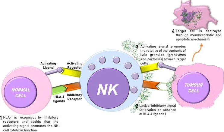
Target choice does not depend on clonal antigen receptors. Instead, each killer cell balances signals from activating and inhibitory surface proteins, scanning target tissues for signs of cellular stress [6]. Activating sensors detect ligands that multiply on host cells during oncogenic stress or viral takeover [8]. When activating cues overpower inhibitory stops, the cell unleashes lethal chemicals against the abnormal target, whereas healthy cells escape attack because their normal surface markers send strong negative signals [1].
A central activating receptor is NKG2D, a lectin-like protein that binds stress ligands like MICA and MICB on damaged epithelial cells and solid tumors, which Bauer and his team showed in Science triggers direct cytolytic destruction [6]. Natural killer cells also deploy natural cytotoxicity receptors called NKp30, NKp44, and NKp46 to bind viral hemagglutinins and tumor-related ligands [4]. They express Toll-like receptors such as TLR2, TLR3, and TLR9. In murine lymphoma experiments, Carmen Maldonado-Bernal and David Sánchez-Herrera reported that synthetic TLR9 ligands cleared tumors in 80% of treated mice [9].
When scanning a candidate target, surface microvilli gather at the contact site to lock against the opposing plasma membrane and establish a stable immunological synapse [4]. Receptors cluster inside this contact zone to control cytolytic granule release [7]. The two cells remain tightly bound until the killer delivers its lethal blow [4].

Missing-Self Recognition and Inhibitory Signals
Target selection relies on missing-self recognition, an essential protective mechanism through which healthy host tissues actively prevent their own destruction by circulating innate killer cells. Most normal nucleated cells display major histocompatibility complex class I proteins, called HLA in humans, on their outer surface [6]. Inhibitory receptors bind these self HLA molecules, delivering dominant negative signals that halt the killing machinery [5]. When a normal cell shows sufficient self HLA, killing cannot begin [1].
In humans, killer-cell immunoglobulin-like receptors, called KIRs, serve as the main inhibitory sensors for classical HLA-A, HLA-B, and HLA-C molecules, while the heterodimer CD94/NKG2A monitors levels of non-classical HLA-E to check overall HLA assembly [11]. Common viruses such as cytomegalovirus downregulate host MHC class I to hide from cytotoxic CD8+ T cells [2]. Tumors use this same evasion trick [10]. When these self markers vanish, the inhibitory brake releases so activating receptors trigger cell death. Catching cells that strip away HLA class I closes an escape route that would otherwise leave the host blind to hidden intracellular infections [6].
Maturing cells learn self-restraint through a training step called licensing in bone marrow and lymph nodes [7]. Cells that bind self HLA gain full killing power while learning to spare healthy self tissue, while unlicensed cells stay quiet to avoid autoimmune reactions against host organs. Self-tolerance is strictly preserved [11].
Granule Exocytosis and Cytotoxic Pathways
When activating cues dominate at the synapse, the cell triggers granule exocytosis. The cell rapidly reorganizes its actin cytoskeleton, steers its microtubule-organizing center directly toward the target, and docks its specialized secretory lysosomes against the contact interface [4]. Granules empty their toxic contents into the intercellular cleft, exposing membrane proteins such as LAMP-1 and CD63, which clinical laboratories use to track degranulation [7].
The main weapon in these granules is perforin [3]. Perforin molecules assemble in a calcium-dependent manner to build circular pores in the lipid bilayer, allowing serine proteases called granzymes to enter the target cytosol [2]. Granzyme B cuts pro-caspases to trigger programmed cell death, while granzyme A causes caspase-independent single-stranded DNA damage [9]. Apoptosis breaks down viral genes inside the dying target so infectious particles cannot leak into nearby tissue [2].

Natural killer cells also kill through surface death receptors [7]. In 1995, Arase and his team demonstrated in The Journal of Experimental Medicine that killer cells eliminate Fas-expressing targets through Fas ligand, while TRAIL provides a parallel apoptotic trigger [6]. When these ligands bind death receptors on a target, intracellular adaptors start caspase cascades that destroy the cell, offering a reliable backup path when abnormal targets resist pore-forming enzymes [7].
Distinct Subsets of NK Cells
Human NK cells split into two main subsets based on surface amounts of CD56 and the Fc receptor CD16 [9]. About 90% belong to the CD56dim subset [7]. They carry high surface levels of CD16 and dense azurophilic granules, making them potent cytolytic killers that execute rapid target lysis upon encountering stressed tissue [3]. They patrol blood vessels to clear circulating tumor cells and virus-infected targets [7].
The other 10% of blood killers, along with most populations in lymph nodes and the liver, belong to the CD56bright subset [4]. In 2001, Cooper and Caligiuri reported in Blood that CD56bright cells serve as the primary source of immunoregulatory cytokines during early innate responses, releasing large quantities of interferon-gamma, TNF-alpha, and GM-CSF when stimulated by monokines like IL-12 and IL-15. Cytokines recruit monocytes and guide dendritic cells. This response speeds up wider immune defenses throughout surrounding tissue [3].
CD56bright cells can mature into CD56dim killers by acquiring CD16 and lytic granules [11]. In chronic HIV infection, Zulu and his team showed that an unusual CD56-negative subset expands in peripheral blood. These exhausted cells upregulate NKG2C while losing Siglec-7, NKG2A, and CD57, showing marked loss of killing power during chronic viremia [9].
Development from Hematopoietic Progenitors
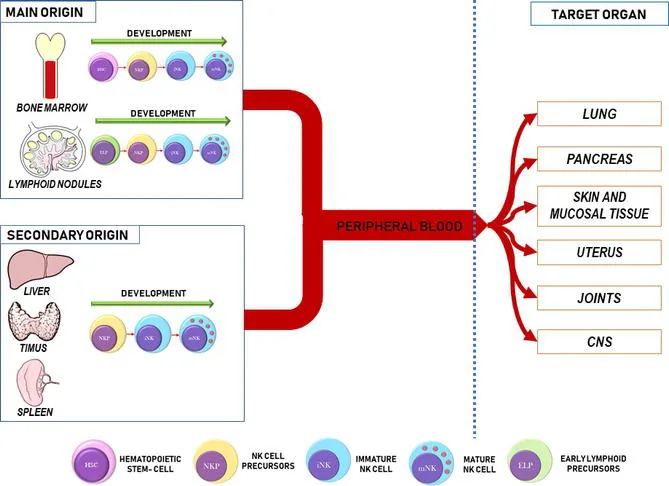
Natural killer development begins in bone marrow [7]. Pluripotent hematopoietic stem cells generate common lymphoid progenitors, which commit to the innate killer lineage after expressing CD122, the beta chain of the IL-2 and IL-15 receptors [3]. Once CD122 appears, developing cells depend on interleukin-15 for survival and growth [7].
Lineage progression follows a clear genetic blueprint. In Frontiers in Immunology, Alex M. Abel and his team described how cytokines and transcriptional regulators orchestrate killer cell maturation in the marrow niche [4]. The adapter protein GAB3 coordinates IL-15 signals. Specific transcription factors take control in turn: Nfil3, Id2, and Tox drive early commitment toward the immature stage, while Eomes and T-bet build cytotoxic machinery and cytokine competence. Terminal maturation requires Zeb2, which equips the cell with full killing power and homing receptors before it enters blood. Genetic flaws cause severe immune defects [7].
Bone marrow does not work alone. Secondary lymphoid organs such as the spleen, tonsils, lymph nodes, and liver also support differentiation. Precursors in these sites acquire activating receptors like NKp80 and settle into resident tissue niches [7]. Dispersed lymphoid tissues create specialized regional defenses throughout the body [9].

How Natural Killer Cells Bridge Immunity
Natural killer cells form a functional bridge between innate defense and the antigen-specific actions of B cells and T cells of adaptive immunity [4]. A central link between these branches is antibody-dependent cellular cytotoxicity, known as ADCC. When antibodies bind antigens on infected or cancerous cells, their Fc tails stick out. The low-affinity IgG receptor CD16 binds these exposed antibody tails, triggering receptor clustering that drives targeted granule release against opsonized target cells [6].
Crosstalk with antigen-presenting cells strengthens early defense. Activated killers secrete interferon-gamma and TNF-alpha, which activate macrophages for phagocytosis and help dendritic cells mature [5]. In turn, mature dendritic cells produce IL-12, IL-15, and IL-18, cytokines that boost killer cell survival and cell killing [4]. Bidirectional signaling accelerates the priming of naive T cells in regional lymph nodes, aligning early innate action with subsequent adaptive responses. The two systems cooperate from the first hours of an infection [3].
Some subsets show traits of immunological memory. After exposure to cytomegalovirus, specific populations expand and persist as memory-like cells that react strongly upon reinfection [7]. True killer cells differ from natural killer T cells, which are unconventional CD1d-restricted T lymphocytes that express semi-invariant TCR chains such as Valpha24-Jalpha18 and recognize glycolipids presented by non-polymorphic CD1d molecules [12]. In Nature Reviews Immunology, Daniel G. Pellicci and Stuart P. Berzins detailed how NKT cells develop in the thymus, depend on the transcription factor PLZF, and respond to alpha-galactosylceramide from the marine sponge Agelas mauritianus [14].
Uterine Populations in Early Pregnancy
During early pregnancy, specialized uterine natural killer cells gather in the decidua basalis at the implantation site and within the mesometrial lymphoid aggregate of pregnancy. These cells expand to form about 70% of all maternal leukocytes in the decidua during the first trimester. They arise from local tissue precursors and circulating CD34+ hematopoietic progenitors, with endometrial CD34+ stem cell fractions rising from 0.69% to 3.97% during implantation [13].
Unlike circulating blood killers, uterine populations lack cytotoxic activity and express CD56 and CD94 without CD16. Instead of attacking fetal tissue, they release angiogenic factors and cytokines like IL-8 and IP-10 to steer the remodeling of maternal spiral arteries. Vascular widening turns narrow, high-resistance vessels into broad conduits that deliver blood and nutrients to the placenta. Mouse studies show that growth factors from these cells support fetal development before the placenta fully forms [13].
Faulty arterial widening brings severe clinical trouble. Poor vascular remodeling starves the placenta of blood, raising the risk of pre-eclampsia, fetal growth restriction, and miscarriage [13]. Uterine white cells show how innate cells can switch roles from cell killing to tissue remodeling and maternal-fetal tolerance [4]. They protect both mother and child during gestation [13].
Therapeutic Applications in Cancer Care
The ability to kill cancer cells without prior sensitization makes natural killer cells valuable tools in oncology. Doctors test adoptive cell therapies using autologous or allogeneic killers expanded in culture. Allogeneic cells from healthy donors or umbilical cord blood offer off-the-shelf treatments without causing graft-versus-host disease, allowing donated cells to destroy malignant tissue safely without matching host HLAs [10].
Genetic engineering has broadened this clinical reach. Researchers build chimeric antigen receptor constructs to yield CAR-NK cells derived from cord blood, induced pluripotent stem cells, or the NK-92 tumor cell line, targeting CD19 on lymphoma and PD-L1 on solid tumors. In clinical trials, ImmunityBio tested the IL-15 superagonist N-803 alongside BCG vaccine in the QUILT-3.032 trial for bladder cancer, achieving complete responses in 71% of patients. Gamida Cell developed GDA-201, while Fate Therapeutics engineered FT516 and FT596. Affimed designed AFM13 to bind CD16 and CD30, guiding killer cells directly to Hodgkin lymphoma targets. NKMax America is testing SNK01 with pembrolizumab in non-small cell lung cancer [10].
Within solid tumors, cancer cells shed decoy ligands, release inhibitory cytokines like TGF-beta, and recruit suppressor cells that block killer cell infiltration into malignant tissue [10]. The protective role of these cells is clearest in natural killer cell deficiency, a rare genetic disorder where the lack of functional cells leaves patients open to severe, recurrent herpesvirus infections. Classical NKD causes absent cells, while functional NKD impairs their killing [8].
- WEBSITE Cleveland Clinic. (2023). What Are Natural Killer Cells (NK Cells)?. [Article Link]
- WEBSITE ScienceDaily. (2026). Natural killer cell. [Article Link]
- WEBSITE MyH Editorial Team. (2024). Natural Killer (NK) Cells: Function, Morphology & Identification. My Hematology. [Article Link]
- WEBSITE Aryal, S., & Sapkota, A. (2022). Natural Killer (NK) Cells- Definition, Structure, Immunity, Functions. Microbe Notes. [Article Link]
- WEBSITE British Society for Immunology. (2026). Natural Killer Cells. BiteSized Immunology. [Article Link]
- WEBSITE Acharya, T. (2026). Natural Killer (NK) Cells: Missing-Self Recognition and Function. Microbe Online. [Article Link]
- WEBSITE Pan, S. (2026). Natural killer (NK) cells – Definition, Structure, Function, Mechanism. Biology Notes Online. [Article Link]
- ONLINE NEWS Simmons, H. (2018). What are Natural Killer Cells?. News-Medical. [Article Link]
- BOOK CHAPTER Maldonado-Bernal, C., & Sánchez-Herrera, D. (2019). Toll-Like Receptors and Natural Killer Cells. In IntechOpen. [Article Link]
- ONLINE NEWS de Lartigue, J. (2021). NK Cell–Targeting Strategies Come Into Their Own. OncLive. [Article Link]
- WEBSITE Wikipedia contributors. (2026). Natural killer cell. Wikipedia, The Free Encyclopedia. [Article Link]
- WEBSITE Wikipedia contributors. (2026). Natural killer T cell. Wikipedia, The Free Encyclopedia. [Article Link]
- WEBSITE Wikipedia contributors. (2026). Uterine natural killer cells. Wikipedia, The Free Encyclopedia. [Article Link]
- ACADEMIC JOURNAL Pellicci, D. G., Koay, H., & Berzins, S. P. (2020). Thymic development of unconventional T cells: how NKT cells, MAIT cells and γδ T cells emerge. Nature Reviews Immunology, 20(12), 756-770. [Article Link]


