






Collagen is the main scaffolding protein in animal tissues. It gives strength and shape to skin, bone, tendons, and organs [1]. What is collagen, and why does animal biology depend on it? The name comes from the Greek word kólla, meaning glue. Early anatomists noticed that fibrous cords bind living structures into bodies [4]. Cells build this material from simple amino acids. Young bodies make new strands easily, but cellular production slows with age. As a result, mature tissue networks become prone to thinning [1].
What Is Collagen?
Collagen is the most plentiful protein family in mammals. In animal bodies, it accounts for 12 to 17 percent of total protein [4]. Clinical data from Cleveland Clinic show that in human bodies, fibrous bundles account for roughly 30 percent of total protein, creating an enduring framework for skeletal bone, articular cartilage, large blood vessels, and the gut wall [1]. Dry skeletal bone contains 25 to 35 percent collagen by mass, forming a tough matrix where calcium phosphate crystals bind to build rigid skeleton parts capable of bearing heavy body loads under gravity. Connective tissue cells called fibroblasts synthesize these matrix strands within intercellular spaces, while in skeletal muscle, delicate sheaths form the endomysium to supply 1 to 2 percent of muscle tissue and roughly 6 percent of muscular weight [4].
The physical behavior of matrix fibers depends on mineral content and cell layout. Bone is hard. In bones, heavy minerals lock fibrils into solid plates that support body weight under gravity. Tendon cords stretch slightly under tension. In joint cartilage, collagen forms a transitional gradient that cushions moving bones, whereas in tendons, fibers align in parallel rows to resist pull from contracting muscles. In skin, cells weave strands into a wavy net that yields under movement yet resists tearing, while similar networks reinforce eye corneas, pulsing arteries, and tooth dentin [1, 4].
Triple Helix Molecular Architecture
At the molecular scale, these strands form a tight triple helix called tropocollagen, built upon a repeating sequence of glycine-proline-hydroxyproline triplets where glycine appears at every third position throughout the entire peptide chain. Glycine accounts for one-third of all amino acid residues. Proline and hydroxyproline take up one-sixth of the sequence. Glycine is tiny. It carries a single hydrogen atom instead of a side chain. Because of this tiny footprint, three peptide strands pack tightly together in a left-handed helix without steric clashes pushing their backbones apart [4].
Extensive hydrogen bonding stitches the three peptide chains into a viscoelastic solid, where tensile strength depends on chain length and cross-links. Short units break and rebuild bonds quickly. Longer units spread tension across a larger span. Biochemical tests on isolated fibrils show that increasing cross-link density from zero to three per molecule raises the maximum tensile stress a strand withstands from 0.5 gigapascals to 6 gigapascals under heavy dynamic strain [4]. Thousands of triple helices aggregate into cylindrical fibrils measuring between 25 and 400 nanometers wide. These fibrils bundle into thick fibers that give living tissue high durability [4, 5].

Five Primary Structural Types
Biologists recognize 28 distinct types in the human body, sorted by their chain shapes [4]. Over 90 percent of all collagen in vertebrates is Type I, encoded by the COL1A1 and COL1A2 genes with a molecular mass of 300,000 g/mol. These thick bands build the dermis, tendons, organ capsules, and the organic matrix of bone and teeth [5].
Type II serves as the main fiber in cartilage, creating fine meshworks that trap proteoglycans and water to cushion everyday bone impacts inside movable joints. Type III consists of delicate reticular fibers that form branching networks in blood vessels and the uterus, sitting next to Type I. Within the heart, dense sheets form the cardiac skeleton around four valve rings, creating an impermeable insulating barrier that prevents atrial fibrillation from propagating directly into ventricular chambers [4].
Type IV builds flat sheets instead of ropes. It forms the basal lamina of basement membranes that anchor epithelial cells across internal linings. Type V sits near hair follicles, sweat glands, and placenta tissue, bridging dermis and epidermis while aiding early wound repair. Other forms include Type VI microfibrils and Type VII anchoring fibers, which carry out specialized support jobs throughout the body [4].
How Cells Build Fibril Networks
Building mature fibers involves a multi-step assembly process that spans both the inside of fibroblasts and the matrix space outside cells. Ribosomes on the rough endoplasmic reticulum translate mRNA sequences, producing pro-alpha1 chains from the COL1A1 gene and pro-alpha2 chains from the COL1A2 gene in a two-to-one ratio [2, 5]. Inside the endoplasmic reticulum, the enzyme prolyl 4-hydroxylase converts proline into hydroxyproline, a step that depends strictly on ascorbic acid (vitamin C) as a cofactor [4, 5]. When vitamin C is missing, nascent precursor chains fail to hydroxylate properly, causing triple-helical structures to fall apart and triggering the severe tissue breakdown characteristic of scurvy [4].
The newly synthesized procollagen helix carries peptide extensions at both ends, which keep the precursor soluble and prevent premature fibril clumping inside secretory vesicles. Once cellular vesicles export procollagen into the matrix space, specific procollagen peptidase enzymes snip off the terminal caps, allowing tropocollagen units to assemble into staggered parallel arrays. The enzyme lysyl oxidase then introduces covalent cross-links between adjacent lysine spots, converting loose fibril aggregates into insoluble, mature fibers capable of bearing heavy loads [5].


Cellular Receptors and Tissue Signaling
Matrix proteins actively direct cell behavior and tissue repair through at least eight dedicated surface receptors that control cell growth, migration, and adhesion. Integrins provide the main bridge between cells and matrix. Among transmembrane integrin pairs, integrin alpha-2-beta-1 acts as the chief receptor on blood platelets and skin cells. It regulates platelet adhesion during vascular repair, activates mast cells, and directs keratinocyte movement over healing wounds. Research by Garnotel R shows that monocytes also bind Type I collagen through integrin alpha-10-beta-2, while integrin alpha-10-beta-1 is expressed on chondrocytes to guide cartilage growth [6].
Discoidin domain receptors DDR1 and DDR2 represent an unusual class of receptor tyrosine kinases that specifically recognize native, triple-helical collagen structures rather than denatured gelatin. Unlike typical cell surface receptors that trigger signaling cascades within minutes, DDR dimers exhibit delayed activation, reaching peak autophosphorylation roughly 18 hours after ligand stimulation. DDR1 regulates mammary gland development, arterial repair, and leukocyte activity, whereas DDR2 promotes chondrocyte growth and induces matrix metalloproteinase expression during skeletal bone growth [6].
On circulating blood platelets, glycoprotein VI pairs with Fc-receptor gamma chains to initiate rapid coagulation when vascular damage uncovers subendothelial matrix strands. This interaction activates intracellular Src kinases Lyn and Fyn, triggering rapid platelet aggregation to seal the wound with a stable hemostatic plug [4, 6]. At the same time, inhibitory receptors like LAIR1 suppress overactive immune responses, and endocytic mannose receptors such as Endo180 internalize damaged fragments for controlled enzymatic recycling [6].
Genetic Conditions Affecting Collagen Assembly
Gene mutations that disrupt fibril assembly cause heritable disorders that impair tissue strength throughout the body [2]. In a 2017 review for the American Journal of Medical Genetics, Brady and Malfait delineated 13 distinct subtypes of Ehlers-Danlos syndromes, expanding beyond the six subtypes recognized in the Villefranche Nosology. These conditions feature joint hypermobility, stretchy skin, and fragile tissues, with causative mutations verified in fibrillar collagen genes or modifying enzymes for all variants except the hypermobile type [7].
The COL1A2 gene directs the synthesis of pro-alpha2 chains for Type I fibers. Heterozygous COL1A2 mutations that delete key exon segments interfere with chain assembly and cause the arthrochalasia subtype of Ehlers-Danlos syndrome, marked by congenital bilateral hip dislocations. Rare homozygous COL1A2 mutations described by Malfait prevent cells from producing normal pro-alpha2 chains, resulting in the cardiac-valvular subtype characterized by heart valve defects and loose skin [2].
Mutations in COL1A1 and COL1A2 also cause osteogenesis imperfecta, a condition marked by fragile bones that fracture easily under minimal trauma. When point mutations replace glycine with bulkier amino acids in the helical domain, abnormal collagen chains severely disrupt mineral deposition during skeletal bone growth [2]. Malfait also reported an overlap syndrome combining brittle bones and Ehlers-Danlos features, while Caffey disease involves a COL1A1 mutation replacing arginine with cysteine at site 836 [5].

Why Tissue Elasticity Declines With Age
As human bodies age, connective tissues undergo progressive structural changes driven by internal biological clocks and external wear [1, 4]. Fibroblasts make fewer matrix fibrils each decade while existing strands break down at a faster pace, causing the dermal layer to thin and lose strength. Female bodies experience a sharp drop in collagen production following menopause, and everyone experiences a marked reduction in synthesis past age 60 [1]. Skin laxity and wrinkling do not stem from collagen loss alone, because neighboring elastin networks provide the recoil that allows dermal tissue to snap back after stretching. Age-related sagging correlates more closely with the degradation of these elastic fibers than with collagen loss alone, so keeping both networks healthy is vital for skin firmness [4].
Unprotected exposure to solar ultraviolet radiation fractures dermal fibrils and causes elastic fibers to degenerate into disorganized clumps, a state dermatologists classify as solar elastosis [4]. Cigarette smoking and diets high in sugar also accelerate tissue damage. Cleveland Clinic clinicians recommend daily application of broad-spectrum sun protection rated at SPF 30 or higher, protective clothing, and sunglasses, while warning that tanning beds accelerate collagen damage. Because blood tests cannot quantify internal collagen levels, doctors monitor structural decline through physical signs such as joint stiffness, thinning skin, and delayed wound repair [1].
Clinical Evidence on Collagen Supplements
The wellness market sells hydrolyzed powders, capsules, and drink formulations designed to counter age-related protein loss. Because the digestive tract cannot absorb whole protein chains intact, supplement makers use enzymatic hydrolysis to break collagen into small peptides or amino acids [1, 3]. A 2019 review in the Journal of Drugs in Dermatology evaluated 11 clinical trials with 805 participants. Doses of 2.5 to 10 grams of hydrolysate, or 3 grams of tripeptides, showed early gains in skin moisture and elasticity [3].
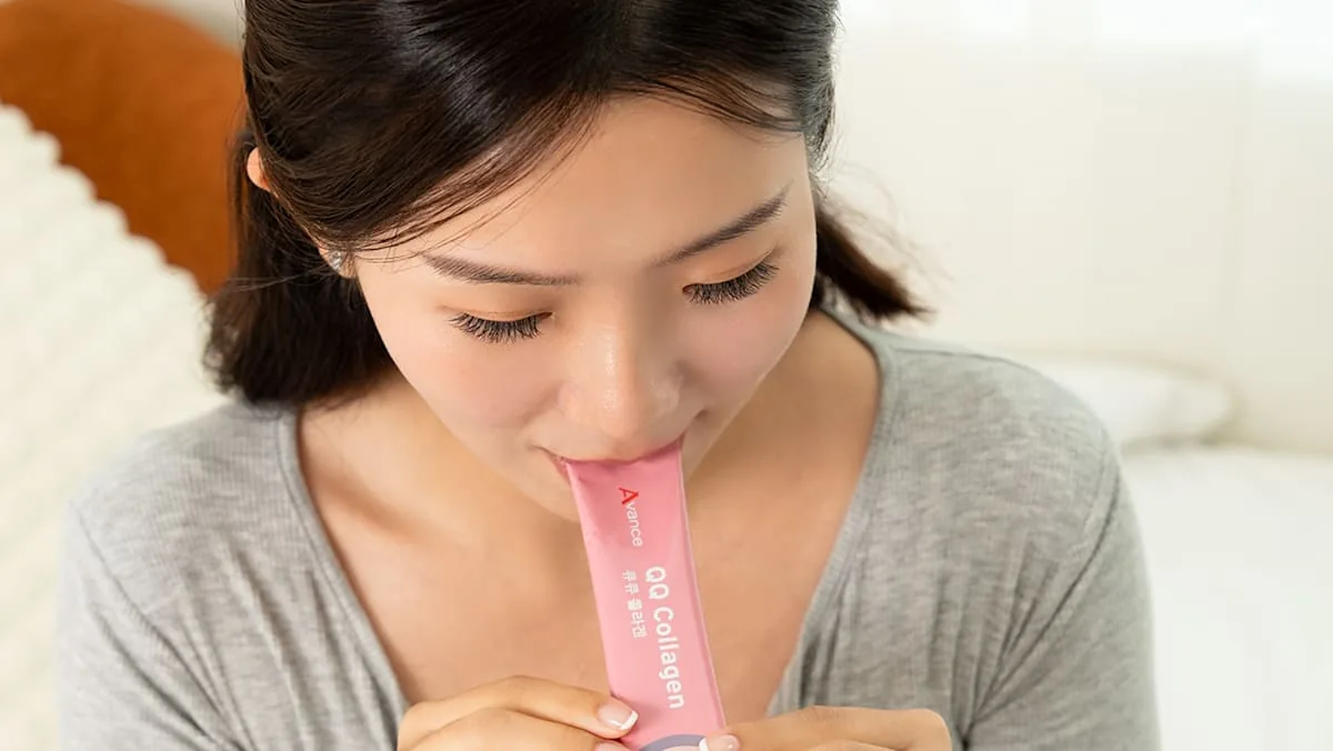
Absorption efficiency depends on peptide size, as shown by Yamamoto and Sakai in the Biological and Pharmaceutical Bulletin. In their trial, participants who took collagen tripeptides with Gly-X-Y sequences absorbed intact Gly-Pro-Hyp and Pro-Hyp into the bloodstream and excreted them in urine [8]. In the Journal of Cosmetic Dermatology, Lin evaluated 75 healthy adults aged 25 to 65 receiving placebo, nonliposomal tripeptides, or liposomal tripeptide formulations over eight weeks. Products like Avance QQ Collagen use 7,500mg fish peptides with liposomal encapsulation, which Lin found produced earlier gains in elasticity and a significant reduction in wrinkle area [3, 9].
Clinicians caution that ingested collagen peptides cannot be directed intentionally to specific target tissues like facial skin or joints. Digested peptides enter general amino acid pools that cells distribute according to physiological priority throughout the body. Cleveland Clinic suggests supporting natural synthesis through a balanced Mediterranean diet containing poultry, beef, fish, eggs, dairy, beans, and citrus fruits rich in vitamin C and zinc [1]. Consumers with known seafood allergies must review product labeling, as commercial supplements often derive from fish [3].
- WEBSITE Cleveland Clinic. (2022). Collagen: What It Is, Types, Function & Benefits. Cleveland Clinic. [Article Link]
- WEBSITE MedlinePlus Genetics. (2019). COL1A2 gene. National Library of Medicine. [Article Link]
- ONLINE NEWS Channel NewsAsia. (2026). Collagen supplements: What matters when choosing one. Mediacorp. [Article Link]
- WEBSITE Wikipedia contributors. (2026). Collagen. Wikipedia, The Free Encyclopedia. [Article Link]
- WEBSITE Wikipedia contributors. (2026). Type I collagen. Wikipedia, The Free Encyclopedia. [Article Link]
- WEBSITE Wikipedia contributors. (2026). Collagen receptor. Wikipedia, The Free Encyclopedia. [Article Link]
- ACADEMIC JOURNAL Malfait, F., Francomano, C., Byers, P., Belmont, J., Berglund, B., Black, J., Bloom, L., Bowen, J. M., Brady, A. F., Burrows, N. P., Castori, M., Cohen, H., Colombi, M., Demirdas, S., De Backer, J., De Paepe, A., Fournel-Gigleux, S., Frank, M., Ghali, N., & Juul-Kristensen, B. (2017). The 2017 international classification of the Ehlers-Danlos syndromes. American Journal of Medical Genetics Part C: Seminars in Medical Genetics, 175(1), 8-26. [Article Link]
- ACADEMIC JOURNAL Yamamoto, S., Deguchi, K., Onuma, M., Numata, N., & Sakai, Y. (2016). Absorption and Urinary Excretion of Peptides after Collagen Tripeptide Ingestion in Humans. Biological & Pharmaceutical Bulletin, 39(3), 428-434. [Article Link]
- ACADEMIC JOURNAL Lin, Y., Liang, C., Huang, B., Lim, S., Lin, Y., Chan, S., Chiang, C., & Mao, Y. (2026). Liposomal Delivery Enhances the Effects of a Collagen Tripeptide-Containing Formulation on Dermal Structure and Optical Skin Parameters: A Randomized, Double-Blind, Placebo-Controlled Trial. Journal of Cosmetic Dermatology, 25(4). [Article Link]


