





Understanding what is cortisol begins with the human endocrine system, where the Adrenal Glands produce this steroid hormone to steer how tissues adapt to daily pressures. As a glucocorticoid, it regulates glucose availability, stabilizes blood pressure, and calms immune reactions when tissues encounter physical injury. Circulating levels rise and fall along a day-night cycle, so healthy organs depend on hormonal feedback loops to keep output within balanced boundaries. Internet videos often blame the substance for facial puffiness, but clinical medicine tracks its actions in the MedlinePlus Medical Encyclopedia to diagnose Cushing Syndrome and Addison Disease [1].
- What Is Cortisol in Human Physiology?
- Steroid Molecular Structure Across Adrenal Tissues
- Metabolic Regulation of Glucose, Proteins, Lipids
- Immune System Modulation Through Cytokine Shifts
- Morning Activation of the Cortisol Awakening Response
- Stress Patterns and Chronic Alcohol Consumption
- Diagnostic Testing in Blood, Saliva, Urine, Hair
- Clinical Role of Hydrocortisone in Cushing Syndrome
What Is Cortisol in Human Physiology?
Cortisol belongs to the glucocorticoid class of steroid hormones and originates within the paired Adrenal Glands located atop each kidney. When the brain perceives a threat, hypothalamic neurons release corticotropin-releasing hormone. This trigger prompts the Anterior Pituitary to discharge adrenocorticotropic hormone (ACTH), which travels through blood vessels to stimulate steroid synthesis inside the Adrenal Cortex [1].
Enzymatic reactions inside the Adrenal Cortex convert cholesterol precursors into cortisol before releasing the completed steroid into systemic circulation [2]. Circulating cortisol regulates its own output through a negative feedback loop that acts on hypothalamic and pituitary binding sites. When blood levels climb, the hormone binds central receptors, slowing the release of CRH and ACTH so that adrenal synthesis drops [4]. Pharmaceutical formulations of this hormone are sold under the name hydrocortisone to treat severe allergies, rashes, and joint swelling [2].
Daily hormone release follows an internal rhythm coordinated by central pacemakers [4]. Blood levels reach their lowest point around midnight, begin rising during the final hours of sleep, and reach a sharp morning peak shortly after waking up [8]. Can everyday stress disrupt this diurnal balance? Prolonged work strain, nighttime light exposure, and frequent shift rotations alter normal secretion curves. This shift leaves people fatigued during daylight hours and restless at night. When ongoing pressure exhausts hormonal reserves, overall adrenal responsiveness declines, leaving patients vulnerable to fatigue and common infections [7].

Steroid Molecular Structure Across Adrenal Tissues
The molecular structure of cortisol contains twenty-one carbon atoms arranged in four fused hydrocarbon rings [2]. Chemical databases catalog the compound under ChEBI 17650 and ChemSpider identifier 5551, classifying the molecule as a trihydroxy steroid bearing functional hydroxyl groups at carbons 11, 17, and 21 across a pregn-4-ene nucleus [3]. This structure gives the molecule a formula of C21H30O5 and a monoisotopic mass of 362.20932 daltons [2].
Because cortisol is fat-soluble, it crosses cell membranes freely to bind glucocorticoid and mineralocorticoid binding sites in the cytoplasm. Upon binding its ligand, the receptor sheds inhibitory heat shock proteins and moves into the cell nucleus. Inside the nucleus, receptor pairs bind specific response elements along DNA to alter gene readout. This genomic timeline explains why systemic steroid effects develop gradually rather than appearing immediately [4].
Beyond slow genomic changes, cortisol triggers rapid actions by interacting with membrane proteins and ion channels in the central nervous system. Electrophysiological studies in brain tissue show that cortisol acts as a biphasic regulator of gamma-aminobutyric acid type A (GABAA) receptors. It functions as a positive modulator at low picomolar levels between 1 and 10 pM, but shifts into a negative modulator at higher nanomolar levels between 10 and 1,000 nM. This concentration shift changes baseline nerve inhibition across limbic circuits, which explains why small morning hormone rises support mental focus, but high stress surges trigger anxiety and poor recall [4].
Metabolic Regulation of Glucose, Proteins, Lipids
Cortisol manages bodily energy distribution by ensuring the brain receives adequate fuel during fasting or acute exertion. It counteracts insulin action by promoting hepatic gluconeogenesis from non-carbohydrate sources. At the same time, it slows glucose uptake in peripheral muscle and fat deposits by limiting the movement of GLUT4 transporter proteins to cell surfaces [4].
Within liver tissue, cortisol promotes both gluconeogenesis and glycogen storage. The organ simultaneously manufactures free glucose and stores glycogen. In skeletal muscle, cortisol collaborates with adrenaline and glucagon to break down intracellular glycogen into glucose-1-phosphate for localized contraction. This cooperative metabolic arrangement guarantees that working muscles gain quick access to stored fuel, but the liver keeps a stable store of glycogen to buffer subsequent drops in blood sugar [4].

When cortisol levels stay high over weeks or months, metabolic adaptations turn harmful, driving tissue breakdown and central fat redistribution. Prolonged glucocorticoid excess speeds up proteolysis in skeletal muscle fibers, breaking down contractile proteins into amino acids that feed ongoing liver glucose production at the cost of muscle wasting [4]. Short hormone spikes break down stored fats, but chronically high cortisol levels provoke high blood sugar that triggers compensatory insulin surges. This hormonal imbalance deposits visceral fat around the abdomen, upper back, and face, but weakens bones by blocking osteoblasts as described in Goldman Cecil Medicine [12].
Immune System Modulation Through Cytokine Shifts
Rather than shutting down bodily defenses entirely, cortisol guides a coordinated shift in cytokine output to prevent runaway inflammation from injuring vital organs. The steroid dials down pro-inflammatory cytokines such as interleukin-12, interferon-gamma, and tumor necrosis factor-alpha. In contrast, it boosts anti-inflammatory chemical messengers including interleukin-4, IL-10, and IL-13 in helper T cells [4].
This cytokine redistribution dampens cellular immunity but supports antibody responses. This shift illustrates why acute stress preserves baseline immune protection. At the cellular level, cortisol prevents helper T cells from multiplying by blocking interleukin-2 and downregulating the surface IL-2 receptor. Furthermore, the hormone dampens the cell-killing power of natural killer cells by downregulating their surface receptors, but prolactin from the Pituitary Gland exerts the opposite action by boosting receptor counts and improving cell lysis [4].
Beyond white blood cells, cortisol stimulates copper enzymes like lysyl oxidase, which cross-links collagen and elastin in healing tissues, and superoxide dismutase, which shields host cells from oxidative injury while permitting superoxides to kill microbes. Certain viral pathogens exploit this neuroendocrine axis to escape host immune detection: influenza viruses, SARS-CoV-1, and SARS-CoV-2 synthesize peptide fragments that closely mimic human adrenocorticotropic hormone. Because these viral mimics lack normal endocrine function, the host immune system makes neutralizing antibodies that attack endogenous ACTH. This viral disruption causes secondary adrenal insufficiency that impairs metabolic stability and weakens host resilience [4].

Morning Activation of the Cortisol Awakening Response
The cortisol awakening response (CAR) is an acute surge in circulating hormone levels that occurs 30 to 45 minutes after waking up in roughly 77% of healthy adults. Salivary cortisol levels increase from roughly 15 nmol/L upon waking to 23 nmol/L at peak, amounting to an average rise between 38% and 75%. Even one hour after getting out of bed, cortisol levels in many people stay heightened by roughly 34% above waking baseline values [8].
Twin studies demonstrate that individual CAR patterns possess notable genetic stability, displaying a heritability estimate of 0.40 for mean morning cortisol increase and 0.48 for the total area under the awakening curve. Unlike baseline circadian fluctuations driven purely by the hypothalamic-pituitary-adrenal axis, the CAR relies heavily on hippocampal modulation. Patients suffering from bilateral hippocampal damage or amnesia show no morning surge at all. This finding confirms that prospective memory and cognitive orientation help guide morning endocrine activation [8].
Environmental conditions and daily schedules exert strong effects on the size and duration of the awakening response. Waking up in lit rooms produces a larger cortisol spike than waking in dark rooms, but taking evening naps between 6:45 p.m. and 8:30 p.m. produces no awakening response at all. What causes shift workers to experience altered morning curves? Hospital nurses waking for early shifts between 4:00 a.m. and 5:30 a.m. display prolonged morning hormone elevations compared to late-day workers. Researchers confirmed that this difference stems from pre-shift worry and poor sleep rather than altered circadian timing alone, but chronic exhaustion and persistent pain flatten the morning curve into a low plateau [8].

Stress Patterns and Chronic Alcohol Consumption
Daily pressures fall into emotional, mental, and environmental categories that each draw on the Adrenal Glands. At Aria Integrative Medicine, Jennifer Bennett explained that acute demands temporarily raise cortisol to mobilize fuel and support muscle action. Once stressful situations end, hormone levels return to baseline, allowing tissues to recover without lasting injury [7].
When stress persists for months, adrenal output struggles to meet demand, depleting hormone reserves over time. Patients with chronic low-grade stress often develop low resting cortisol levels, leaving them prone to viral illness and post-stress flare-ups of autoimmune diseases. Bennett observed that individuals with low reserves often experience symptom flare-ups following major life shocks, such as the death of a family member, because falling hormone levels remove normal immune suppression [7].
Chronic alcohol intake disrupts the hypothalamic-pituitary-adrenal axis by damaging normal inhibitory control mechanisms. In a 1966 clinical trial, Mendelson and colleagues demonstrated that both drinking and non-drinking subjects developed elevated cortisol while consuming alcohol, with dependent drinkers reaching their highest levels during withdrawal. Recent assessments confirm that heavy drinkers show high overnight urinary cortisol and reduced heart rate variability governed by the autonomic nervous system. These changes promote arterial hypertension and fragmented sleep. Analysis of human hair samples reveals that drinking subjects carry hair cortisol concentrations three to four times higher than non-drinkers, but values return to normal following sustained sobriety [13].
Diagnostic Testing in Blood, Saliva, Urine, Hair
Clinicians measure cortisol across multiple bodily fluids to tell apart acute stress spikes from chronic endocrine diseases. In clinical guidelines updated by Sandeep Dhaliwal and reviewed by David Dugdale and Brenda Conaway in the MedlinePlus Medical Encyclopedia, female reference ranges stay below 45 mcg/24 hours, or 124 nmol/24 hours, but healthy male excretion caps at 60 mcg/24 hours, corresponding to 165 nmol/24 hours. Endocrine specialists also consult reference chapters by Richard Auchus in the Williams Textbook of Endocrinology to interpret these values [1].
Salivary testing provides an easy, non-invasive method for tracking daily curves because salivary hormone levels reflect unbound free cortisol in serum. A clinical study published by Xie and Lu in Clinical Chemistry and Laboratory Medicine confirmed that cortisol levels in 1-cm hair segments correlate closely with multi-week salivary averages, proving hair analysis works as a reliable biomarker of chronic HPA axis activity [15]. In addition, Russell and colleagues published in Therapeutic Drug Monitoring that sweat cortisol levels range from 8.16 to 141.7 ng/mL following heavy exercise, opening new avenues for non-invasive testing [14].

Cortisol tracking is increasingly valuable in psychiatric medicine for assessing symptom severity in patients with mood disorders and psychoses [16]. Writing in Psychological Medicine, researcher Herbert stated that disturbed diurnal rhythms and exaggerated awakening surges help identify specific subtypes of major depressive disorder, offering objective markers to guide clinical psychiatric treatment [6]. Cortisol evaluation has even extended to environmental ecology and aquaculture. In the Journal of Fish Biology, Sadoul and Geffroy documented non-invasive protocols measuring cortisol in fish skin mucus, scales, water, and fins, allowing researchers to monitor stress in wild and captive populations without inflicting the physical trauma associated with needle blood draws [17].
Clinical Role of Hydrocortisone in Cushing Syndrome
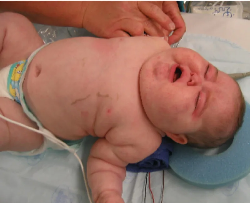
Cushing Syndrome describes a clinical state resulting from chronic exposure to excessive glucocorticoid levels. Typical features include central obesity, thin skin that bruises easily, purple abdominal striae, and rounded facial contours known as moon facies. The most common cause is iatrogenic, developing when patients take synthetic steroids like prednisone for autoimmune conditions or asthma [12]. When hypercortisolemia stems from an ACTH-secreting pituitary tumor, doctors designate the condition as Cushing Disease [1].
Critical illness also alters cortisol kinetics through reduced metabolic clearance. In the New England Journal of Medicine, Boonen and Berghe evaluated 158 intensive care patients, demonstrating an 83% increase in cortisol production alongside a 50% drop in steroid clearance during systemic trauma [5]. Outside intensive care, medical steroid treatments can induce swift physical changes. Actress Amy Schumer publicly disclosed her Cushing Syndrome diagnosis after receiving high-dose steroid injections for endometriosis, which produced noticeable facial changes that sparked public speculation [9].
On social media platforms, the trending hashtag #CortisolFace has accumulated over 11,000 videos claiming that routine work stress causes facial bloating [11]. In New York, dermatologist Samer Jaber of Washington Square Dermatology told CBS News that stress stimulates oil glands and triggers acne rather than facial swelling [9]. In Boston, medical reporter Mallika Marshall at CBS Boston clarified that everyday stress does not elevate cortisol high enough or long enough to cause moon face [10]. At CBS Pittsburgh, Natalie Gentile of the Cleveland Clinic noted that natural fluctuations occur daily, and Marshall pointed out that temporary puffiness usually stems from poor sleep, salty meals, or alcohol intake [9].
- WEBSITE Dhaliwal, S. K., Dugdale, D. C., & Conaway, B. (2025). Cortisol urine test. MedlinePlus Medical Encyclopedia. [Article Link]
- WEBSITE European Bioinformatics Institute. (2023). Cortisol (CHEBI:17650). Chemical Entities of Biological Interest. [Article Link]
- WEBSITE Royal Society of Chemistry. (2026). Cortisol (ChemSpider ID 5551). ChemSpider. [Article Link]
- WEBSITE Wikipedia contributors. (2026). Cortisol. Wikipedia, The Free Encyclopedia. [Article Link]
- ACADEMIC JOURNAL Boonen, E., Vervenne, H. B., Meersseman, P., Andrew, R., Mortier, L., Declercq, P. E., Vanwijngaerden, Y., Spriet, I., Wouters, P. J., Perre, S. V., Langouche, L., Vanhorebeek, I., Walker, B. R., & Berghe, G. V. d. (2013). Reduced Cortisol Metabolism during Critical Illness. New England Journal of Medicine, 368(16), 1477-1488. [Article Link]
- ACADEMIC JOURNAL Herbert, J. (2012). Cortisol and depression: three questions for psychiatry. Psychological Medicine, 43(3), 449-469. [Article Link]
- WEBSITE Bennett, J. (2020). Cortisol and Autoimmune Diseases. Aria Integrative Medicine. [Article Link]
- WEBSITE Wikipedia contributors. (2026). Cortisol awakening response. Wikipedia, The Free Encyclopedia. [Article Link]
- ONLINE NEWS Moniuszko, S. (2024). What is cortisol? How to lower levels amid concerns over “cortisol face”. CBS News. [Article Link]
- ONLINE NEWS Marshall, M. (2024). What is the trending TikTok topic “cortisol face” and what causes it? CBS Boston. [Article Link]
- ONLINE NEWS Ismail, D. (2026). What Is Cortisol Face? And Can I Get Rid of It? GQ Middle East. [Article Link]
- WEBSITE Lecturio. (2025). Cushing Syndrome. Lecturio Concise Medical Knowledge. [Article Link]
- WEBSITE Wikipedia contributors. (2026). Alcohol and cortisol. Wikipedia, The Free Encyclopedia. [Article Link]
- ACADEMIC JOURNAL Russell, E., Koren, G., Rieder, M. J., & Uum, S. H. M. V. (2013). The Detection of Cortisol in Human Sweat. Therapeutic Drug Monitoring, 36(1), 30-34. [Article Link]
- ACADEMIC JOURNAL Xie, Q., Gao, W., Li, J., Qiao, T., Jin, J., Deng, H., & Lu, Z. (2011). Correlation of cortisol in 1-cm hair segment with salivary cortisol in human: hair cortisol as an endogenous biomarker. Clinical Chemistry and Laboratory Medicine (CCLM), 49(12), 2013-2019. [Article Link]
- ACADEMIC JOURNAL Dziurkowska, E., & Wesołowski, M. (2021). Cortisol as a Biomarker of Mental Disorder Severity. Journal of Clinical Medicine, 10(21), 5204. [Article Link]
- ACADEMIC JOURNAL Sadoul, B. C., & Geffroy, B. (2019). Measuring cortisol, the major stress hormone in fishes. Journal of Fish Biology, 94(4), 540-555. [Article Link]


