




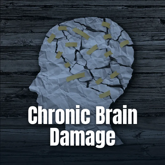
Chronic traumatic encephalopathy symptoms emerge years or decades after an athlete or soldier sustains repetitive head impacts during contact sports or military combat. While acute concussions draw immediate public attention during televised sports, researchers at the BU CTE Center emphasize that cumulative subconcussive blows—repetitive head impacts that produce no immediate clinical symptoms—drive underlying neurodegeneration over decades [10]. This condition causes abnormal tau proteins to accumulate around small blood vessels in the brain, gradually destroying adjacent brain cells. Patients face severe mood changes, memory loss, and cognitive decline years after their athletic careers or military deployments conclude [3].
- Early Boxing Origins of CTE
- How Repetitive Head Impacts Trigger Tau Pathology
- The Four Neuropathological Stages of CTE
- Chronic Traumatic Encephalopathy Symptoms Over Time
- Motor Deficits and Parkinsonian Traits
- Biomarker Advances in Fluid Diagnostics
- Trauma Exposure in Collision Sports
- Preventive Measures for Long-Term Protection
Early Boxing Origins of CTE
Chronic traumatic encephalopathy is a distinctive tauopathy that doctors originally identified in prize fighters during the late 1920s under the popular term dementia pugilistica. For decades, clinicians regarded the syndrome as an uncommon condition confined to veteran boxers who absorbed countless punches to the head during lengthy ring careers [8].
The condition differs from other degenerative illnesses like Alzheimer’s disease in its specific distribution through cerebral tissue. In Alzheimer’s patients, tau tangles spread diffusely through the hippocampus alongside amyloid-beta plaques. In contrast, the pathognomonic lesion of this trauma-linked disease consists of hyperphosphorylated tau aggregates clustering in the depths of cerebral sulci around small blood vessels. In October 2022, the National Institutes of Health updated its formal guidance following an open letter signed by 41 leading medical researchers, officially acknowledging that repetitive sports head impacts cause neurodegenerative disease. That revision brought the NIH into agreement with the Centers for Disease Control and Prevention, which reached that same conclusion in 2019 [9].
A detailed analysis of an NFL study modeling chronic traumatic encephalopathy highlights how risk correlates with total years of exposure in collision athletics. Christopher Nowinski of the Concussion Legacy Foundation pointed out that the primary driver stays environmental exposure through recurring hits to the skull. Because millions of young people participate in amateur sports, understanding this degenerative process is a critical public health goal [4].
How Repetitive Head Impacts Trigger Tau Pathology
The cellular damage begins with rotational acceleration and sudden shearing forces that deform brain tissue. Over 70 years ago, physicist A. H. S. Holbourne showed that the brain’s relative incompressibility makes rotational acceleration and twisting shear strains far more damaging to delicate microscopic axons than direct linear head impacts. Rapid head turns stretch cellular membranes and microscopic axons, producing mild traumatic brain injury, or TBI [11].
Under normal biological conditions, tau proteins stabilize internal microtubules that act as transport highways carrying vital cellular cargo along nerve fibers. When repeated rotational forces twist and pull cellular axons, an imbalance between intracellular kinases and phosphatases triggers hyperphosphorylation, prompting tau molecules to detach from microtubules and assemble into toxic neurofibrillary tangles. The free tau proteins then misfold into insoluble neurofibrillary tangles and glial tangles that accumulate in sulcal depths. Over months and years, accumulating tau deposits provoke persistent cellular inflammation, cell death, and widespread brain atrophy [2]. The spreading pathology gradually severs connections between neighboring cortical areas [11].
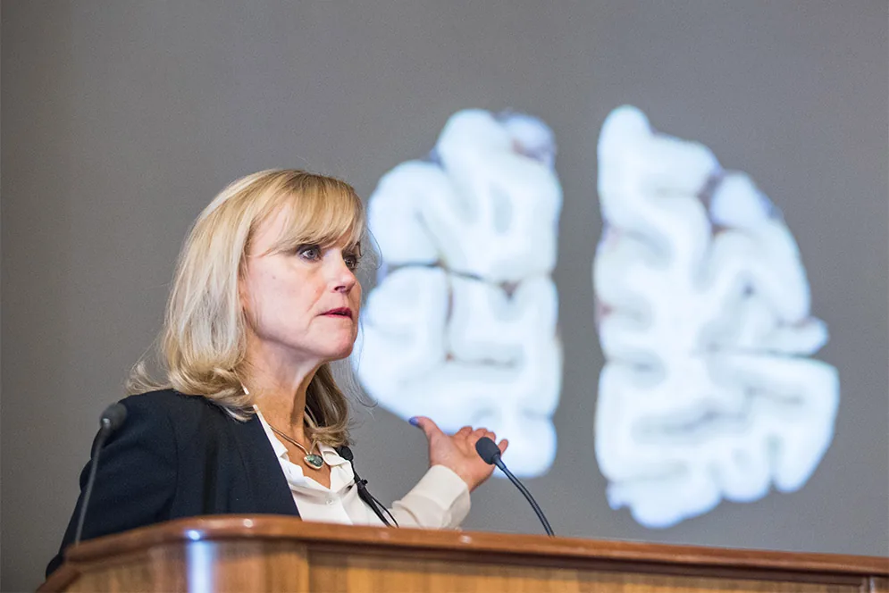
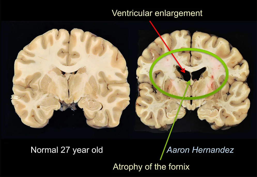
Tissue destruction was evident in former New England Patriots tight end Aaron Hernandez, who died in prison at age 27. Boston University neuropathologist Dr. Ann McKee found Stage 3 disease in Hernandez’s brain, with frontal lobe damage and ventriculomegaly. Dr. McKee stated that “we’ve never seen…in our 468 brains, except for individuals very much older” [13]. Hernandez had absorbed thousands of hits during his football career.
The Four Neuropathological Stages of CTE
Neuropathologists classify disease progression using a four-tier staging system developed by Dr. Ann McKee and her colleagues at the BU CTE Center [1]. In Stage I, abnormal tau tangles remain confined to isolated epicenters located at the depths of cortical sulci within the frontal cortex. Many people at this initial point experience only subtle headaches. Owen Thomas showed early Stage 1 changes at age 21 [11].
In Stage II, tau pathology spreads into adjacent sulcal depths and penetrates superficial cortical layers across frontal and temporal lobes, causing mood swings, irritability, and uncharacteristic behavioral outbursts. As tau pathology advances into Stage III, neurofibrillary tangles infiltrate the temporal lobes, amygdala, and hippocampus, leading to ventriculomegaly, marked fornix shrinkage, and progressive cognitive decline that undermines daily independence. The gross brain exhibits dilated ventricles, thinning of the corpus callosum, and marked shrinkage of the fornix. These changes reflect extensive neuronal death, impairing intellectual functioning [1].
In Stage IV, neurofibrillary tangles infiltrate the cerebral cortex, medial temporal lobe, and brainstem tegmentum. Severe cortical thinning and marked brain atrophy produce full-blown dementia. Pathologists also identify comorbid molecular changes in late-stage brains. Beta-amyloid plaques appeared in 43% of autopsied brains. Phosphorylated TDP-43 protein inclusions occur in most advanced specimens, worsening the clinical course [2].
Chronic Traumatic Encephalopathy Symptoms Over Time
Clinical symptoms typically emerge years or decades after an athlete retires from contact sports. In a landmark 2013 study of 36 deceased athletes published in Neurology, Dr. R. A. Stern and co-authors showed that patients with early behavioral symptoms developed problems around age 35, whereas cognitive-variant patients presented in their late 50s [3]. Junior Seau died by suicide in 2012 [11].
The early behavioral presentation appears in a person’s 20s or 30s, featuring emotional instability, impulsivity, depression, and explosive anger outbursts. Relatives frequently describe a fundamental personality change where patient empathy vanishes and interpersonal relationships suffer severe disruption. Because emotional volatility and explosive anger wax and wane during early years, family members and doctors often mistake these subtle personality shifts for marital friction, depression, or standard midlife psychological stress. Many affected people turn to alcohol or substance misuse in an attempt to self-medicate their persistent sleep issues and mental distress. Over time, these emotional disturbances become continuous as tau lesions spread across frontal and limbic networks [11].
The cognitive variant typically emerges in a person’s late 50s or 60s, beginning with short-term memory loss and executive dysfunction. As cortical neurodegeneration expands across temporal lobes, individuals frequently encounter severe speech impairments and language disruptions linked to aphasia that compromise daily communication. Patients struggle with planning, organizing, and multitasking before progressing toward profound disorientation and dementia. Many patients also lose insight into their own cognitive deficits [11].


Motor Deficits and Parkinsonian Traits
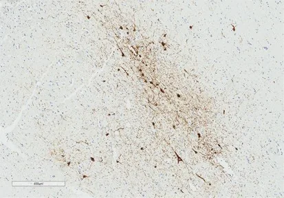
Motor disturbances also afflict many athletes suffering from head trauma. Approximately 24.7% to 25% of donors with diagnosed postmortem CTE exhibited parkinsonism, displaying resting tremors, muscle rigidity, and abnormal slowness of movement. These motor features historically led early clinicians to describe retired boxers as punch drunk or staggered in gait [11].
In April 2025, corresponding author Dr. Thor Stein, MD, PhD, and his colleagues evaluated 481 deceased athletes in JAMA Neurology, discovering that 76% of donors with parkinsonism showed cell loss in the substantia nigra without Lewy bodies. Examining subjects who donated brains to the Understanding Neurologic Injury and Traumatic Encephalopathy (UNITE) brain bank, Dr. Stein stated: “We were surprised to find that most individuals with CTE and parkinsonism did not have Lewy body pathology” [12]. Dr. Stein explained that symptoms were driven by severe cell loss in the substantia nigra, a movement-control center in the brainstem. The risk of motor symptoms rose with every additional decade of contact sport play.
Other athletes develop motor neuron disorders resembling amyotrophic lateral sclerosis. When former Philadelphia Eagles player Kevin Turner died at age 46, an autopsy revealed that his motor cortex was heavily damaged by tau tangles rather than classic amyotrophic lateral sclerosis. Doctors diagnosed Kevin Turner with ALS at age 40 [8]. Dr. Ann McKee reported 17 cases exhibiting combined signs of CTE and motor neuron disease in her brain bank. Motor slowing and physical frailty often complicate cognitive decline, echoing broader medical findings where muscle loss with age signals dementia risk across geriatric populations.
Biomarker Advances in Fluid Diagnostics
Can doctors detect the condition while an athlete is still alive? For decades, definitive diagnosis required postmortem brain dissection, leaving clinicians to diagnose living patients with traumatic encephalopathy syndrome based solely on symptom histories. This diagnostic barrier prevented researchers from launching targeted clinical drug trials for affected patients [14].
In late 2026, Dr. A. E. Miner and colleagues evaluated plasma endogenously cleaved microtubule-binding region tau in JAMA, discovering that donors with high-stage neuropathology averaged 0.021 pg/mL compared to just 0.001 pg/mL in low-stage disease [5]. Known as eMTBR-tau, the candidate marker was measured in 21 former football players from Boston University. Plasma concentrations correlated strongly with cumulative tau burden across 11 brain regions. These associations persisted after excluding donors with coexisting Alzheimer’s disease, proving marker specificity for trauma-induced tau tangles. The test represents the first blood biomarker capable of identifying advanced postmortem tau tangles in living donors [14].
In a companion study published in JAMA, Dr. K. Horie and team demonstrated that fluid eMTBR-tau243 distinguished advanced traumatic tauopathy from healthy controls with a high diagnostic score of 0.97 across multicenter patient cohorts. Combining eMTBR-tau with phosphorylated tau 217 enables physicians to separate traumatic encephalopathy from Alzheimer’s disease during clinical practice [14].
Trauma Exposure in Collision Sports
Public concern regarding head trauma expanded following autopsy reports of famous professional football players. Boston University and VA researchers identified CTE in 99% of donated brains from former NFL players and in 96 percent of National Football League players examined in earlier cohorts [11]. Brain banks suffer from substantial ascertainment bias [4].
To measure disease rates across an unbiased population, Dr. Kevin F. Bieniek, PhD, evaluated 2,566 autopsies from the Mayo Clinic Tissue Registry, identifying 300 former athletes and 450 non-athletes in Brain Pathology. Dr. Bieniek, now director at the Glenn Biggs Institute at UT Health San Antonio, led the team in screening tissue in a blinded manner, finding CTE pathology in 42 individuals, or 5.6% of the cohort. American football players showed the highest disease frequency at 15% [4]. Non-athletes accounted for 15 cases, proving that falls or physical abuse can also trigger tau pathology. Dr. Bieniek noted that the 6% rate provides a realistic population baseline.

Military veterans also experience significant trauma-related brain degeneration from battlefield explosives. At Boston University, Dr. Lee Goldstein and Dr. Ann McKee proved that an improvised explosive device blast wind reaching 330 miles per hour causes rapid rotational head motion, resulting in persistent learning and memory impairments. Holding the head secure during explosive testing stopped the cognitive and memory impairments that otherwise developed following shockwaves [13].
Preventive Measures for Long-Term Protection
Because modern medicine cannot reverse established tau tangles, primary prevention remains the single most effective tool against chronic traumatic encephalopathy. Public health advocates argue that reducing repetitive head impacts in youth sports is essential to protect young brains. To safeguard youth participants from long-term neurological harm, Concussion Legacy Foundation cofounder Christopher Nowinski has recommended that collision sports leagues eliminate tackle football and soccer heading for children before age 14 [8].
The United Kingdom National Health Service outlines supportive multidisciplinary care pathways modeled after dementia programs, providing patients with occupational therapy, physical therapy, and speech therapy to maintain functional mobility and communication. Patients benefit from occupational therapy to adapt home environments, physical therapy to address balance issues or tremors, and speech therapy for cognitive communication deficits. A dedicated care team including a general practitioner, dementia specialist, and social services coordinates long-term support for the patient and family. Involving mental health professionals early helps mitigate emotional distress, impulsive behaviors, and domestic friction before relationship crises occur [10].
Pharmacological interventions focus on easing mood and sleep symptoms rather than attempting to dissolve tau tangles. Neither ALS nor CTE has a medical cure [8]. Physicians cautiously prescribe mood stabilizers, antidepressants, and sleep aids to manage irritability and insomnia [10]. Meanwhile, validated blood biomarkers like eMTBR-tau will soon enable earlier enrollment in clinical drug trials [14]. Protecting youth athletes and military personnel through strict safety reforms provides the only permanent solution to this neurodegenerative disease [9].
- ACADEMIC JOURNAL McKee, A. C., Stein, T. D., Nowinski, C. J., Stern, R. A., Daneshvar, D. H., Alvarez, V. E., Lee, H., Hall, G. F., Wojtowicz, S. M., Baugh, C. M., Riley, D., Kubilus, C. A., Cormier, K., Jacobs, M. A., Martin, B. M., Abraham, C. R., Ikezu, T., Reichard, R. R., Wolozin, B. L.,. Cantu, R. C. (2012). The spectrum of disease in chronic traumatic encephalopathy. Brain, 136(1), 43-64. [Article Link]
- ACADEMIC JOURNAL McKee, A. C., Stein, T. D., Kiernan, P. T., & Alvarez, V. E. (2015). The Neuropathology of Chronic Traumatic Encephalopathy. Brain Pathology, 25(3), 350-364. [Article Link]
- ACADEMIC JOURNAL Stern, R. A., Daneshvar, D. H., Baugh, C. M., Seichepine, D. R., Montenigro, P. H., Riley, D., Fritts, N. G., Stamm, J. M., Robbins, C. A., McHale, L., Simkin, I., Stein, T. D., Alvarez, V. E., Goldstein, L. E., Budson, A. E., Kowall, N. W., Nowinski, C. J., Cantu, R. C., & McKee, A. C. (2013). Clinical presentation of chronic traumatic encephalopathy. Neurology, 81(13), 1122-1129. [Article Link]
- ACADEMIC JOURNAL Bieniek, K. F., Blessing, M. M., Heckman, M. G., Diehl, N. N., Serie, A. M., Paolini, M. A., Boeve, B. F., Savica, R., Reichard, R. R., & Dickson, D. W. (2020). Association between contact sports participation and chronic traumatic encephalopathy: a retrospective cohort study. Brain Pathology, 30(1), 63-74. [Article Link]
- ACADEMIC JOURNAL Miner, A. E., Stein, T. D., McKee, A. C., Mez, J., Asken, B. M., Singh, A., Hanley, D., Zetterberg, H., Goostrey, K., Nosek, S. B., Labonte, J., Nowinski, C. J., Daneshvar, D. H., Tanner, J. A., Tripodis, Y., Young, A., Abdolmohammadi, B., Morisaki, K., Roebuck, M.,. Reiman, E. M. (2026). Plasma eMTBR-tau as a Biomarker for Chronic Traumatic Encephalopathy Tau Tangle Neuropathology. JAMA. [Article Link]
- ACADEMIC JOURNAL Ling, H., Morris, H. R., Neal, J. W., Lees, A. J., Hardy, J., Holton, J. L., Revesz, T., & Williams, D. D. R. (2017). Mixed pathologies including chronic traumatic encephalopathy account for dementia in retired association football (soccer) players. Acta Neuropathologica, 133(3), 337-352. [Article Link]
- ACADEMIC JOURNAL Mez, J., Daneshvar, D. H., Kiernan, P. T., Abdolmohammadi, B., Alvarez, V. E., Huber, B. R., Alosco, M. L., Solomon, T. M., Nowinski, C. J., McHale, L., Cormier, K., Kubilus, C. A., Martin, B. M., Murphy, L., Baugh, C. M., Montenigro, P. H., Chaisson, C. E., Tripodis, Y., Kowall, N. W.,. Stern, R. A. (2017). Clinicopathological Evaluation of Chronic Traumatic Encephalopathy in Players of American Football. JAMA, 318(4), 360-360. [Article Link]
- ONLINE NEWS Swetlitz, I. (2016). Head trauma link to ALS strengthened with findings of NFL player Kevin Turner’s autopsy. STAT. [Article Link]
- ONLINE NEWS Bull, A. (2022). US health body rules collision sports cause CTE in landmark change. The Guardian. [Article Link]
- WEBSITE National Health Service. (2017). Chronic traumatic encephalopathy. nhs.uk. [Article Link]
- WEBSITE Wikipedia contributors. Chronic traumatic encephalopathy. Wikipedia, The Free Encyclopedia. [Article Link]
- WEBSITE Boston University Chobanian & Avedisian School of Medicine. (2025). Study Reveals Link Between Playing Contact Sports, Parkinsonism in Individuals with Chronic Traumatic Encephalopathy. [Article Link]
- WEBSITE Boston University. (2012). Study Shows First Case Series of Chronic Traumatic Encephalopathy in Blast-Exposed Military Service Personnel and Mechanism of Injury in Blast Neurotrauma. [Article Link]
- ONLINE NEWS Zarour, R. (2026). New CTE Blood Biomarker Detects Late-Stage Brain Pathology in Life. European Medical Journal. [Article Link]


