





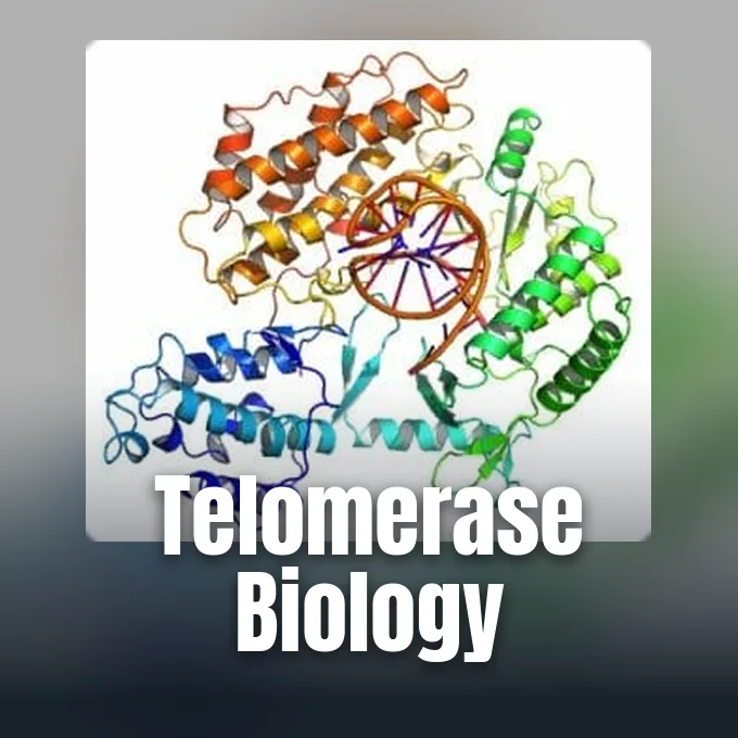
Every dividing cell must duplicate its chromosomes without losing genetic data from their tips, a biological puzzle that explains what is telomerase. Telomerase is a ribonucleoprotein enzyme that adds repetitive DNA sequences to chromosome ends, replenishing the caps called telomeres. By halting the sequence loss that happens in normal replication, this enzyme shapes human cell lifespan [2]. In 2009, the Nobel Assembly at Karolinska Institutet awarded Elizabeth Blackburn, Carol W. Greider, and Jack W. Szostak the Nobel Prize in Physiology or Medicine for discovering how telomeres and telomerase protect chromosomes from degradation [4]. Normal adult cells keep the enzyme quiet [2]. Embryonic cells, male germ cells, and adult stem lineages use it to divide.
- What Is Telomerase?
- The End-Replication Challenge in Linear Chromosomes
- Subunits of the Telomerase Ribonucleoprotein
- Mechanism of Repeat Synthesis and Translocation
- Shelterin Coordination at Chromosome Ends
- Cellular Senescence at the Hayflick Limit
- Why Cancer Cells Reactivate Telomerase
- Clinical Interventions Targeting Telomerase
What Is Telomerase?
Linear chromosomes in an eukaryotic cell end in nucleoprotein structures termed telomeres, which shield genetic data from degradation, end-to-end joining, or false recognition as broken strands. In human cells, these caps consist of tandem TTAGGG repeats bound to shelterin proteins. Standard replication machinery cannot copy the lagging strand end, so chromosomes lose telomeric DNA during each division. Telomerase acts as a reverse transcriptase that adds new repeats onto the three-prime overhang, restoring sequence and keeping chromosomes stable [2]. Human telomerase works as a dimer where two units join together [1].
Soviet biologist Alexey Olovnikov first proposed this mechanism in 1973 as he studied the growth limits of human cells [6]. Olovnikov recognized that DNA polymerases cannot fully duplicate terminal sequences on linear templates, predicting that cells need a compensatory enzyme to counter progressive chromosomal attrition. He suggested that telomere loss acts as a clock governing cellular aging, cancer growth, and brain disorders [3]. His hypothesis anticipated laboratory discoveries by a decade.
In 1984, Carol W. Greider and Elizabeth Blackburn identified enzymatic telomere elongation in the ciliate Tetrahymena thermophila. Greider, then a graduate student at the University of California, Berkeley who was born in San Diego, detected terminal transferase activity on Christmas Day in 1984. Blackburn, born in Hobart, Tasmania, had earned her doctorate from the University of Cambridge before setting up her laboratory at Berkeley. Working with Jack W. Szostak at Harvard Medical School and Cornell University, Blackburn showed that isolated CCCCAA sequences from Tetrahymena protected linear minichromosomes inside yeast cells. Greider later moved to Cold Spring Harbor Laboratory and Johns Hopkins University School of Medicine, and Szostak continued his work in Boston [4].
The End-Replication Challenge in Linear Chromosomes
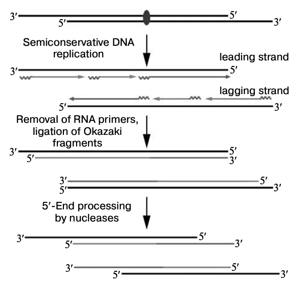
DNA replication needs an RNA primer to start synthesis because DNA polymerases only assemble incoming nucleotides in the five-prime to three-prime direction. Because conventional DNA polymerases can only assemble incoming nucleotides in the five-prime to three-prime direction from an existing primer, the lagging strand inevitably surrenders terminal sequences during each subsequent cell division. Leading strands synthesize continuously toward the chromosome terminus, but lagging strands assemble discontinuously through short Okazaki fragments [2]. Once cellular enzymes remove the final terminal RNA primer, DNA polymerase has no upstream hydroxyl group to initiate synthesis. A short gap remains uncopied at the chromosome tip.
Biologists describe this natural constraint as the end-replication problem [2]. In the 1930s, Hermann Muller, who later won the Nobel Prize in 1946, and Barbara McClintock, who earned the Nobel Prize in 1983, observed that natural chromosome ends prevent chromosomes from sticking together [4]. Most eukaryotes use telomerase to fix this issue. The fruit fly Drosophila melanogaster lacks the enzyme and uses retrotransposons instead [6]. Mammals depend on telomerase to add back lost terminal repeats [2].
Uncapped chromosome ends look like double-strand breaks to surveillance enzymes in the nucleus. When telomeres erode completely, non-homologous end joining pathways fuse separate chromosomes together at their bare tips. Fused chromosomes form dicentric bridges that tear apart during anaphase, scattering gene deletions, broken fragments, and severe structural abnormalities across daughter cells during division [6]. This recurring breakage-fusion-bridge cycle leads to cell death or malignant genomic chaos. Non-coding TTAGGG buffers absorb replication loss, and stop repair enzymes from gluing distinct chromosome ends together [4].
Subunits of the Telomerase Ribonucleoprotein
Human telomerase is a ribonucleoprotein complex formed by catalytic proteins, an RNA scaffold, and helper subunits [2]. Its catalytic core is telomerase reverse transcriptase, known as TERT or hTERT in human cells. Located on chromosome 5, the human hTERT gene contains 16 coding exons with 15 intervening introns across a 35-kilobase stretch while encoding a protein of 2627 amino acids. The core promoter of hTERT spans 330 base pairs upstream of the AUG translation start site and exhibits a high GC content lacking conventional TATA boxes [8]. Human TERT contains distinct finger, palm, thumb, and RNA-binding domains [6]. X-ray crystallography from the Wistar Institute shows that TERT motifs 2 and B prime position the RNA template close to catalytic aspartate residues [5].
The RNA component is TERC, or hTR, encoded on chromosome 3 [3]. Human TERC spans 451 nucleotides. The protein subunit translates in the cytoplasm, but TERC folds in the nucleus into a pseudoknot domain and an H/ACA motif [7]. These RNA structures provide the template for DNA synthesis and bind helper proteins.
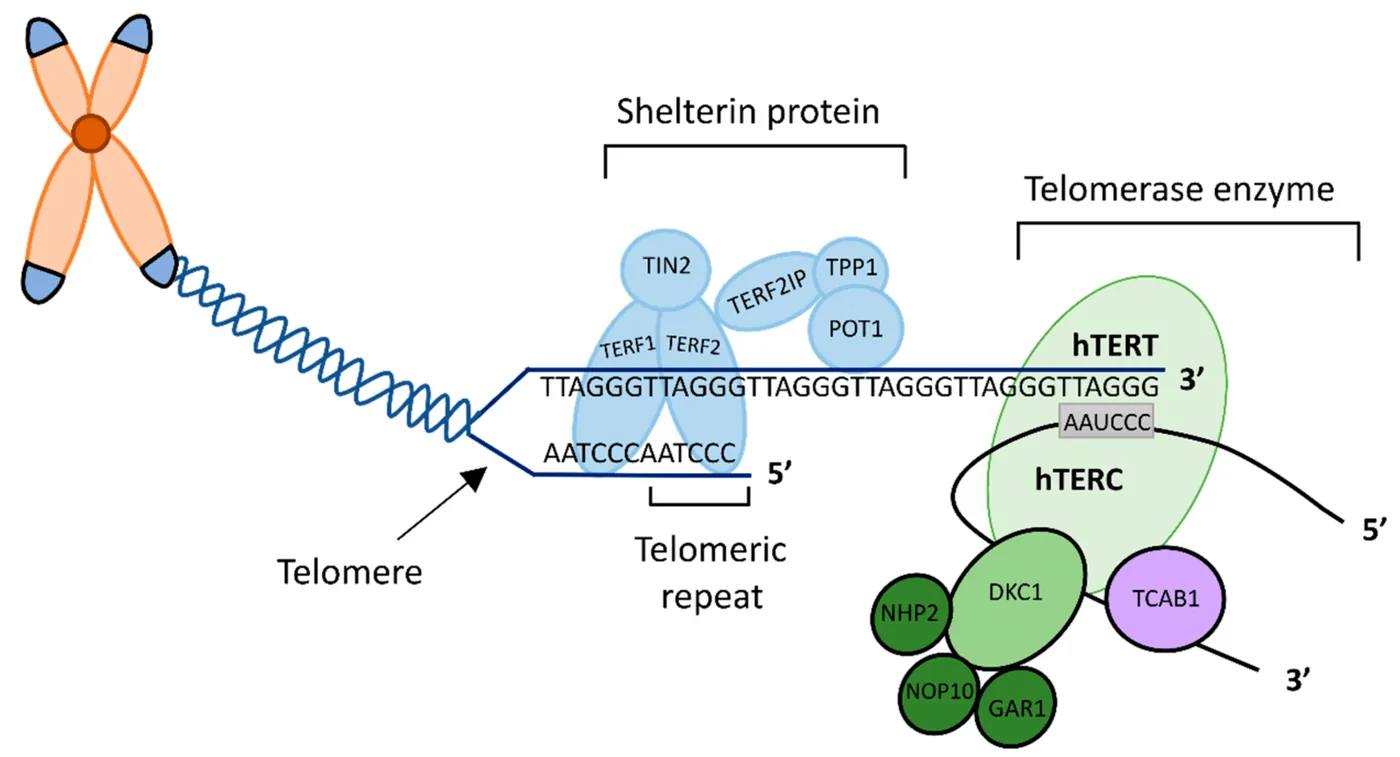
Four accessory proteins bind TERC to form a stable complex: dyskerin (encoded by DKC1), NHP2, NOP10, and GAR1 [2]. In 2018, researchers at the University of California, Berkeley resolved the architecture of human telomerase using cryo-electron microscopy, revealing that the functional ribonucleoprotein complex incorporates two copies of the H/ACA subcomplex [6]. Another helper protein, TCAB1, binds the CR7 domain of TERC [7]. TCAB1 escorts the ribonucleoprotein into nuclear Cajal bodies where final assembly occurs before delivering the complex to chromosome tips during S phase [2].
Mechanism of Repeat Synthesis and Translocation
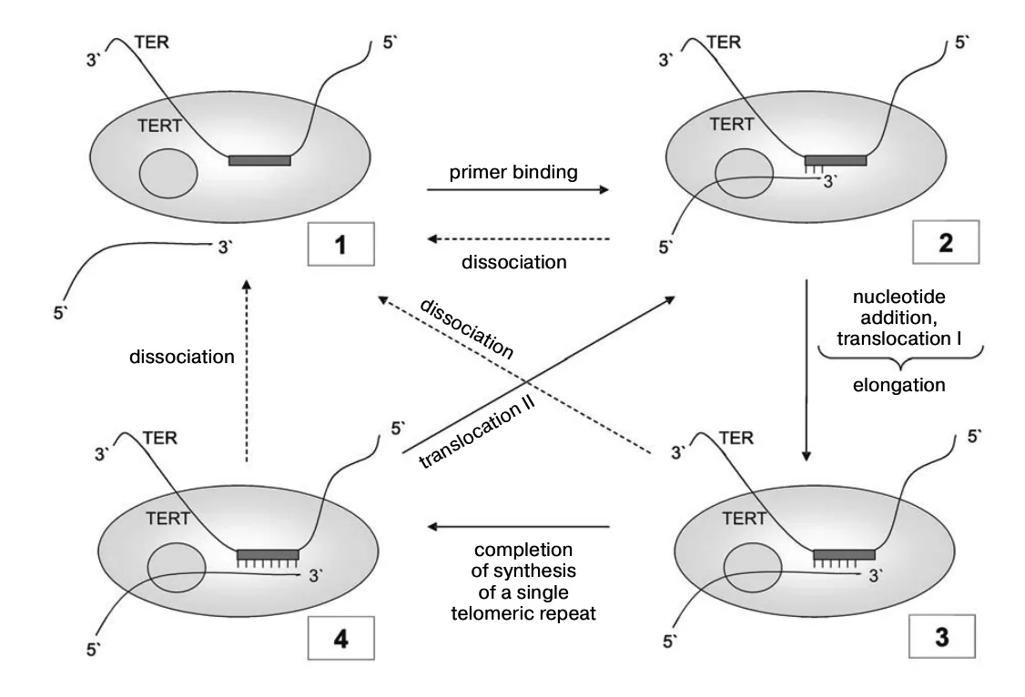
Telomerase operates through a reiterative catalytic cycle that sets it apart from ordinary polymerases, repeatedly copying a short internal RNA template without ever dissociating from terminal chromosomal overhangs [3]. In human cells, the template sequence inside TERC spans eleven nucleotides: three-prime CAAUCCCAAUC five-prime [6]. Synthesis starts when the single-strand overhang of telomeric DNA pairs with the complementary alignment region of the RNA template. This docking aligns incoming nucleotides with catalytic aspartate residues in TERT [2]. In the parasite Trypanosoma brucei, the template sequence is instead three-prime CCCAAUCCC five-prime [6]. Each species uses a specific template to direct synthesis.
TERT adds nucleotides step by step to assemble the canonical repeat 5′-TTAGGG-3′ [6]. The enzyme pauses. Without detaching from the substrate, telomerase shifts forward along the DNA primer to realign the newly synthesized three-prime terminus with the beginning of the internal template for another synthesis round. Enzymologists call this translocation [3].
Through high repeat processivity, a single telomerase complex adds multiple hexanucleotide copies before releasing the chromosome tip [3]. Structural studies by Skordalakes at the Wistar Institute in Philadelphia confirmed that TERT holds the RNA-DNA hybrid firmly inside its interior cavity during each cycle of repeat synthesis [5]. Once telomerase detaches, conventional DNA polymerase alpha and primase synthesize the complementary five-prime strand through lagging-strand mechanics [3]. This combined action rebuilds the double-stranded telomere and keeps a short three-prime single-strand overhang for shelterin binding.


Shelterin Coordination at Chromosome Ends
Telomeres do not float as naked DNA in the nucleus, but remain bound and guarded by a six-protein protective shield named the shelterin complex [2]. Human shelterin contains TRF1, TRF2, POT1, TIN2, TPP1, and RAP1. While TRF1 and TRF2 bind double-stranded TTAGGG repeats, POT1 selectively coats the single-stranded three-prime overhang to prevent the formation of inhibitory G-quadruplex structures during synthesis [3]. TIN2 acts as a central structural linchpin that physically interconnects TRF1, TRF2, and the TPP1-POT1 heterodimer to regulate telomere length and safeguard chromosome ends against unwanted repair [2].
Shelterin loops the chromosome tip [2]. Within this conformation, known as a T-loop, the single-stranded overhang invades preceding double-stranded DNA tracts to assemble a displacement D-loop that buries terminal sequences away from ATM kinase [6]. This tuck prevents checkpoint sensors from mistaking natural chromosome ends for catastrophic double-strand breaks [4].
Shelterin also controls when telomerase can reach chromosome tips. During the synthetic phase of the cell division cycle, TPP1 directly contacts the TEN domain of the catalytic protein, recruiting the ribonucleoprotein complex from nuclear Cajal bodies to restore critically depleted chromosome ends. The subunit TPP1 binds directly to the TEN domain of TERT, recruiting telomerase to telomeres and boosting its synthesis rate [2]. In yeast, proteins like Rap1p, Rif1p, and Rif2p manage a similar length balance. Long telomeres with dense arrays of TRF1 and TRF2 block telomerase from docking [3]. Feedback channels telomerase toward short telomeres and leaves longer repeats untouched.
Cellular Senescence at the Hayflick Limit
Most normal human somatic cells turn off the TERT gene after embryonic development, leaving little enzyme activity in adult tissues. As a result, somatic cells lose telomeric DNA with each division, functioning as a biological timer that marks cellular age. In 1961, Leonard Hayflick discovered that cultured human fetal fibroblasts can only divide 50 to 70 times before stopping permanently. This ceiling is the Hayflick limit. Cells reaching this boundary enter senescence, staying alive and metabolically active without ever dividing again [6]. Replicative arrest guards organisms against unchecked cell growth.
Critically short telomeres cause the protective T-loop to unravel. When progressive division shortens terminal repeats past a critical threshold, chromosomes lose their protective loop architectures, exposing bare DNA ends that prompt cellular senescence through p53 and p21 signaling pathways. Exposed ends trigger DNA damage responses that activate p53 and turn on the inhibitor p21 [2]. Cells with mutated p53 and retinoblastoma pathways skip this checkpoint and enter crisis, where fused chromosomes cause massive cell death [6].
Tissue levels vary [3]. While differentiated adult somatic cells lack significant enzyme activity, high levels persist in male germline lineages and embryonic cells to preserve chromosomal lengths across successive generations. Adult stem cell compartments in the bone marrow and skin maintain moderate telomerase activity to support ongoing tissue renewal. Differentiated human skeletal muscle telomeres remain remarkably stable from ages 23 to 74, demonstrating that non-dividing tissues do not undergo progressive telomere shortening as individuals age [6].
Why Cancer Cells Reactivate Telomerase
Senescence and crisis stop damaged cells from turning into malignant tumors [6]. To form an aggressive cancer, transforming cells must bypass the Hayflick limit and achieve unlimited division. Because telomere exhaustion triggers massive genomic crisis in pre-malignant cells, surviving clones almost universally require an active telomere maintenance mechanism to establish continuous proliferative capacity [2]. About 90 percent of all human tumors achieve immortality by reactivating telomerase, stabilizing their short telomeres so they can divide indefinitely. HeLa cells, isolated from a cervical tumor in 1951, have divided continuously in cell culture for decades because their telomerase stays active [6]. Without telomerase or an equivalent mechanism, developing tumor clones would perish in crisis.
Promoter mutations on chromosome 5 drive hTERT reactivation [6]. Spanning 330 base pairs upstream of the translation start site, the GC-rich core promoter lacks conventional TATA boxes but responds to multiple regulatory factors across diverse tissue environments. While oncogenes like c-Myc and Sp1 stimulate transcription, tumor suppressors including p53 and Menin repress promoter activity in healthy tissues. Factors like HIF-1, AP2, and WT1 also bind [8].
Tumors that do not turn on telomerase rely on Alternative Lengthening of Telomeres, known as ALT. Found in about 10 percent of cancers, such as osteosarcomas and glioblastomas, the ALT pathway uses homologous recombination instead of reverse transcription to maintain telomeres [6]. Many ALT tumors carry loss-of-function mutations in chromatin remodeling genes like ATRX and DAXX [10]. Both strategies confirm that escaping telomere shortening is necessary for cancer growth.
Clinical Interventions Targeting Telomerase
Because telomerase activity appears in about 90 percent of human malignant tumors but remains low in most normal somatic tissues, the enzyme represents a compelling target for oncological drug development [10]. Biotechnology company Geron Corporation cloned the human RNA and catalytic subunits while developing the TRAP assay to measure telomerase activity in malignant patient tissues across diverse clinical tumor samples. Geron also designed imetelstat (GRN163L), a lipid-conjugated oligonucleotide that binds the template region of TERC to block enzyme access [3]. Researchers have also evaluated peptide vaccines in clinical trials to direct cytotoxic T cells against TERT-producing tumor cells [4].
Genetic defects in telomerase cause severe inherited conditions called telomere biology disorders [6]. Germline mutations in DKC1, TERC, or TERT cause dyskeratosis congenita, leading to bone marrow failure and nail dystrophy [2]. Mutations in hTERT are also linked to Cri du chat syndrome and familial pulmonary fibrosis [8]. Premature aging syndromes like Werner, Bloom, and Fanconi anemia also feature accelerated telomere shortening [6].
Dual clinical roles have led scientists to test compounds that stimulate telomerase for anti-aging therapies. Studies showed that overexpressing the catalytic subunit extended lifespan in cancer-resistant mice. The commercial supplement TA-65, derived from Astragalus membranaceus, contains purified cycloastragenol and produces a measurable 2.2-fold boost in telomerase activity across treated human cell cultures. Extracts of Centella asiatica show an 8.8-fold increase, and oleanolic acid yields a 5.9-fold rise in vitro [6]. Scientists warn that boosting telomerase in humans could increase cancer risk by helping mutated cells survive [1].
- WEBSITE BD Editors. (2017). Telomerase: Definition, Function, Structure and Cancer. Biology Dictionary. [Article Link]
- WEBSITE Najid, M. (2025). Telomerase: Structure, Function, and Its Role in Cancer and Aging. Cancer Biology Research. [Article Link]
- WEBSITE Pan, S. (2024). Telomerase – Structure, Mechanism, Regulation, Functions. Biology Notes Online. [Article Link]
- PRESS RELEASE Nobel Assembly at Karolinska Institutet. (2009). The Nobel Prize in Physiology or Medicine 2009: How Chromosomes Are Protected by Telomeres and the Enzyme Telomerase. NobelPrize.org. [Article Link]
- REPORT Mitchell, M., Gillis, A., Futahashi, M., Fujiwara, H., & Skordalakes, E. (2010). Structure of the catalytic subunit of telomerase bound to its RNA template and telomeric DNA (PDB ID: 3KYL). Worldwide Protein Data Bank. [Article Link]
- WEBSITE Wikipedia Contributors. (2026). Telomerase. Wikipedia, The Free Encyclopedia. [Article Link]
- WEBSITE Wikipedia Contributors. (2026). Telomerase RNA component. Wikipedia, The Free Encyclopedia. [Article Link]
- WEBSITE Wikipedia Contributors. (2026). Telomerase reverse transcriptase. Wikipedia, The Free Encyclopedia. [Article Link]
- WEBSITE School of Molecular Sciences. (2026). Telomerase Database. Arizona State University. [Article Link]
- ACADEMIC JOURNAL Jafri, M., Ansari, S. A., Al-Qahtani, M. H., & Shay, J. W. (2016). Roles of telomeres and telomerase in cancer, and advances in telomerase-targeted therapies. Genome Medicine, 8(1), 69. [Article Link]


