





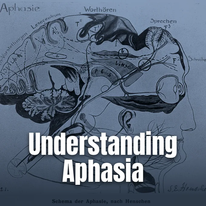
To understand what is aphasia, doctors examine how focal injury to cortical networks impairs language without reducing a person’s intelligence [1]. Aphasia is an acquired neurogenic communication disorder that disrupts how people formulate and understand spoken sentences, read printed text, and write messages after brain injury [2]. It happens when localized lesions harm brain areas that control word retrieval and grammar, most often after an acute stroke [3]. This disorder alters the shared code people use to express thoughts, but it leaves their life experience and logical reasoning intact [5]. Recognizing these symptoms early allows families and medical teams to start therapy without delay.
What Is Aphasia?
Aphasia disrupts the daily exchange of words and ideas, turning familiar talk into an exhausting struggle for millions of adults around the world. According to the National Aphasia Association, roughly 100,000 to 180,000 people acquire the condition each year in the United States, and between 2 million and 4 million people live with its lasting effects [1]. The disorder develops when focal head trauma, vascular blockage, or progressive disease injures left-hemisphere centers that govern language execution [2]. It touches multiple channels of expression, including verbal fluency, auditory listening, reading, and writing [1]. People who acquire these deficits may struggle to find basic nouns or follow rapid group dialogue, but their memory, perception, and general intelligence remain unharmed [5]. The National Aphasia Association tracks these national figures [1].
The core symptom across every clinical presentation is anomia, which means persistent difficulty finding words during everyday speech or formal naming tests [1]. When anomia occurs, a speaker knows the intended meaning but cannot retrieve the spoken label, often resorting to roundabout descriptions like calling a pencil a “thing used to write” [5]. Some people substitute related words like arm for art, or produce invented sounds [19]. Other speakers retain automatic phrases, singing “Happy Birthday” but struggling to request water [5].
Language barriers bring mental distress and social isolation for stroke patients who face high rates of post-stroke depression when speech falters during daily interactions with loved ones [1]. Patience from family members gives people the time they need to express thoughts, and caring partners help restore speaking confidence [2, 19].
How Brain Networks Support Language
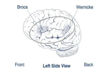
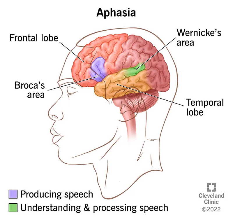
Language processing requires coordinated activity among cortical hubs and deep white matter tracts throughout the brain. For decades, classical neurology relied on the Wernicke-Lichtheim model, which placed expressive speech in the frontal lobe and receptive understanding in the posterior temporal lobe during verbal tasks. Neuroscientists now recognize that speech functions rely on distributed cerebral circuits rather than isolated compartments [4].
Modern neurological models describe speech through a dual-stream architecture proposed by Hickok and Poeppel. In a 2017 review in Brain, researcher J. Fridriksson explained that speech signals travel through two distinct cortical pathways. A ventral stream courses through temporal regions to translate acoustic signals into semantic representations, allowing listeners to recognize vocabulary, grammar, and sentence meaning during fast conversation. Meanwhile, a dorsal stream connects the posterior superior temporal gyrus to frontal premotor regions through the arcuate fasciculus, mapping sound patterns directly to motor articulations for speech production. Damage along this dorsal route undermines repeating words and syntactic formulation, but ventral lesions harm auditory understanding. Both pathways cooperate to sustain natural human dialogue [4].
Most people rely on left perisylvian cortex for language, where the middle cerebral artery supplies oxygenated blood [2]. When right-handed people develop language loss after right-hemisphere injury, neurologists diagnose crossed aphasia [1]. Deep subcortical structures like the thalamus, the internal capsule, and the caudate nucleus also coordinate lexical retrieval and executive language control during complex daily conversations. When subcortical circuits break, cortical hubs lose regulatory signals needed for dialogue [5].
Primary Triggers Behind Brain Damage
Vascular accidents represent the most frequent cause of language loss in clinical neurology [2]. As Cleveland Clinic outlines in its medical guidance on what causes aphasia, strokes cut off blood flow and deprive neural tissue of oxygen within minutes [3]. Data from the American Speech-Language-Hearing Association show that 25% to 50% of stroke patients develop language problems during acute hospital care. Age heavily influences vulnerability, with clinical records showing that 15% of people under age 65 develop speech deficits after a first ischemic stroke compared to 43% for patients aged 85 and older [1]. A transient ischemic attack (TIA) or mini stroke can cause speech loss for a few hours without leaving permanent damage [2]. Quick action limits brain damage [3].
Head trauma is another common trigger of speech disorders. Data from veterans of the conflicts in Iraq and Afghanistan show that 1% of service members with combat trauma developed aphasia. In civilian hospital settings, trauma studies report rates between 13% and 19%. Epidemiologist Nina Simmons-Mackie calculated that between 64,653 and 1,228,421 people in the United States live with trauma-related language loss after collisions, falls, or combat concussions. Mechanical impact damages fragile axonal connections between distant cortical lobes [1].
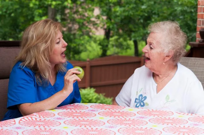
Primary brain tumors cause speech issues in 30% to 50% of patients as growing tissue compresses perisylvian zones [1]. In primary progressive aphasia (PPA), speech declines gradually due to frontotemporal dementia or Alzheimer’s disease [2]. Infections like herpes simplex encephalitis also harm temporal language tissue [5].
Distinguishing Fluent from Nonfluent Forms
Clinicians evaluate speech rate, phrase length, and grammatical structure to separate language disorders into fluent and nonfluent categories [1]. Damage to the posterior inferior frontal gyrus causes Broca’s aphasia [6]. In nonfluent presentations, patients drop small grammatical words like is, and, and the, producing effortful, telegraphic phrases such as “Walk dog” while preserving basic communicative intent during daily conversation [2]. Right-sided body weakness often accompanies this frontal damage. Unlike patients with sensory comprehension deficits, people with Broca’s aphasia monitor their own output closely, recognizing speaking mistakes immediately and experiencing profound frustration as documented by French physician Paul Broca. Clinical records describe patients struggling through dental visits with fragmented phrases [6].
In contrast, fluent presentations exhibit effortless speech production and preserved rhythm despite severe semantic deficits. Individuals with Wernicke’s aphasia, named after German physician Carl Wernicke, speak in long, grammatically intricate sentences that convey little clear meaning following damage to the left superior temporal cortex. Their speech often contains paraphasias and neologisms, such as saying “dorflur” for shoe or uttering phrases like “smoodle pinkered” [2, 7]. A central feature is anosognosia, which leaves patients completely unaware of their own spoken mistakes [7].
Other syndromes present unique communicative profiles. In global aphasia, broad damage across frontal and temporal zones wipes out expressive and receptive language. Conduction aphasia leaves fluency intact but impairs repeating words [2]. Transcortical aphasias preserve repeating phrases but limit spontaneous speech or auditory understanding [17].
Key Differences Between Aphasia and Dysarthria
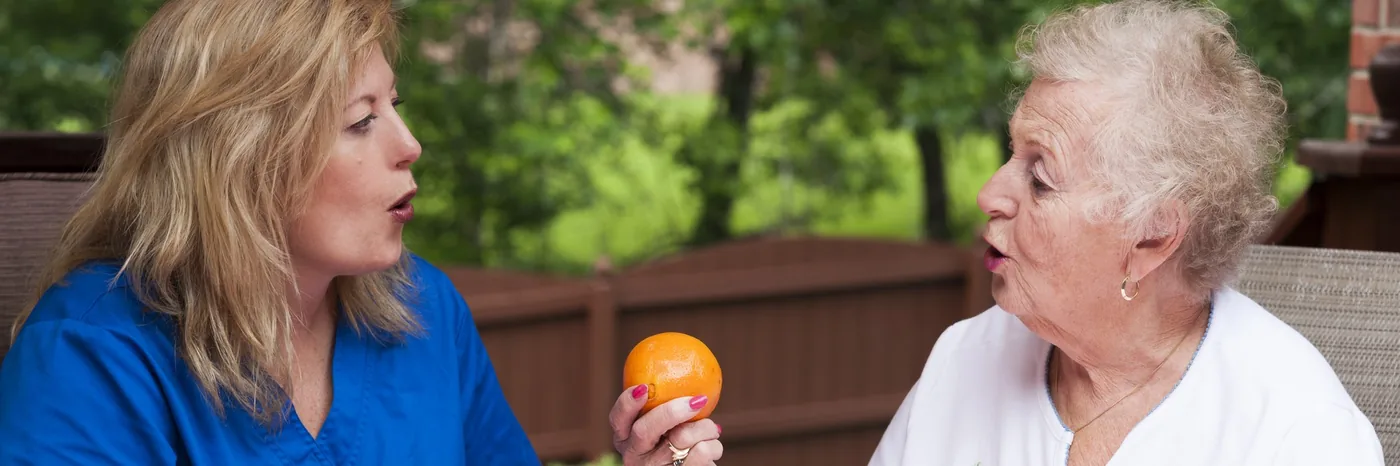
Because stroke and head trauma impair speech production, observers often confuse language processing deficits with neuromuscular articulation problems that affect the physical muscles of the mouth. But aphasia is a cognitive language deficit, whereas dysarthria is a motor speech disorder caused by muscle weakness or poor coordination [8]. A person with aphasia struggles to access linguistic symbols, organize syntax, or comprehend dialogue [9]. In contrast, an individual with dysarthria knows exact words and understands all spoken conversation, but cannot control the tongue, lips, vocal cords, or diaphragm to pronounce sounds clearly. Dysarthria produces slurred, quiet, or choppy speech without disturbing reading comprehension or written spelling. Johns Hopkins Medicine notes that distinguishing these conditions requires careful clinical assessment [8].
Neurologists classify dysarthria into distinct physical categories based on neural lesion sites, distinguishing flaccid dysarthria from lower motor neuron injury, spastic speech from upper motor neuron damage, and ataxic dysarthria from cerebellar coordination disruption. Cadence turns irregular. Parkinson’s disease, amyotrophic lateral sclerosis (ALS), and Bell palsy frequently trigger motor speech problems along with severe swallowing difficulties [8, 9].
Clinicians also distinguish both conditions from apraxia of speech, which is a motor planning breakdown where the brain cannot sequence articulatory movements despite normal muscle strength [1, 6]. In apraxia, patients struggle to position their lips and tongue intentionally during speech [1]. Muscle exercises aid dysarthria, and cognitive speech therapies restore language circuits in aphasia [8].
Clinical Tests and Brain Scans
Medical evaluation starts immediately when an acute neurological emergency occurs [1]. Emergency physicians first check basic communication at the bedside by testing whether the patient follows simple commands, identifies familiar objects, and answers questions [2]. If stroke symptoms appear—such as facial drooping, arm weakness, or slurred words—calling 911 immediately limits permanent tissue loss by restoring cerebral blood flow as fast as possible [3]. Once medical stability returns, a licensed SLP conducts detailed testing across speaking, listening, reading, and writing domains [1]. Standard diagnostic instruments include the Boston Diagnostic Aphasia Examination and the Western Aphasia Battery-Revised, which calculates an overall aphasia quotient to measure expressive and receptive severity [4, 10]. These formal assessments help doctors map language deficits accurately [1].
Specialists also use the Aphasia Diagnostic Profiles, an impairment-based tool designed by Nancy Helm-Estabrooks in 1992 under World Health Organization assessment principles to evaluate conversational speech. The battery features nine subtests that evaluate conversational speech, phrase repetition, reading, and writing using an informal conversational format. Results yield standardized scores that establish impairment severity and guide therapy goals. Nancy Helm-Estabrooks introduced this tool in 1992 [11]. Computerized testing on Cinderella narratives in AphasiaBank also detects subtle discourse deficits in high-functioning patients [1].
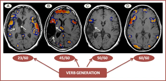
Neuroimaging tests confirm the site and size of brain damage. CT scans rapidly detect acute bleeding, and MRI provides clear structural views of ischemic stroke damage [10]. Functional MRI tracks blood oxygen changes during language tasks [5]. Combining imaging with SLP testing ensures accurate clinical diagnosis [1].
Speech Therapy Options for Aphasia
Speech-language therapy serves as the primary intervention for restoring functional communication after focal brain damage [10]. Major clinical evidence regarding treatment dosage emerged from the RELEASE study, a network meta-analysis led by M. C. Brady in 2022 that analyzed 174 studies across 28 countries covering nearly 6,000 individuals [12, 13]. Researchers concluded that recovery was strongest when therapy started within 28 days of aphasia onset, providing 20 to 50 total hours of therapy delivered over 4 to 5 days weekly. People under 55 years made the fastest gains, but adults aged 75 and older still improved. Noticeable gains in speech comprehension required intensive regimens of 9 or more hours per week. Home practice tasks produced the greatest gains [12].
Therapists use specific behavioral techniques to target individual language weaknesses [10]. For severe nonfluent speech, Melodic Intonation Therapy uses rhythm and musical pitch to stimulate latent language capacities in the right hemisphere [14]. When treating anomia, doctors deliver confrontation naming drills with phonemic and orthographic cues. A clinical trial by Karen Sage at the University of Manchester demonstrated that non-intensive therapy yielded superior one-month word retention compared to intensive schedules for stroke patients [15]. These findings influenced clinical stroke guidelines across the United Kingdom, Australia, and New Zealand [12]. Programs like Promoting Aphasic Communicative Effectiveness (PACE) and Computerized Script Training (CST) also build conversational confidence [10].
Medical teams also investigate medications to assist cognitive recovery. A scientific review by M. L. Berthier showed that combining intensive constraint-induced language therapy with donepezil or memantine may support neuroplasticity. Clinical guidelines from NICE and the European Stroke Organisation prioritize structured behavioral therapy from a licensed SLP [10, 12, 16].
Practical Communication Strategies at Home
Long-term recovery relies on supportive communication within home environments and local communities [1]. When natural speech remains limited, speech-language pathologists introduce augmentative and alternative communication (AAC) tools. AAC options range from picture books and alphabet boards to digital speech-generating apps on tablets and mobile phones [2, 19]. Rather than replacing spoken words, nonverbal communication reduces emotional frustration and frequently prompts spontaneous language retrieval by providing alternative pathways to express meaning [19]. Clinicians encourage family members to welcome drawing, pointing, and natural gestures [2]. People with aphasia naturally produce more iconic gestures than typical speakers, using hand movements to convey meaning when words stall [1]. These adaptive methods keep individuals actively connected to loved ones [19].
Partner training improves home dialogue [1]. Care partners should minimize background noise from televisions and radios while speaking at a normal conversational volume in short, direct sentences, asking simple yes-or-no questions to avoid communicative overload. Family members should never finish sentences prematurely. Listeners must avoid interrupting or correcting minor speech slips, treating the adult as an intelligent conversation partner to preserve mutual trust and self-esteem throughout daily recovery [10, 19].
Community support groups provide emotional comfort and social engagement for stroke patients [2]. Support charities like Say Aphasia, Aphasia Reconnect, The Brain Charity, and the Stroke Association offer community groups and helplines to help patients regain social connection [18]. Local stroke clubs meet across the country. With persistent therapy and supportive families, people with aphasia can rebuild fulfilling lives [1, 2].
- WEBSITE American Speech-Language-Hearing Association. (2023). Aphasia. [Article Link]
- REPORT National Institute on Deafness and Other Communication Disorders. (2025). Aphasia. [Article Link]
- WEBSITE Cleveland Clinic. (2026). What Causes Aphasia?. [Article Link]
- ACADEMIC JOURNAL Fridriksson, J., den Ouden, D. B., Hillis, A. E., Hickok, G. S., Rorden, C., Basilakos, A., Yourganov, G., & Bonilha, L. (2017). Anatomy of aphasia revisited. Brain, 141(3), 848-862. [Article Link]
- WEBSITE Wikipedia. (2026). Aphasia. [Article Link]
- WEBSITE Wikipedia. (2026). Expressive aphasia. [Article Link]
- WEBSITE Wikipedia. (2026). Receptive aphasia. [Article Link]
- WEBSITE MedicineNet. (2022). What is the difference between aphasia and dysarthria?. [Article Link]
- WEBSITE MedlinePlus. (2026). Speech impairment in adults. U.S. National Library of Medicine. [Article Link]
- ONLINE NEWS Medical News Today. (2017). Aphasia: Symptoms, diagnosis, and learning to communicate again. [Article Link]
- BOOK CHAPTER Patterson, J. (2011). Aphasia Diagnostic Profiles. In J. S. Kreutzer, J. DeLuca, & B. Caplan (Eds.), Encyclopedia of Clinical Neuropsychology. Springer. [Article Link]
- REPORT Cassidy, S. (2023). Speech and language therapy for aphasia after a stroke. NIHR Evidence. [Article Link]
- ACADEMIC JOURNAL Brady, M. C., Ali, M., VandenBerg, K., Williams, L. J., Williams, L. R., Abo, M., Becker, F., Bowen, A., Brandenburg, C., Breitenstein, C., Bruehl, S., Copland, D. A., Cranfill, T. B., di Pietro-Bachmann, M., Enderby, P., Fillingham, J., Galli, F. L., Gandolfi, M., Glize, B.,. Howard, D. (2022). Complex speech-language therapy interventions for stroke-related aphasia: the RELEASE study incorporating a systematic review and individual participant data network meta-analysis. Health and Social Care Delivery Research, 10(28), 1-272. [Article Link]
- ACADEMIC JOURNAL Albert, M. L., Sparks, R. W., & Helm, N. A. (1973). Melodic intonation therapy for aphasia. Archives of Neurology, 29(2), 130-131. [Article Link]
- ACADEMIC JOURNAL Sage, K., Snell, C., & Lambon Ralph, M. A. (2011). How intensive does anomia therapy for people with aphasia need to be?. Neuropsychological Rehabilitation, 21(1), 26-41. [Article Link]
- ACADEMIC JOURNAL Berthier, M. L., Pulvermüller, F., Dávila, G., Casares, N. G., & Gutiérrez, A. (2011). Drug therapy of post-stroke aphasia: A review of current evidence. Neuropsychology Review, 21(3), 302-317. [Article Link]
- WEBSITE WebMD. (2026). Aphasia: What to know. [Article Link]
- WEBSITE National Health Service. (2025). Aphasia. [Article Link]
- WEBSITE American Speech-Language-Hearing Association. (2026). Aphasia. Helping You Communicate. [Article Link]
- WEBSITE LaDonna. (2026). What is aphasia. Lingraphica. [Article Link]
Cover image: Wikimedia Commons, CC BY 4.0


