





An enzyme-linked immunosorbent assay, known as an ELISA, is a plate-based laboratory method designed to detect and quantify soluble substances such as proteins, peptides, antibodies, and hormones in biological samples [2]. Medical teams at Cleveland Clinic and research facilities rely on the technique as a testing standard for diagnosing infections, evaluating endocrine functions, and screening blood donations [1], [9]. Learning how elisa tests work involves understanding how specific antibody-antigen binding pairs with an enzyme-catalyzed reaction to generate a measurable optical signal across laboratory test samples [1]. When target proteins bind to antibodies immobilized inside plate wells, attached reporter enzymes convert colorless chemical substrates into colored reaction products [6]. The intensity of this resulting color change corresponds to the level of the target analyte present in the fluid sample [7].
- Biochemical Principles of Enzyme-Linked Immunosorbent Assay Kits
- How Enzyme Immunoassay Methods Replaced Radioactivity
- Comparing Direct and Indirect Detection Geometries
- Sandwich versus Competitive Plate Designs
- Explaining How ELISA Tests Work in Medical Diagnosis
- Solid Support Plastics, Surface Blocking, Reporter Enzymes
- Calibrating Standard Curves for Optical Signal Analysis
- Interference Factors in Clinical Fluid Analysis
Biochemical Principles of Enzyme-Linked Immunosorbent Assay Kits
Plate assays depend on two coupled events: solid-phase binding and enzymatic signal amplification, as described in Elabscience research protocols [3]. Polystyrene plates from Thermo Fisher Scientific, such as MaxiSorp and MultiSorp modules, provide the physical platform where target antigens or capture antibodies bind securely to plastic surfaces. Coating occurs through passive adsorption driven by hydrophobic interactions between non-polar protein residues and the synthetic polymer matrix. Laboratory protocols published by Cell Signaling Technology introduce protein solutions dissolved in an alkaline buffer, such as phosphate-buffered saline at pH 7.4 or carbonate-bicarbonate buffer at pH 9.4, at levels between 2 and 10 micrograms per milliliter [2], [4]. These plates incubate for several hours at room temperature or overnight at 4 degrees Celsius to establish stable surface adhesion [2], [7].
Unoccupied plastic sites bind proteins non-specifically if left untreated [2]. Technicians add bovine serum albumin (BSA) or casein blockers to coat remaining bare areas [3], [5]. This step keeps background noise low. Washes with Tween-20 buffer remove loose sample debris [7], [11].
Enzymatic conversion delivers high testing reach during optical evaluations. In fact, one conjugated enzyme molecule can turn hundreds of clear substrate units into colorful products every second. This rapid catalytic amplification lets hospital laboratory teams register trace amounts of target analyte that would otherwise escape optical detection during routine bench testing [5], [16]. Once color development reaches its target window, technicians add sulfuric acid stop solution to halt catalysis and stabilize signal readings for microplate readers [3], [7].
How Enzyme Immunoassay Methods Replaced Radioactivity
Before enzyme-based detection entered medical laboratories, clinical researchers had only one reliable tool for measuring trace hormones and serum proteins: the radioimmunoassay [9],. First described in 1960 by Rosalyn Sussman Yalow and Solomon Berson, radioimmunoassays measured biological targets by tracking competitive displacement between unlabeled antigens and radioactively tagged molecules. While radioimmunoassay achieved impressive sensitivity, radioactive isotopes carried serious hazards [16]. Radiated reagents decayed rapidly, demanded strict radiation safety licenses, and exposed clinic staff to potential health risks during waste disposal [9], [16]. Medical teams at Danaher Life Sciences and research hospitals needed a stable, non-radioactive testing platform that could match that precision without ionizing rays [9], [12], [16].
Breakthroughs in Sweden and France solved these radiation problems. In 1966, Jerker Porath and Leif Wide built solid-phase immunosorbents. Stratis Avrameas and G. B. Pierce then linked active enzymes directly to antibodies. These methods held targets firmly while removing radiation risks [16].
In 1971, Peter Perlmann and Eva Engvall at Stockholm University in Sweden published their pioneering method combining enzyme conjugates with solid-phase adsorption, coining the name enzyme-linked immunosorbent assay [2], [9],. Simultaneously in the Netherlands, Anton Schuurs and Bauke van Weemen published an enzyme immunoassay method based on similar principles [16]. As documented by Microbe Notes, researchers introduced competitive formats in 1976, sandwich assays in 1977, and indirect tests in 1978 [11]. These plate-based systems quickly replaced radioimmunoassays worldwide [9], [11].
Comparing Direct and Indirect Detection Geometries
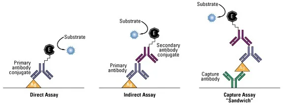
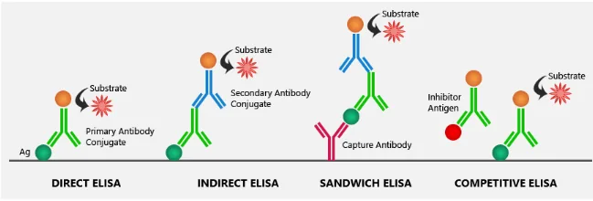
Direct ELISA represents the simplest and quickest setup among plate-based immunoassays [11]. Target antigens adsorb directly onto microplate wells, followed by incubation with an enzyme-labeled primary antibody that binds straight to the target [7], [11]. Because direct formats require fewer incubation and washing steps, technicians complete assays faster while avoiding cross-reactivity from second antibodies [9]. Laboratory teams observing how ELISA tests work note that direct immobilization lacks target selectivity, because crude proteins in raw samples compete aggressively for plastic binding space. Low-abundance antigens struggle to bind when mixed with abundant background proteins [16]. Teams at Biology Notes Online note that direct assays work best for rapid viral screening and epitope mapping [7].
Indirect assays split capture and detection between two distinct antibodies [5], [9]. Known antigens first capture primary antibodies from patient blood [7], [11]. An enzyme-linked secondary antibody, such as anti-human IgG, then binds the primary antibody [6], [7]. This two-stage setup multiplies the final signal [6], [9].
Reagent flexibility makes indirect testing popular in commercial diagnostic centers. Instead of linking enzymes to dozens of custom primary antibodies, a clinical lab uses one standardized second conjugate to detect multiple primary binders from the same animal host [6]. This versatility makes indirect testing ideal for serological surveys, vaccine titer monitoring, and immune evaluations [7]. Common diagnostic uses include screening blood donations for antibodies produced against rubella and dengue [11].
Sandwich versus Competitive Plate Designs
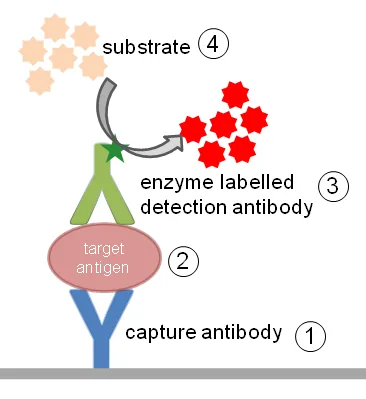
Sandwich ELISA serves as the primary format for clinical testing and biomedical research because it delivers superior specificity and sensitivity [2], [5]. Rather than coating crude sample proteins directly onto plastic, the method immobilizes a specific capture antibody on the well surface [2],. When technicians add blood serum or cell lysates to the wells, the capture antibody selectively pulls target antigens out of the fluid mixture without requiring prior purification [6], [16]. A second detection antibody then binds to a separate, non-overlapping epitope on the captured antigen to form a molecular sandwich [2],. Because two distinct antibodies must recognize separate binding sites on the same molecule, background noise drops sharply [4], [6].
Matched antibody pairs must bind distinct epitopes without steric clashes [4], [12]. The target protein must also be large enough to accommodate two bulky immunoglobulins simultaneously [5], [12]. Labs rely on sandwich kits to measure thyroid-stimulating hormone, interferon-gamma, and viral HIV p24 core antigen [5], [12], [13]. Small molecules cannot fit this sandwich geometry [2], [12].
Competitive ELISA provides a clever alternative for measuring small molecules, haptens, and single-epitope drugs [2], [12]. Unlabeled target molecules from patient samples compete with enzyme-labeled reference antigens for a limited number of coated antibody sites [2], [6]. An inverse relationship governs signal output: high target levels in patient blood displace labeled competitors, yielding less color after substrate addition and enzymatic cleavage [5], [6]. Conversely, faint sample target levels allow labeled antigens to bind freely, producing intense optical signal [5], [6].


Explaining How ELISA Tests Work in Medical Diagnosis
Clinical pathology departments apply enzyme immunoassays across an expansive variety of bodily fluids, analyzing serum, plasma, urine, saliva, and cerebrospinal fluid (CSF) [1], [9], [12]. Explaining how ELISA tests work in diagnostic clinics highlights their value in identifying viral, bacterial, and parasitic pathogens [1], [11]. Physicians routinely order ELISA panels to detect blood-borne markers for hepatitis B, hepatitis C, syphilis, and Lyme disease caused by Borrelia burgdorferi [5],. The assay also detects antibodies against childhood illnesses such as varicella-zoster virus, which triggers chickenpox and later herpes zoster, as well as vector-borne threats like Zika virus and Rocky Mountain spotted fever [8].
Autoimmune and allergy panels rely heavily on plate immunoassays. Labs measure autoantibodies in patients suspected of having systemic lupus erythematosus or rheumatoid arthritis. Tests also quantify allergen-specific IgE antibodies. These blood assays replace uncomfortable skin prick tests [9].
Hospital screening panels require secondary confirmation before physicians make a definitive diagnosis [1], [8]. False-positive readings can occur if cross-reactive antibodies or interfering proteins bind non-specifically during testing [1],. In addition, finding antibodies shows past infection or vaccination rather than active disease [5], [8]. For infections like human immunodeficiency virus (HIV), clinical protocols mandate that reactive ELISA results undergo secondary verification using Western blot or nucleic acid tests [5], [8], [10].
Solid Support Plastics, Surface Blocking, Reporter Enzymes
Standard laboratory assays take place in rigid polystyrene plates manufactured in 96-well or 384-well microplate formats [2], [7]. Plates designed for colorimetric reading feature transparent flat bottoms that let light pass unobstructed through the sample liquid to an optical detector [2], [16]. Black or white opaque plates prevent optical cross-talk during fluorogenic or chemiluminescent detection. Quality assay plates from brands like NUNC Immuno maintain a protein-binding capacity of at least 400 ng/cm2 with a coefficient of variation below 5%, ensuring uniform protein adsorption across all wells [2], [13]. Bench assays demonstrating how ELISA tests work rely on these strict physical manufacturing standards to deliver repeatable diagnostic measurements [2].
Special pre-coated surfaces help researchers orient antibodies properly. Protein A, Protein G, or Protein A/G coatings bind antibody Fc regions, pointing binding arms outward. Streptavidin plates capture biotinylated targets. Glutathione surfaces bind glutathione S-transferase (GST) fusion proteins [2].
Signal development begins when enzyme-conjugated reagents meet chromogenic substrates [6],. Horseradish peroxidase (HRP) and alkaline phosphatase (AP) serve as the primary enzyme labels [15]. When paired with HRP, 3,3′,5,5′-tetramethylbenzidine (TMB) produces a blue color that turns yellow after adding acid, absorbing light at 450 nm [7],. As documented by Thermo Fisher Scientific, OPD substrate yields a yellow-orange product reading at 492 nm, while ABTS yields a green product reading at 410 nm [15]. Alkaline phosphatase hydrolyzes p-nitrophenyl phosphate (pNPP) into a soluble yellow product [7], [11].
Calibrating Standard Curves for Optical Signal Analysis
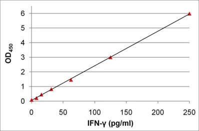
Microplate spectrophotometers measure light transmission through each well to calculate optical density (OD) values after stopping the enzymatic reaction [5], [7]. Data output takes three analytical forms for in vitro diagnostics: qualitative, semi-quantitative, or fully quantitative [6],. Qualitative assays produce a binary positive or negative call by comparing sample absorbance against a cut-off threshold, typically set two or three standard deviations above negative controls [16]. Semi-quantitative assays assess relative signal differences to track changing antibody titers over time [6]. Fully quantitative assays determine exact analyte concentrations by comparing sample absorbance directly against a known reference curve generated from serial dilutions of purified standards [3], [6].
Standard curves require serial dilutions of purified reference proteins [3], [13]. Analysts plot standard concentrations along the X-axis against background-corrected optical density on the Y-axis. Most quantitative assays fit a four-parameter logistic (4-PL) mathematical curve. Valid curves require a correlation coefficient (R-value) above 0.99 [3].

Quality control guidelines from Elabscience establish clear benchmarks for assay acceptance. In sandwich assays, blank control wells should yield an optical density at or below 0.1, while maximum standards should exceed 1.2. Labs assess well-to-well consistency by checking the coefficient of variation (CV%), which should remain below 10% across duplicate wells. If a patient sample produces an optical density reading that exceeds the highest standard point, the sample must be diluted and retested [3].
Interference Factors in Clinical Fluid Analysis
Chemical and biological interferences can distort experimental findings despite the high sensitivity of enzyme immunoassays [5], [9]. One prominent challenge is antibody cross-reactivity, where detection reagents bind to structurally similar non-target antigens, generating false-positive signals [5], [9]. In sandwich formats, extremely high analyte concentrations can trigger the high-dose hook effect [2], [5]. When target molecules vastly outnumber available antibodies, free antigens saturate capture and detection antibodies separately, preventing the formation of complete sandwich complexes and causing falsely depressed optical readings [2], [5]. Field studies tracking how ELISA tests work show that diluting concentrated samples prevents this hooking error effectively [2], [5].
Sample matrices also cause analytical interference. Hemolyzed blood, lipids, or heterophile antibodies in patient serum distort binding kinetics [5]. Incomplete washing leaves unbound proteins on well walls, raising background absorbance [4],. Labs run negative, positive, and blank controls on every plate to catch these errors [5].
Modern innovations continue expanding immunoassay formats beyond standard 96-well plates [17], [18]. Multiplex assays described by Tighe and colleagues allow researchers to quantify dozens of cytokines simultaneously in single microplate wells [17]. In low-resource settings, Whitesides and Verma developed sliding-strip paper microfluidic devices that detect C-reactive protein (CRP) across concentrations of 1 to 100 ng/mL in diluted whole blood [18]. These technologies illustrate how core enzyme-linked immunoassay principles adapt to meet portable point-of-care medical needs [17], [18].
- WEBSITE Cleveland Clinic. (2023). ELISA Technique. [Article Link]
- WEBSITE Thermo Fisher Scientific. (2016). ELISA Assay: Principles, Types, & Applications. [Article Link]
- WEBSITE Elabscience. (2026). ELISA Results Interpretation and Standard Curve Analysis Guide. [Article Link]
- WEBSITE Cell Signaling Technology. (2025). Principle of ELISA (Enzyme-linked Immunosorbent Assay) Test. [Article Link]
- WEBSITE Healthy Beginnings Magazine. (2026). How Does Elisa Work Principle Steps And Types? Key Facts. [Article Link]
- WEBSITE Laboratoryinfo.com. (2022). ELISA: Principle, Procedure, Types, Applications and Animation. [Article Link]
- WEBSITE Biology Notes Online. (2026). ELISA test – Definition, Principle, Procedure, Types, Steps, Uses. [Article Link]
- WEBSITE Healthline Media. (2012). ELISA: Purpose, Procedure, and Results. [Article Link]
- WEBSITE Wyndly. (2026). ELISA Test: Types, Technique, and Results Explained. [Article Link]
- WEBSITE MedicineNet. (2022). ELISA Test Steps, Results, Types, Risks, Full Form, HIV. [Article Link]
- WEBSITE Microbe Notes. (2022). Enzyme-linked immunosorbent assay (ELISA). [Article Link]
- WEBSITE Danaher Life Sciences. (2023). What is ELISA? Types & Advantages. [Article Link]
- WEBSITE British Society for Immunology. (2022). Enzyme-linked immunosorbent assay (ELISA). [Article Link]
- WEBSITE Cell Signaling Technology. (2025). What is an ELISA Assay. [Article Link]
- WEBSITE Thermo Fisher Scientific. (2026). ELISA Enzyme Substrates. [Article Link]
- WEBSITE Wikipedia. (n.d.). ELISA. [Article Link]
- ACADEMIC JOURNAL Tighe, P. J., Ryder, R. R., Todd, I., & Fairclough, L. C. (2015). ELISA in the multiplex era: Potentials and pitfalls. PROTEOMICS – CLINICAL APPLICATIONS, 9(3-4), 406-422. [Article Link]
- ACADEMIC JOURNAL Verma, M. S., Tsaloglou, M., Sisley, T. A., Christodouleas, D. C., Chen, A. C., Milette, J., & Whitesides, G. M. (2017). Sliding-strip microfluidic device enables ELISA on paper. Biosensors and Bioelectronics, 99, 77-84. [Article Link]


