





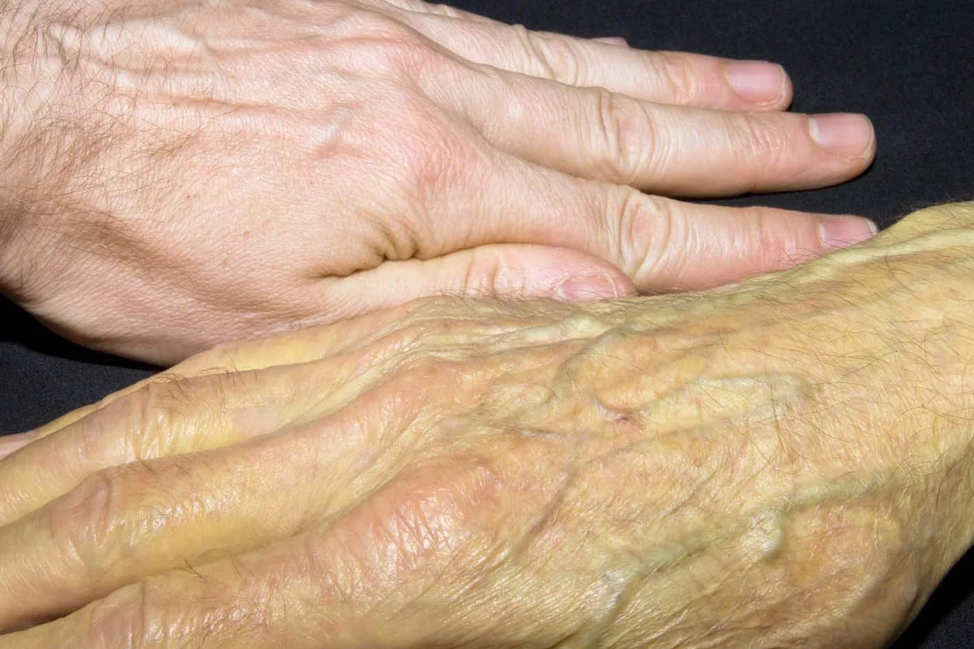
What is jaundice? In clinical medicine, jaundice describes the yellow tint of the skin, tissues, and eyes that develops when bilirubin builds up in the blood to abnormal levels [1]. Healthcare providers at the Cleveland Clinic and the NHS call this state hyperbilirubinemia or icterus [2, 8]. The yellow pigment forms naturally when the body breaks down old red blood cells, which the liver filters and prepares for clearance through the gut [3]. When liver cells cannot process this pigment, or when blocked bile ducts stop its flow, it leaks into the bloodstream and binds to elastic body tissues [10]. Although neonatal jaundice affects roughly 60 percent of full-term newborn infants and often clears on its own, adult cases always point to an underlying health problem that needs clinical care [2].
What Is Jaundice?
Jaundice is a visible sign of altered pigment balance rather than an independent illness [1]. In healthy people, normal total bilirubin in the blood stays below 1.0 mg/dL, which equals about 17 micromoles per liter [11]. Jaundice becomes visible to examining doctors once blood pigment levels climb above 2.0 to 3.0 mg/dL, which equals roughly 34 to 51 micromoles per liter during routine clinical testing in adult patients [8]. French doctors named it jaune [7].
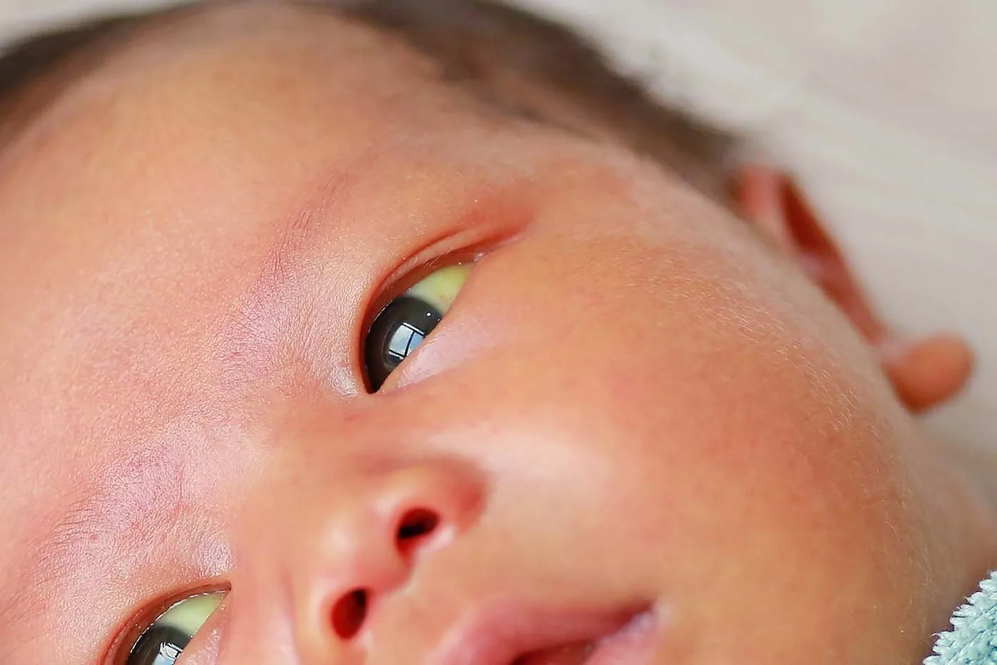
The whites of the eyes, known as sclerae, often show yellow tones before the skin changes color [11]. Bilirubin binds strongly to elastic tissue, which exists in high amounts within the eye conjunctiva [10]. Although medical texts often use the term scleral icterus, the pigment actually settles within the thin conjunctival layer over the eye rather than inside the deeper white wall itself [11]. In people with dark skin tones, yellowing is harder to spot under dim indoor lighting. Dr. Whitney Casares, a pediatrician writing for the American Academy of Pediatrics, noted that checking the gums, inner lips, palms, and soles gives a more reliable assessment when evaluating darker-skinned infants for subtle changes in skin color. Bright daylight makes these yellow tones much easier to see during checks [2].
As blood pigment levels climb, other body symptoms often accompany the changing skin color [8]. Bile salts settling in skin layers irritate sensory nerves, provoking intense pruritus, or severe itching [1, 10]. Patients often notice that urine turns a tea-like brown shade, while stools become pale, resembling grey or clay-colored bowel movements without normal brown tones [2, 8]. Greenish skin tones can develop from biliverdin [11].
How Bilirubin Cycles Through the Body
The creation of bilirubin starts inside white cells of the spleen and liver. Mature red blood cells live for about 120 days before being engulfed and broken down [7, 11]. The enzyme heme oxygenase opens the heme ring to yield iron, carbon monoxide, and biliverdin, which biliverdin reductase promptly turns into indirect, or unconjugated, bilirubin [11]. The human body makes about 4 mg of bilirubin per kilogram of weight daily. Roughly 80 percent comes from broken red cells, while 20 percent comes from faulty red cell production in the marrow and the turnover of muscle heme like myoglobin [7]. Because newly formed unconjugated bilirubin is fat-soluble, it cannot dissolve in watery blood and must bind tightly to albumin for safe transport to the liver [8, 11].
Liver cells take up albumin-bound pigment through their outer walls as blood filters through liver tissue, where the enzyme uridine diphosphate-glucuronyl transferase, or UGT, binds it to glucuronic acid to produce water-soluble direct bilirubin [7, 8, 11]. Cells pump direct bilirubin into tiny channels [8, 11]. Canalicular export is the slowest step [11].
Bile travels through liver ducts to the bile sac before emptying into the upper gut [7, 8]. In the bowel, friendly bacteria change bilirubin into colorless urobilinogen compounds. Most urobilinogen oxidizes into stercobilin, the natural pigment that gives healthy stool its brown color before leaving the body [10, 11]. Kidneys filter any leftover urobilinogen into urobilin, tinting urine yellow [11].
Prehepatic Breakdown and Blood Disorders
Prehepatic jaundice, also called hemolytic jaundice, develops when rapid red blood cell breakdown makes bilirubin faster than liver cells can process it, swamping clearance pathways despite normal liver health [7, 11, 12]. Because liver cells still work, the pigment in the blood consists almost completely of unconjugated bilirubin [11, 12]. Bound to albumin, this fat-soluble form cannot pass through kidney filters into urine [7, 8]. Total bilirubin in pure hemolysis rarely tops 5 mg/dL [7].
Faulty red blood cells are a common cause of prehepatic jaundice [7, 12]. In hereditary spherocytosis, genetic flaws in cell wall proteins force red cells to become stiff spheres instead of flexible discs. These fragile spheres get trapped in the spleen, where scavenger cells promptly destroy them. Gallstones often develop early. Affected patients suffer from anemia, jaundice, and an enlarged spleen, while glucose-6-phosphate dehydrogenase deficiency leaves red cells vulnerable to acute oxidative stress triggered by infections or exposure to drugs like sulfa medicine and nitrofurantoin [6, 7, 8]. Sickle cell disease and thalassemia also speed up red cell destruction, causing steady baseline jaundice that spikes during sudden pain crises [7, 12].

Acquired blood conditions destroy cells [7, 12]. In autoimmune hemolytic anemia, rogue antibodies target red blood cell surface antigens to cause rapid extravascular breakdown and positive Coombs tests, while in thrombotic thrombocytopenic purpura, reduced activity of the enzyme ADAMTS13 causes microvascular clots that mechanically fragment passing erythrocytes [12]. Reabsorbing a large internal hematoma after an injury also floods the liver with excess heme [7].
Hepatic Causes Inside Liver Tissue
Hepatic jaundice develops when injury to liver cells impairs the uptake, coupling, or export of bilirubin across hepatocytes [7, 8]. Because pumping pigment into bile channels is the slowest step, liver inflammation makes both unconjugated and conjugated bilirubin spill into circulating blood, producing a mixed pattern on standard laboratory blood panels during acute illness [8, 11]. Swollen liver cells burst [7]. These swollen, sick cells also press on neighboring bile channels, adding local drainage blocks while water-soluble conjugated bilirubin filters into urine, turning it a dark amber or tea-colored shade early in illness [8, 11]. Liver transaminase enzymes jump during active injury [7].
Viral infections and toxic chemical exposures are top causes of acute liver death [7, 8]. Hepatitis viruses A, B, and C kill liver cells, pushing alanine aminotransferase and aspartate aminotransferase levels above 400 to 1,000 U/L [7, 10]. Heavy alcohol use leads to fatty liver, alcoholic hepatitis, and cirrhosis, where tough scar tissue replaces soft liver tissue [1, 7]. Tylenol, known as acetaminophen, destroys liver cells when taken in high doses [7].
Inherited liver traits make up a distinct group of intrahepatic jaundice cases [7, 8]. Gilbert syndrome is a mild, harmless genetic trait found in 3 to 7 percent of the population [7, 10]. People with this trait have lower UGT enzyme activity, showing mild yellowing during fasting, hard exercise, stress, or a simple flu [7, 8]. Liver function stays normal, and the trait needs no medicine [7]. Crigler-Najjar syndrome causes severe enzyme loss, while Dubin-Johnson syndrome and Rotor syndrome block the export of processed bile pigment [8, 10].



Posthepatic Obstruction in Bile Ducts
Posthepatic jaundice, commonly called obstructive jaundice, happens when a physical blockage in bile ducts prevents bile from reaching the gut, forcing conjugated pigment into liver lymph and blood channels under high duct pressure [7, 8, 11]. This causes a sharp rise in direct bilirubin that filters into urine, producing a dark tea-like tint [8, 11]. Bile cannot reach the gut [8].
Gallstones are the leading cause of blocked bile ducts in adult patients [7, 10]. When a stone slips from the gallbladder and wedges inside the common bile duct—a state known as choledocholithiasis—it halts the flow of bile into the intestine [7, 11]. A stuck stone can trigger ascending cholangitis, a dangerous bacterial infection recognized by Charcot’s triad of high fever, belly pain under the right ribs, and jaundice [7, 8]. Without prompt drainage of the blocked duct, septic shock can follow [8]. Other benign causes of blocked ducts include surgical scars, chronic pancreatitis pressing on bile tubes, and parasitic liver flukes like Opisthorchiidae and Fasciolidae [7, 11]. Alkaline phosphatase and GGT enzymes climb to more than three times normal limits when bile flow stops [8, 10].
Tumors of the pancreas and bile system are urgent concerns when older adults develop obstructive jaundice [7, 10]. A tumor in the head of the pancreas often squeezes the main bile duct, causing slow, painless jaundice along with weight loss and poor appetite [7, 8]. An enlarged, painless bile sac felt on exam—called Courvoisier’s sign—points to a tumor rather than gallstones [7, 10]. Stools turn pale, clay-colored, or grey [8].
Neonatal Hyperbilirubinemia in Newborn Infants
Neonatal hyperbilirubinemia affects roughly 60 percent of term newborn infants and up to 80 percent of preterm babies during the first week of life. Newborns make bilirubin at more than twice the adult rate because they have more red blood cells and a shorter cell lifespan of 80 to 90 days [9, 11]. At the same time, the newborn liver is still developing with low UGT enzyme levels that slow conjugation, while the sterile infant gut contains mucosal beta-glucuronidase, an enzyme that uncouples bilirubin and sends the free pigment back into portal circulation. Most cases clear within two weeks [4, 9]. The American Academy of Pediatrics provides detailed guidance for every newborn infant born at 35 or more weeks of gestation [9].
Feeding patterns directly affect how fast a newborn gets rid of bilirubin. Suboptimal intake jaundice happens in the first week when low milk volume slows bowel movements, letting the gut absorb more unconjugated bilirubin back into the blood [4, 9]. In contrast, breast milk jaundice shows up after day seven, occasionally leading doctors to suggest brief bottle feeds for 12 to 48 hours to confirm the diagnosis. Pediatricians recommend feeding babies 8 to 12 times a day to produce bowel movements that carry pigment out in stool [4, 9].
High levels of indirect bilirubin can harm a newborn’s brain. Unbound fat-soluble bilirubin slips past the blood-brain barrier, settling in brainstem nuclei and basal ganglia to cause kernicterus [9, 11]. This brain injury leads to cerebral palsy, nerve deafness, and damaged tooth enamel [6, 9]. Nurse Jean Ward discovered phototherapy at Rochford General Hospital in England in 1965, inspiring classic trials by Dr. Lucey [11].
Visible Signs of Icterus
Checking for icterus begins with a careful look in natural daylight [8]. Jaundice spreads down a baby’s body in order, starting on the face, moving down the chest and belly, and reaching the feet last [9, 11]. Pressing gently on the forehead or nose blanches skin blood, making the yellow tint easier to see [2, 9]. In adults, yellowing shows first in the eyes when bilirubin reaches 3 mg/dL [8].
Doctors must tell true icterus apart from diet-related skin changes [5, 8]. Eating lots of foods packed with beta-carotene—like carrots, squash, pumpkin, and sweet potatoes—can cause carotenemia. In carotenemia, extra plant pigment builds up in the fatty outer layer of skin, turning the palms, soles, and nose folds yellow-orange. But carotenemia never turns the whites of the eyes yellow and never raises blood bilirubin [5, 7]. A clear eye exam easily proves that the patient has safe food pigment buildup rather than liver disease or rapid blood cell breakdown [5, 8]. Certain chemicals like dinitrophenol and quinacrine pills can also stain skin yellow without causing jaundice [8].

Some symptoms with jaundice signal an urgent medical emergency [8, 10]. In adults, high fever, belly pain, and shaking chills warn of infected bile ducts needing fast hospital care [7, 8]. Late liver failure causes asterixis—a flapping hand tremor—plus confusion, swollen fluid in the belly, and bleeding veins in the gullet, while spider angiomas on the chest indicate chronic cirrhosis [8, 10]. The NHS advises calling 999 or seeking emergency care if an infant is floppy or difficult to wake [2].
Diagnostic Testing and Clinical Management
A workup begins by measuring total, direct, and indirect bilirubin in the blood [7, 8]. High indirect bilirubin points to red cell breakdown or coupling flaws, while high direct bilirubin signals liver cell damage or blocked ducts, guiding doctors to order specific enzyme panels that reveal the exact pattern of tissue injury [7, 10]. Acute viral or toxic injury drives ALT and AST transaminases above 400 to 1,000 U/L, whereas blocked bile ducts push alkaline phosphatase and GGT to three times normal limits [8, 10]. Prothrombin time checks protein synthesis [7, 8]. Blood counts show whether red cell loss is driving pigment production [7].
Imaging tests find blockages along bile paths. An ultrasound scan is the first test used to spot gallstones, thick bile walls, and swollen ducts. Magnetic resonance cholangiopancreatography (MRCP) gives clear pictures of bile paths, while endoscopic retrograde cholangiopancreatography (ERCP) lets doctors take out stones and place stents [7, 10]. A liver biopsy is kept for unclear liver tissue disease [7].
Because jaundice is a symptom, care targets the underlying cause [1, 7]. For babies with high bilirubin, blue-green light turns pigment into lumirubin, a water-soluble form that leaves the body in bile and urine without needing liver processing. When levels near dangerous brain-damage limits, an exchange transfusion swaps the baby’s blood with donor blood to wash out pigment and maternal antibodies [9, 11]. In adults, care includes taking out gallstones, starting antiviral drugs, or stopping toxic medicine [1, 7]. Questran powder, which contains cholestyramine, binds bile acids in the gut to calm severe skin itching [7, 8].
- WEBSITE Cleveland Clinic. (2023). Adult Jaundice. Cleveland Clinic. [Article Link]
- WEBSITE National Health Service. (2024). Jaundice. NHS. [Article Link]
- WEBSITE National Library of Medicine. (2006). Jaundice. MedlinePlus. [Article Link]
- WEBSITE Centers for Disease Control and Prevention. (2025). Jaundice and Breastfeeding. CDC Breastfeeding Special Circumstances. [Article Link]
- WEBSITE Medscape. (2026). Carotenemia: Background, Etiology, Pathophysiology. Medscape Reference. [Article Link]
- WEBSITE Genetic and Rare Diseases Information Center. (2013). Hereditary Spherocytosis. National Institutes of Health. [Article Link]
- ACADEMIC JOURNAL Roche, S. P., & Kobos, R. (2004). Jaundice in the adult patient. American Family Physician, 69(2), 299-304. [Article Link]
- WEBSITE Tholey, D. (2025). Jaundice. Merck Manual Professional Edition. [Article Link]
- WEBSITE Dysart, K. (2024). Neonatal Hyperbilirubinemia. Merck Manual Professional Edition. [Article Link]
- WEBSITE Patient.info. (2026). Jaundice. Patient.info Professional Reference. [Article Link]
- WEBSITE Wikipedia contributors. (2026). Jaundice. Wikipedia, The Free Encyclopedia. [Article Link]
- WEBSITE Wikipedia contributors. (2026). Hemolytic Jaundice. Wikipedia, The Free Encyclopedia. [Article Link]


