




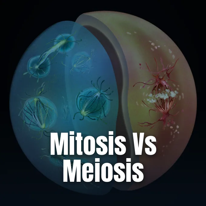
Every living cell needs nuclear division to grow tissue and pass hereditary information to new generations. The core difference in mitosis vs meiosis centers on chromosome counts and genetic mixing: mitosis makes two exact diploid body cells for growth and tissue repair, while meiosis yields four unique haploid sex cells for sexual breeding. Robert Hooke first observed cork cell walls in London in the seventeenth century, and Rudolf Virchow later showed in Berlin that all living cells arise exclusively from pre-existing cells [6]. This division work relies on tiny protein motors that keep genetic blueprints safe.
- How Mitosis vs Meiosis Governs Living Cells
- Why Mitosis Is Called Equational Division
- How Meiosis Halves the Diploid Genome
- Mitosis and Meiosis Follow Ordered Steps
- Genetic Variation Through Crossing Over
- Cell Cycle Checkpoints and Molecular Regulators
- Evolutionary Diversity in Nuclear Division
- Comparing Mitosis vs Meiosis Biological Roles
How Mitosis vs Meiosis Governs Living Cells
An adult human body holds roughly 100 trillion cells that originate from a single fertilized egg cell [3]. Body tissues use mitosis to replace dying cells in blood, intestinal lining, and skin, while germline cells use meiosis to produce eggs and sperm, which cuts 46 chromosomes to 23 and keeps ploidy stable after fertilization [2].
Tissues show varied division rates in different bodily organs. Brain neurons and heart muscle cells stop dividing and rest in a stage called G0, while bone marrow stem cells divide daily [6]. OpenStax biology authors Mary Ann Clark, Matthew Douglas, and Jung Choi explain in Biology 2e that living cells balance mass growth with nuclear splits [1]. The basic rules of eukaryotic cell organization demand this coordination so daughter cells do not shrink after division. Cells shrink if division outpaces growth.
Baker’s yeast, Saccharomyces cerevisiae, finishes a full division cycle in 90 to 120 minutes, while human cells need about 24 hours to double. Interphase occupies approximately 23 hours of this mammalian schedule for growth and DNA copying, while mitosis and cytokinesis conclude in only one hour [6]. In rich culture broth, rod-like Escherichia coli cells divide in 20 minutes by binary fission using divisome rings assembled from FtsZ proteins [9]. Complex eukaryotic cells require far more intricate structural management because they must segregate hundreds of millions of base pairs packaged into multiple discrete chromosomes without tangling, strand breakage, or unequal chromosome loss during cytokinesis. Each chromosome must separate cleanly.

Why Mitosis Is Called Equational Division
Scientists call mitosis equational division because each daughter cell gets an exact duplicate of parental chromosomes. In humans, a diploid cell with 46 chromosomes copies its DNA during the synthetic S phase to form pairs of sister chromatids joined by cohesin proteins [1]. In his research lectures at the University of California, San Francisco, David Morgan showed how spindle fibers guide chromosome separation with extreme accuracy [4]. The cell distributes one chromatid to each pole and preserves 46 chromosomes in each nucleus, ensuring that both daughter cells match the parent cell [2].
Over a normal lifetime, a human body goes through about 10 quadrillion cell divisions to replace shed cells in skin and gut. When skin suffers mechanical abrasion, local stem cells divide by mitosis to heal the gap, while gardeners use plant cuttings to grow whole crops from cloned cells without altering genetic profiles [2].
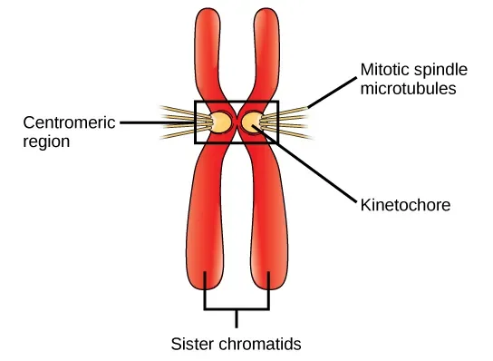
Assembled at the centromere of each chromatid, kinetochores bind microtubules that emerge from opposing centrosomes during prometaphase [1]. Spindle fibers pull both sides until all chromosomes line up on the equatorial plate. If even one kinetochore fails to latch securely onto spindle fibers, biochemical switches halt progression until the defect is repaired, shielding daughter cells from harmful chromosome errors [4]. OpenStax authors Mary Ann Clark and Matthew Douglas note that without this mechanical checkpoint, daughter cells receive unequal chromosome numbers, a condition that can cause cell death or trigger tumors, which explains why cell cycle fidelity remains central to cancer research [3].
How Meiosis Halves the Diploid Genome
Meiosis uses two rounds of division after a single round of DNA copying to turn one diploid cell into four haploid sex cells. In meiosis I, matching partner chromosomes find each other and pair up on the spindle apparatus [2]. The spindle then pulls these paired partners to opposite cell poles while keeping sister chromatids joined at their centromeres. Because this first split cuts chromosome numbers from diploid (2n) to haploid (1n), biologists classify meiosis I as a reductional division.
The second round, meiosis II, proceeds without DNA replication and looks like standard mitosis. Sister chromatids line up and pull apart into separate haploid nuclei [2]. New clues from nematode research into chromosome segregation and fertility show how chromosome mistakes during meiosis lead to failed embryos. Cytokinesis then seals four haploid cells.
If gametes were produced by regular mitosis rather than meiosis, the fusion of egg and sperm would double chromosome counts in every generation, creating genomic instability that would destroy the species. Animals rely on gametic meiosis to generate functional eggs and sperm directly within reproductive organs. Many mosses and ferns use sporic meiosis instead and produce haploid spores that develop into a small leafy plant called a gametophyte before generating sex cells. Both biological pathways maintain stable chromosome counts over deep evolutionary time, proving that sexual reproduction across the planet relies on reductional splits to safeguard species identity and maintain genomic constancy across generations [2].
Mitosis and Meiosis Follow Ordered Steps
Cells run through four core phases: prophase, metaphase, anaphase, and telophase. In mitotic prophase, loose DNA coils into tight visible rods with help from condensin proteins, the nucleolus fades, and centrosomes move to opposite cell sides. Meiotic prophase I takes longer because matching chromosome partners must find each other and form bivalents. The nuclear envelope breaks into tiny vesicles so spindle fibers can reach chromosomes. OpenStax authors Mary Ann Clark and Matthew Douglas highlight how early spindle fibers extend between migrating centrosomes to set cell polarity [1].
In mitotic metaphase, individual chromosomes form a single row on the plate. In meiotic metaphase I, partner pairs sit side by side. In anaphase, cohesin breaks down and spindle fibers pull chromatids to the poles as the dividing cell elongates into an oval shape [1]. Polar microtubules push the poles apart.
New nuclear membranes form around the two chromosome clusters at each end of the cell during telophase, while condensed DNA relaxes back into thin chromatin threads. Spindle fibers dissolve into tubulin building blocks that daughter cells reuse for their own cytoskeletons. Cytokinesis then physically partitions the cytoplasm into two separate cellular bodies. In animal cells, an actin and myosin ring pinches the middle of the membrane to create a cleavage furrow. In plant cells, Golgi vesicles fuse along the equatorial plane to build a sturdy cell plate guided by a specialized microtubule framework called the phragmoplast, which eventually merges with existing cell walls [1].


Genetic Variation Through Crossing Over
Comparing genetic outcomes in mitosis vs meiosis shows how each process treats cellular DNA. Mitosis blocks genetic change so every body cell stays identical to its parent. Meiosis does the opposite by mixing parental genes through two steps: homologous recombination and random chromosome sorting. These steps make every gamete unique and supply the genetic variety that natural selection uses to help species adapt to changing environments across generations [2].
Crossing over happens during prophase I of meiosis when matching partner chromosomes swap reciprocal pieces of DNA. A key enzyme called Spo11 protein cuts both strands of the DNA backbone using a catalytic method similar to topoisomerase. When cellular repair enzymes fix these cuts using the matching partner chromosome as a template, maternal and paternal gene variants become permanently linked into new combinations on a single strand [2].
In meiotic metaphase I, partner chromosome pairs line up by chance, so maternal and paternal chromosomes head to either pole without preset instructions. In human cells with 23 pairs, this independent assortment creates over eight million possible chromosome sets in eggs or sperm. Extra care from protective telomere caps at chromosome ends shields these newly swapped DNA arms from enzyme damage during repeated cycles of division. Due to this dual mixing mechanism of crossover and random assortment, no two human siblings ever receive the exact same genome from their parents, with the sole exception of identical twins [2]. Every gamete carries a fresh genome.
Cell Cycle Checkpoints and Molecular Regulators
Cells move through division under the control of biochemical checkpoints. At the Fred Hutchinson Cancer Research Center in Seattle, Washington, Leland H. Hartwell isolated mutated yeast strains with damaged division genes, discovering over one hundred CDC genes including CDC28, which controls the crucial start step in G1. Hartwell demonstrated that checkpoints pause the cycle when DNA gets damaged, giving cells adequate time to fix broken genes before replication resumes [3].
Working at the Imperial Cancer Research Fund in London, Paul M. Nurse studied fission yeast, Schizosaccharomyces pombe, and identified the cdc2 gene, later cloning human CDK1 to show that cyclin-dependent kinases govern division in all complex life. Timothy Hunt discovered cyclins in sea urchins, Arbacia, while conducting experiments at the Marine Biological Laboratory, proving that cyclins accumulate and degrade periodically to turn kinases on and off. For these foundational discoveries, Hartwell, Nurse, and Hunt shared the 2001 Nobel Prize in Physiology or Medicine from the Karolinska Institutet in Stockholm, Sweden. Their work revealed how CDK enzymes act as cellular engines while cyclins operate as regulatory gearboxes that drive the cell cycle forward [3].
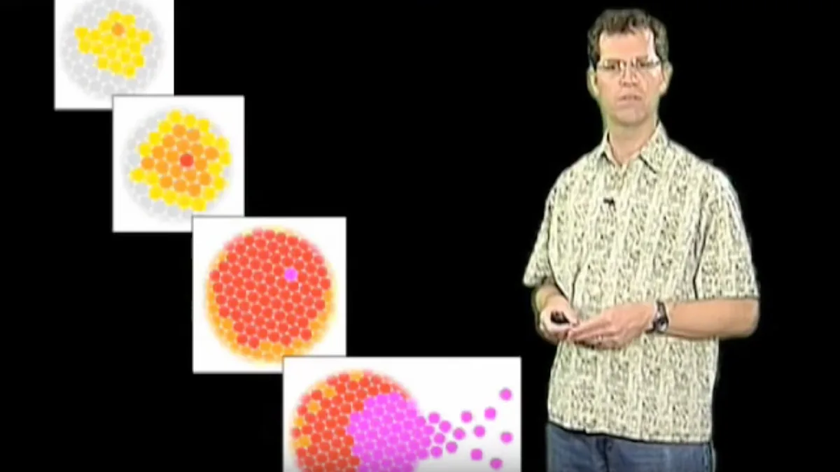
At metaphase, a molecular switch triggers anaphase. David Morgan at the University of California, San Francisco, who completed doctoral research under Richard Roth and postdoctoral work with William Rutter, showed that unattached kinetochores send stop signals that inhibit the anaphase-promoting complex [4]. When chromosomes line up under tension, this brake turns off and allows the cell to destroy securin. Securin degradation frees the enzyme separase, which promptly cuts cohesin rings and lets single chromosomes migrate toward opposite spindle poles [2].
Evolutionary Diversity in Nuclear Division
Eukaryotes divide their nuclei in surprisingly varied ways. In typical animal cells, open mitosis dissolves the outer nuclear membrane during prometaphase so the growing spindle can capture chromosomes. In contrast, most fungi use closed mitosis, keeping their nuclear wall intact while building spindle poles inside the nucleus [7].
Gautam Dey at EMBL Heidelberg and Omaya Dudin at the Swiss Federal Institute of Technology in Lausanne, Switzerland, investigated Ichthyosporea protists with Yannick Schwab and post-doctoral fellow Hiral Shah. Collaborating with Eelco Tromer from the University of Groningen in the Netherlands and Iva Tolic from the Ruđer Bošković Institute in Zagreb, Croatia, they analyzed cell division in two distinct marine species. One species, Sphaeroforma arctica, relies on closed mitosis like fungi during multinucleate life cycle stages. The other species, Corallochytrium perkinsii, uses open mitosis like animal embryos during mononucleate development. Their research in Nature proved that animal-like open mitosis evolved hundreds of millions of years before true animal life appeared on Earth [7].
Packaging vast amounts of genetic material creates substantial cellular challenges. A single human cell holds 1.8 metres of DNA coiled tightly into 46 chromosomes. At EMBL Heidelberg in Germany, Jan Ellenberg and Felipe Mora-Bermúdez found that chromosomes undergo super-condensation during late anaphase under control of Aurora kinase. This extra shortening prevents trailing chromosome arms from being severed when the cleavage furrow divides the cell body in two, safeguarding genomic structure [5].
Comparing Mitosis vs Meiosis Biological Roles
Looking at the biological roles of mitosis vs meiosis highlights how each division path meets different life needs. Mitosis happens in somatic body cells throughout life, making two exact diploid cells from one parent. This process preserves gene copies for body growth, tissue renewal, and wound healing. Meiosis happens only in germline cells, running two divisions to produce four varied haploid gametes for sexual breeding [2].
Mitosis has one split with no partner pairing and no crossing over. Meiosis requires homologous partner pairing and crossing over in prophase I to generate novel gene arrangements. Chromatids split in mitotic anaphase, while meiosis separates partner chromosomes in anaphase I and sister chromatids only during anaphase II [2].
Cell growth requires biomolecule synthesis driven by Myc transcription factors and the TOR kinase pathway, which activates ribosomal S6K and translation factor eIF4E to promote cell mass enlargement. When nutrient supplies drop, cells pause division and slip into resting G0 states [8]. Partitioning during cytokinesis also distributes low-abundance cellular molecules by chance. Mathematical modeling by D. Huh and J. Paulsson in the Proceedings of the National Academy of Sciences showed that random segregation of low-copy RNAs causes significant partitioning variations between daughter cells [10]. By balancing growth with chromosome distribution through mitosis vs meiosis, living cells keep bodies healthy while passing fresh gene combinations to the next generation [2].
- BOOK Clark, M. A., Douglas, M., & Choi, J. (2018). 10.2 The Cell Cycle. Biology 2e. OpenStax. [Article Link]
- WEBSITE Wikipedia contributors. Cell division. Wikipedia, The Free Encyclopedia. [Article Link]
- PRESS RELEASE The Nobel Assembly at Karolinska Institutet. (2001). The Nobel Prize in Physiology or Medicine 2001. NobelPrize.org. [Article Link]
- WEBSITE Morgan, D. (2020). Controlling the Cell Cycle. iBiology. [Article Link]
- ONLINE NEWS European Molecular Biology Laboratory. (2007). Researchers Shed Light On Shrinking Of Chromosomes. ScienceDaily. [Article Link]
- ONLINE NEWS Davidsecko. (2006). The Cell Cycle: A Universal Cellular Division Program. The Science Creative Quarterly. [Article Link]
- ONLINE NEWS Ghosh, S. (2024). Exploring diversity in cell division. European Molecular Biology Laboratory News. [Article Link]
- WEBSITE Wikipedia contributors. Cell growth. Wikipedia, The Free Encyclopedia. [Article Link]
- ACADEMIC JOURNAL Egan, A. J. F., & Vollmer, W. (2012). The physiology of bacterial cell division. Annals of the New York Academy of Sciences, 1277(1), 8-28. [Article Link]
- ACADEMIC JOURNAL Huh, D., & Paulsson, J. (2011). Random partitioning of molecules at cell division. Proceedings of the National Academy of Sciences, 108(36), 15004-15009. [Article Link]


