





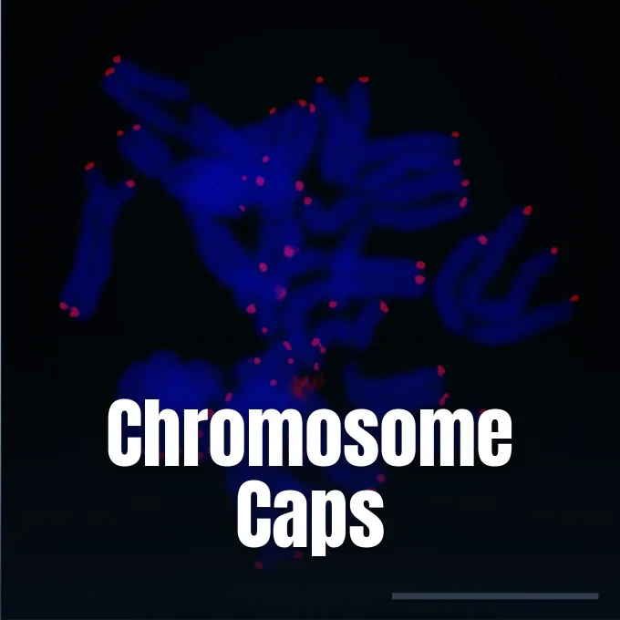
Every time a human cell prepares to divide, it faces a physical barrier built into its genetic copying machinery. DNA polymerases can copy long genetic strands with high fidelity, but they cannot duplicate the terminal tips of linear chromosomes due to the unidirectional nature of replication. If left unprotected, chromosomes would lose functional genes with each reproductive cycle or fuse together into destructive combinations that trigger rapid cellular death. This biological puzzle explains what are telomeres: repetitive nucleotide sequences bound by protective proteins that cap chromosome ends like the plastic tips on shoelaces. They act as disposable buffers that absorb inevitable copying erosion, shielding vital genetic information while establishing a finite counting mechanism for cellular lifespan [6].
- What Are Telomeres in Human Cells?
- Why Telomeres Shorten During DNA Replication
- How the Shelterin Complex Protects Telomeric DNA
- Telomerase Activity in Dividing Cells
- Oxidative Stress Triggers for Senescence
- Telomere Length Dynamics Across Early Childhood
- Lifestyle Influence on Biological Aging
- Cancer Immortality and Targeted Therapies
What Are Telomeres in Human Cells?
In human chromosomes, telomeres consist of repetitive hexanucleotide strings reading TTAGGG repeated thousands of times along the outer edge of each linear strand. Geneticists Hermann Joseph Muller and Barbara McClintock first deduced their existence during the late 1930s while studying irradiated chromosomes in fruit flies and maize plants. Muller coined the name from the Greek telos, meaning end, and meros, meaning part. He observed that intact natural chromosome ends possessed unique physical properties that prevented them from sticking together or forming abnormal ring chromosomes during division. The terminal cap preserves the linear identity of each chromosome so that cellular repair pathways do not mistake natural tips for broken fragments [1].
These repetitive sequences do not encode functional proteins. Instead, their duty lies in preserving structural stability so the genome stays intact during nuclear division. When chromosomes break internally, cellular repair systems fuse the exposed ends together. Telomeres block this reaction at chromosome tips, preventing catastrophic end-to-end fusions [1].
Telomere length varies widely across the tree of life, as documented in the TeloBase database compiled by researchers at Masaryk University [7]. While single-celled yeasts carry short arrays of roughly 300 base pairs, human somatic cells begin life with telomeres spanning 10 to 15 kilobases. At the outermost tip of every human telomere, the guanine-rich strand extends beyond its complement, creating a single-stranded 3′ overhang between 75 and 300 nucleotides long that burrows back into the duplex DNA [9].

Why Telomeres Shorten During DNA Replication
The progressive shortening of chromosome ends is rooted in the fundamental mechanics of semiconservative DNA replication. DNA polymerases synthesize new strands exclusively in a 5′ to 3′ direction and require a pre-existing RNA primer to begin synthesis. While the leading strand copies smoothly to the chromosome terminus, the lagging strand relies on discontinuous Okazaki fragments, each initiated by its own short RNA primer. When the replication fork reaches the physical boundary of the chromosome, the final RNA primer on the lagging strand gets removed by exonucleases, but no upstream template exists to allow DNA polymerase to fill the resulting gap [1]. Consequently, each cell division leaves a short uncopied segment at the terminal end.
In the early 1970s, Soviet theorist Alexey Olovnikov recognized this end-replication problem and realized its connection to limited cellular lifespan [5]. He built upon the discoveries of Leonard Hayflick, who demonstrated in 1961 that normal human cells can divide only 40 to 60 times before halting. Olovnikov formulated his marginotomy theory, predicting that chromosomes lose terminal sequences until reaching a threshold where division ceases, with cultured human cells dropping roughly 50 to 100 base pairs per division [9].
Once telomeres reach a short boundary, the protective cap collapses. Cellular surveillance machinery recognizes the exposed tip as an unrepairable double-strand break, triggering permanent cell cycle arrest known as the Hayflick limit [1]. The senescent cell remains metabolically active, yet it permanently loses the capacity to replicate [10].
How the Shelterin Complex Protects Telomeric DNA
Telomeric DNA does not float unprotected inside the nucleus; it relies on a six-protein mantle known as the shelterin complex. Identified in human cells as TRF1, TRF2, TIN2, POT1, TPP1, and RAP1, these protective proteins form a physical barrier that prevents DNA repair enzymes from attacking chromosome ends. TRF1 and TRF2 bind double-stranded TTAGGG repeats directly, while POT1 binds the single-stranded 3′ overhang to coordinate end capping [9].
The shelterin complex facilitates the structural remodeling of the chromosome terminus into a protective loop known as the T-loop. Here, the single-stranded 3′ overhang curves backward and physically invades the double-stranded telomeric tract, displacing one strand to generate a triple-stranded displacement loop, or D-loop [9]. This lasso configuration tucks the exposed terminal sequence away from the surveillance mechanisms of ATM and ATR kinases, suppressing the accidental initiation of non-homologous end joining or homologous recombination. If components like TRF2 become disrupted, chromosome ends undergo widespread fusions that tear chromosomes apart during mitosis [1].

Beyond the T-loop, guanine-dense telomeric repeats can also fold into four-stranded structures known as G-quadruplexes. These non-canonical DNA arrangements provide extra protection against nucleolytic degradation. However, they also create physical obstacles that replication enzymes must unwind to prevent replication forks from stalling [9].
Telomerase Activity in Dividing Cells
If cells possessed no countermeasure against continuous shortening, all linear genomes would vanish over evolutionary time. In 1978, molecular biologists Elizabeth Blackburn and Joseph Gall examined the ciliated protozoan Tetrahymena thermophila, uncovering tandem hexanucleotide repeats at its chromosome ends. In 1982, Blackburn and Jack Szostak demonstrated that these Tetrahymena sequences protected linear minichromosomes from enzymatic degradation inside yeast, proving that telomere stability relies on conserved biological mechanisms that bridge evolutionary kingdoms [6].
On Christmas Day in 1984, doctoral student Carol Greider and Blackburn detected the enzymatic activity responsible for building these terminal repeats de novo. They isolated and named the enzyme telomerase, demonstrating that it functions as a ribonucleoprotein reverse transcriptase carrying its own non-coding RNA template [6]. The internal RNA component provides the complementary pattern needed to synthesize telomeric repeats onto chromosome ends without requiring an external DNA primer [1]. Szostak showed that yeast lacking telomerase suffered progressive telomere shortening and eventual growth arrest, confirming the enzyme’s central role in maintaining replicative immortality. For these discoveries, Blackburn, Greider, and Szostak received the 2009 Nobel Prize in Physiology or Medicine [6].
In humans, telomerase remains repressed in most somatic tissues, functioning only within germline cells, embryonic stem cells, and select proliferating lymphocytes [9]. Somatic cells lack sufficient telomerase to counteract replication losses, ensuring that progressive attrition acts as a built-in timer for cellular lifespan [1].



Oxidative Stress Triggers for Senescence
While the end-replication problem accounts for progressive erosion, oxidative damage can trigger telomere dysfunction far more rapidly than normal cell division. Because guanine possesses the lowest oxidation potential among nucleic acid bases, the TTAGGG repeats of telomeric DNA are particularly susceptible to oxidation. Reactive oxygen species produced by cellular respiration and inflammation readily generate 8-oxo-guanine (8oxoG) lesions across chromosome ends, creating structural vulnerabilities within the genome [2].
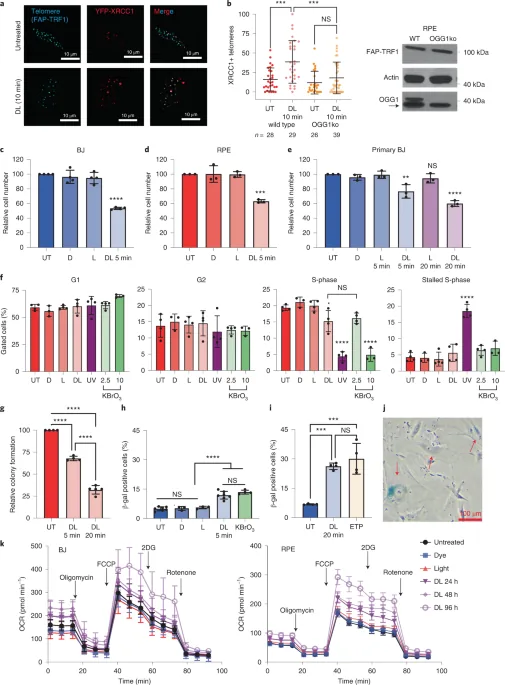
A 2022 investigation led by Ryan Barnes and colleagues at the University of Pittsburgh and published in Nature Structural & Molecular Biology examined this mechanism directly using a precision chemoptogenetic tool. By activating a fluorogen-activating peptide targeted to TRF1 with far-red light, they produced singlet oxygen and 8-oxo-guanine lesions exclusively at telomeres in non-diseased human fibroblasts and epithelial cells. A single five-minute induction proved sufficient to trigger permanent cell cycle arrest and multiple hallmarks of p53-dependent senescence within four days, even though telomeres make up roughly 0.025% of the total human genome. Crucially, this rapid senescence occurred without any physical shortening of telomeres or loss of shelterin proteins, driven instead by acute replication fork stalling and localized DNA damage signaling [2].
Cells exposed to telomeric 8-oxo-guanine developed cytoplasmic chromatin fragments that activated the cGAS immune sensor, inducing a senescence-associated secretory phenotype. This work revealed that oxidative base lesions can trigger cellular aging directly through replication stress, disproving the notion that telomeres influence senescence solely through physical shortening [2].
Telomere Length Dynamics Across Early Childhood
Telomere shortening does not proceed at a constant, linear pace across a person’s lifespan. Pediatric research conducted by the Columbia Center for Children’s Environmental Health at Columbia University Mailman School of Public Health demonstrates that the fastest telomere loss occurs during the first three years of life. The study, published in Psychoneuroendocrinology, followed 224 children from birth through age 9 by measuring leukocyte telomeres isolated from umbilical cord blood and serial blood draws [4].
Led by senior author Julie Herbstman and first author Whitney Cowell, the researchers found that telomere attrition is steepest between birth and age 3, before entering a stabilizing plateau leading up to puberty. The team observed that maternal telomere length measured at delivery predicts newborn telomere length, which in turn correlates with the child’s telomere measurements into pre-adolescence. Herbstman said that this early attrition window makes telomeres especially sensitive to external influences, including environmental pollutants and air quality, potentially shaping lifetime health trajectories. Curiously, several children in the cohort exhibited temporary telomere lengthening, a phenomenon also observed in select animal models [4].
Technological advances have revealed even greater nuance in chromosomal dynamics. A 2024 study published in Science by Carol Greider evaluated 147 individuals using a method called Telomere Profiling, showing that specific chromosome ends maintain distinct lengths set at birth. Rather than existing as uniform caps, telomeres on individual chromosome arms shorten at predictable, end-specific rates throughout life [8].
Lifestyle Influence on Biological Aging
Because leukocyte telomere length reflects cumulative biological wear rather than chronological time, public health researchers have examined whether lifestyle habits influence chromosomal aging. A major 2025 study published in Neurology investigated 356,173 participants from the UK Biobank with an average age of 56. The research team, led by Tamara Kimball of Massachusetts General Hospital, analyzed how leukocyte telomere length and the McCance Brain Care Score interact to influence the long-term risk of age-related brain diseases like stroke and dementia [8].
The UK Biobank analysis revealed that individuals with both short telomeres and low Brain Care Scores experienced significantly higher incidence of stroke and dementia over time. However, participants who possessed short telomeres but maintained high Brain Care Scores, reflecting healthy diet, regular exercise, and minimal alcohol intake, did not show a higher risk for these cognitive conditions. Kimball said that adopting healthy lifestyle choices can mitigate the risks associated with shorter telomeres, proving that biological aging remains modifiable even when cells show signs of advanced wear. As medical director Joshua Berkowitz said, telomere length alone is not a definitive predictor of longevity, as environmental factors and metabolic health play key roles [8].

Everyday behaviors show measurable correlations with telomere preservation. Studies indicate that walking roughly 8,000 steps daily reduces all-cause mortality by 51% compared to walking 4,000 steps, while nutrient-rich regimens like the Mediterranean diet help shield cells against systemic inflammation. Genetic engineer Sebnem Unluisler of the London Regenerative Institute explained that while shorter telomeres track cellular aging, lifestyle interventions support the body’s overall regenerative balance [8].
Cancer Immortality and Targeted Therapies
The finite lifespan dictated by telomere shortening serves as a protective safeguard against cancer by halting cells before they acquire dangerous mutational loads. When normal cells exhaust their telomeres, replicative senescence stops further division permanently [1]. In 1989, molecular biologist Gregg Morin discovered telomerase activity in human cancer cells, revealing how malignant lineages bypass this proliferative ceiling. Today, scientists know that telomerase becomes reactivated in over 90% of human cancers, allowing malignant clones to maintain their chromosome caps indefinitely and achieve cellular immortality [5].
However, having exceptionally long telomeres carries its own severe trade-offs. Research led by oncologist Mary Armanios at Johns Hopkins University demonstrated that individuals carrying inherited mutations in the POT1 gene develop unusually long telomeres, yet face higher risks for melanoma and lymphomas. Because their telomeres fail to shorten normally, aging cells carrying accumulated somatic mutations avoid senescence and continue dividing, creating fertile ground for malignant transformation. Armanios said that telomere length reflects an evolutionary balance: short telomeres drive degenerative organ failure and tissue exhaustion, while excessively long telomeres increase susceptibility to cancer by permitting damaged cells to survive [8].
To exploit the cancer dependency on telomere maintenance, researchers are pursuing targeted telomerase inhibitors such as GRN163L, a lipid-modified oligonucleotide that binds the enzyme’s RNA template to block elongation. In 1994, Jerry Shay and Woodring Wright introduced the Telomeric Repeat Amplification Protocol (TRAP) to quantify telomerase activity in clinical samples. Because telomerase inhibitors require several weeks of cell division to erode telomeres down to critical thresholds, oncologists are testing them alongside standard therapies to prevent tumor recurrence [5].
- ACADEMIC JOURNAL Varela, E., & Blasco, M. A. (2010). 2009 Nobel Prize in Physiology or Medicine: telomeres and telomerase. Oncogene, 29(11), 1561-1565. [Article Link]
- ACADEMIC JOURNAL Barnes, R. P., De Rosa, M., Thosar, S. A., Detwiler, A. C., Roginskaya, V., Van Houten, B., Bruchez, M. P., Stewart-Ornstein, J., & Opresko, P. L. (2022). Telomeric 8-oxo-guanine drives rapid premature senescence in the absence of telomere shortening. Nature Structural & Molecular Biology, 29(7), 667-677. [Article Link]
- WEBSITE Genetic Science Learning Center. (2026). Are Telomeres the Key to Aging and Cancer? Learn.Genetics, University of Utah. [Article Link]
- PRESS RELEASE Columbia University Mailman School of Public Health. (2021). Telomere Length, a Longevity Marker, May Be Determined Early in Life. [Article Link]
- WEBSITE Olovnikov Lab. (2009). Library Index: Telomeres and Telomerase: From Breakthrough Discovery to Clinical Revolution. [Article Link]
- PRESS RELEASE Nobel Media AB. (2009). The Nobel Prize in Physiology or Medicine 2009: How Chromosomes Are Protected by Telomeres and the Enzyme Telomerase. [Article Link]
- ACADEMIC JOURNAL Lyčka, M., Bubeník, M., Závodník, M., Peska, V., Fajkus, P., Demko, M., Fajkus, J., & Fojtová, M. (2024). TeloBase: a community-curated database of telomere sequences across the tree of life. Nucleic Acids Research, 52(D1), D311-D321. [Article Link]
- ONLINE NEWS Lang, K. (2024). Are telomeres really the key to living longer, youthful lives? Medical News Today. [Article Link]
- WEBSITE Wikipedia contributors. (2026). Telomere. Wikipedia, The Free Encyclopedia. [Article Link]
- WEBSITE Wikipedia contributors. (2026). Relationship between telomeres and longevity. Wikipedia, The Free Encyclopedia. [Article Link]
- WEBSITE Wikipedia contributors. (2026). Mega-telomere. Wikipedia, The Free Encyclopedia. [Article Link]
- WEBSITE Central European Institute of Technology. (2022). Chromatin Molecular Complexes: Research Group of Jiri Fajkus. [Article Link]


