



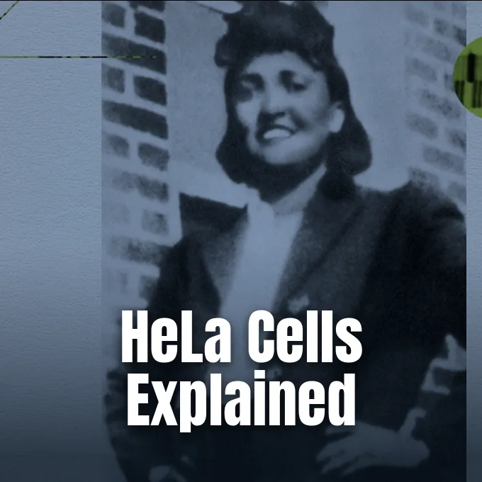
HeLa cells represent the oldest and most widely used continuous human cell line in biological science. In early 1951, doctors in Baltimore collected cervical tissue from a young Black tobacco farmer named Henrietta Lacks without her knowledge or consent during treatment for a malignant tumor [3]. Most human tissue samples die in glass dishes within days, but these tumor cells multiplied indefinitely. With HeLa cells explained through seven decades of laboratory study, this durable line became a foundation for polio vaccines, gene mapping, and modern medical standards.
The 1951 Discovery in Baltimore
In January 1951, thirty-year-old Henrietta Lacks felt severe abdominal pain and noticed unusual spotting several months after giving birth to her fifth child in Virginia. She sought medical treatment at Johns Hopkins Hospital in Baltimore, which stood as one of the few Maryland facilities that treated Black patients during an era of strict racial segregation [3]. Gynecologist Howard Jones Jr. examined her cervix and diagnosed a malignant adenocarcinoma that had developed with uncommon speed [8]. To understand how this line developed, one must examine the clinical origin of HeLa cells explained through early oncology records in Baltimore.
During her first radium treatment, surgeon Lawrence Wharton Jr. cut small tissue samples from her cervix without her knowledge, following standard hospital procedures of that era [8]. Wharton sent the fresh biopsy to George Otto Gey, head of tissue culture research at Johns Hopkins University, who had spent decades attempting to grow human cells in test tubes without success [3]. Gey handed the tissue to his assistant Mary Kubicek. She placed the cellular fragments into glass tubes filled with nutrient broth and loaded them onto a mechanical roller-tube apparatus designed to keep liquid culture moving. Earlier tissue specimens from surgical patients had always died within days, but these cells doubled in number every 20 to 24 hours [8].
Kubicek labeled the glass culture tubes using the first two letters of the patient’s first and last names to produce the designation HeLa [8]. The malignant cancer spread quickly through Henrietta’s body despite repeated radium treatments, and she died on October 4, 1951, at age 31 [3]. Gey freely shared samples of her surviving cells with research teams across North America to advance human cellular biology [8].
Why the HeLa Cell Line Never Dies
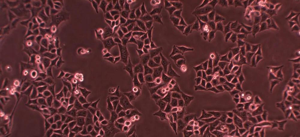
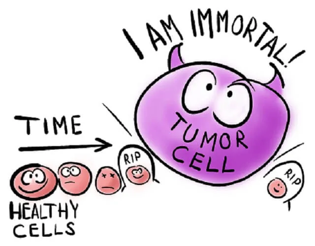
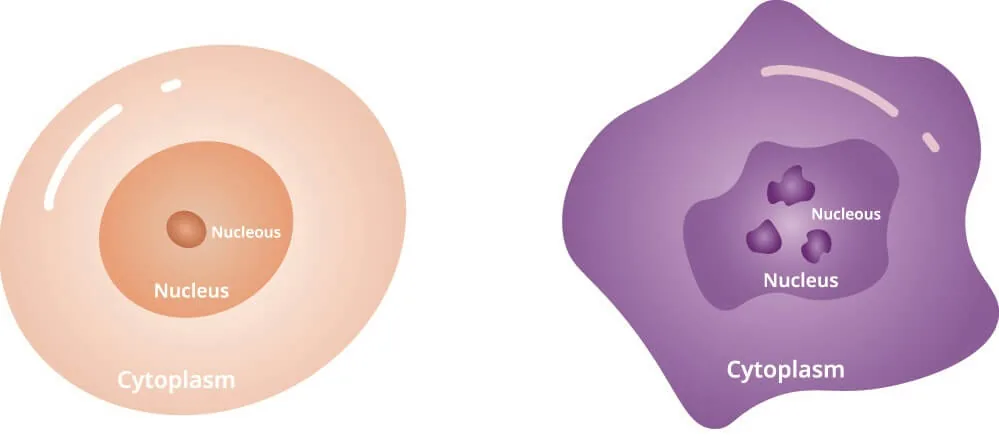
Healthy human cells follow clear biological timers that limit how many times they can divide in a dish. Normal somatic cells maintain forty-six chromosomes and eventually enter senescence, stopping all replication when their genetic limits arrive. Cancer cells break these internal rules. Gene mutations disable natural cell cycle checkpoints. These mutations let the cells divide continuously as long as laboratory technicians supply basic nutrients [2].
HeLa cells differ markedly from normal human tissue because they possess an unstable, mutated genome with between 76 and 80 chromosomes, frequently showing 82 chromosomes in detailed karyotype counts [7]. This massive chromosomal abnormality stemmed in part from infection by human papillomavirus (HPV-18), which integrated viral genes into Henrietta’s cellular DNA and knocked out natural tumor suppressor mechanisms. The viral proteins prevented the host cell from repairing damaged genetic code. As a result, mutations accumulated unchecked during every division cycle [8]. Syphilis had also weakened her immune system before she developed cancer, permitting the malignant cells to proliferate with unusual speed [3].
Why do these abnormal cells survive without end in laboratory culture? The secret rests in the protective caps that sit at the ends of chromosomes. In normal human tissue, these caps wear down with each cellular division until the cell can no longer reproduce. HeLa cells escape this fate by repairing the caps after every cycle. This constant repair keeps their genetic code stable indefinitely [2].
Telomerase Action in Cancer Tissue
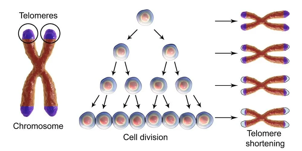
Telomeres are repetitive non-coding DNA sequences positioned at the ends of chromosome arms, functioning like plastic aglets that prevent shoelaces from fraying. They shield core genes against chemical damage caused by reactive oxygen species during cellular metabolism. Every time a normal cell divides, its copying machinery fails to reach the absolute tip of the DNA strand, causing telomeres to shorten slightly. After roughly fifty divisions, this loss stops further growth [2]. The cellular longevity of HeLa cells explained at the molecular level centers on the mechanics of chromosome maintenance.
HeLa cells overcome this natural limit by producing an active enzyme called telomerase, which rebuilds the missing telomere sequences after each division cycle. Telomerase consists of a protein reverse transcriptase component known as TERT and an accompanying RNA template designated TERC. Healthy adult cells keep the TERT gene switched off, causing somatic tissues to age and eventually die. In malignant HeLa cells, genetic mutations keep both TERT and TERC continuously active. The active enzyme adds fresh protective sequences to chromosome ends after every round of DNA replication. This constant repair preserves chromosome integrity. The cell line divides through thousands of generations without entering cellular senescence [2].
Curiously, HeLa cells do not carry the longest telomeres in laboratory biology. Lines such as U2OS, derived from a teenage patient with osteosarcoma, have much longer caps. What matters for immortality is not total telomere length, but steady telomerase activity [2]. Discoveries linking telomerase to cellular aging and cancer progression earned researchers a Nobel Prize in Physiology or Medicine in 2009 [3, 4].
Vaccine Research with HeLa Cells Explained
During the early 1950s, epidemic poliomyelitis paralyzed tens of thousands of children in North America and Europe, creating an urgent medical push to evaluate candidate vaccines. Jonas Salk developed an inactivated polio vaccine, but testing its safety and potency required massive quantities of human cells. Animal tissues were expensive, difficult to maintain, and produced variable results. HeLa cells provided researchers with a uniform and fast-growing testing ground [8].
To mass-produce cells for vaccine testing, the National Foundation for Infantile Paralysis (NFIP) established a major culture facility at Tuskegee University in Alabama in the spring of 1953. Black scientists, technicians, and staff at the Tuskegee laboratory produced trillions of HeLa cells each week. Staff packaged the cells in sturdy glass tubes and shipped them to laboratories across the country. Poliovirus easily infected HeLa cells, causing visible cell breakdown that allowed technicians to measure virus neutralization with extreme accuracy. The facility proved that human tissue cultures could be standardized for commercial pharmaceutical testing [8].
The availability of mass-produced HeLa cells allowed Salk to begin human clinical trials within a year. The vaccine gained nationwide approval in 1955. Having HeLa cells explained and standardized across research centers removed the need to kill thousands of monkeys for polio tests. This production system laid the technical foundation for modern virology and cell-based drug screening [8].

Role in Virology and Gene Mapping
In 1955, biophysicists Theodore Puck and Philip I. Marcus at the University of Colorado in Denver developed a technique to clone individual HeLa cells, growing uniform single-cell colonies in laboratory dishes for the first time [5, 8]. This cloning method enabled researchers to study genetically uniform lines rather than mixed cell populations, opening the door to systematic human cytogenetics and somatic cell genetics. Puck and Marcus followed this breakthrough in 1956 by measuring how X-rays damage mammalian cells, showing that human tissues are far more sensitive to radiation than single-celled bacteria [4, 5]. Their quantitative survival curves established that doses as low as 96 roentgens cut cell survival to 37 percent, providing fundamental safety data that guided clinical radiation therapy [5].
Virologists at Columbia University, including Professor Vincent Racaniello, later used HeLa cultures to study infectious diseases such as polio and influenza. Molecular biologist Richard Axel engineered HeLa cells by inserting the gene for the human CD4 protein. This modification let the human immunodeficiency virus (HIV) bind to and infect the cells. This genetic modification allowed scientists to study how HIV infects human targets, providing a stable laboratory platform to screen antiretroviral medications [8]. Giannella and colleagues also used HeLa cultures to demonstrate how Salmonella typhimurium bacteria penetrate human epithelial layers during intestinal infections [10].
In the mid-1980s, German researcher Harald zur Hausen analyzed HeLa cells and discovered integrated DNA sequences from human papillomavirus strain 18. His work confirmed that HPV infection triggers cervical cancer, earning zur Hausen a Nobel Prize in Physiology or Medicine. His findings led directly to HPV diagnostic tests and preventative vaccines. In low- and middle-income nations, the HPV vaccine prevents an estimated 11.24 deaths per 1,000 vaccinated people [8].
Cross-Contamination Risks in the HeLa Cell Line
Because HeLa cells multiply so aggressively and endure harsh conditions, they represent an notorious source of laboratory contamination. Microscopic cells can float through air currents, cling to unwashed pipettes, or transfer on technician gloves to colonize adjacent dishes [8]. In 1974, cytogeneticist Walter Nelson-Rees alerted the scientific world that HeLa had silently contaminated dozens of supposedly distinct cell lines across global repositories [7].
Nelson-Rees used karyotype analysis to prove that cell lines cataloged as breast, prostate, or liver tissue actually carried the telltale genetic markers and 82-chromosome count of HeLa. Later surveys confirmed that 10 to 20 percent of all cell lines maintained in international biological repositories had been overgrown by HeLa cells. This hidden contamination compromised thousands of published studies and wasted millions of research dollars, because scientists spent decades studying what they believed were specific tissue models when they were actually experimenting on cervical cancer. The controversy forced laboratories worldwide to adopt routine cell authentication protocols and genetic fingerprinting standards [7].
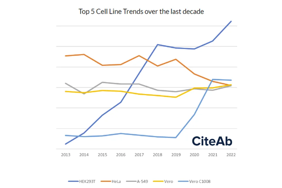
These persistent contamination risks and new biological tools gradually shifted laboratory preferences toward alternative human cell models. A 2022 analysis by Rhys Bowen Morris at CiteAb revealed that the embryonic kidney line HEK293T took over first place in 2018, holding a 6.22 percent citation share. The popular line from Henrietta dropped to fourth place overall. Meanwhile, the monkey kidney line Vero C1008, derived from a line established in 1962 by Yoshihiro Yasumura at Chiba University, rose to second place with a 3.36 percent share, fueled by heavy use in coronavirus studies during the COVID-19 pandemic [6].
Henrietta Lacks and the Ethics of Consent
While pharmaceutical firms and laboratory supply companies sold HeLa cultures around the globe for large profits, Henrietta’s family lived in complete ignorance of her medical legacy [3, 4]. Henrietta was buried in an unmarked grave in Clover, Virginia, leaving behind five young children who grew up in deep poverty without access to regular medical care. Her husband, Day Lacks, and her children only learned that her cells were alive in 1973, when researchers from Johns Hopkins contacted them seeking blood samples. The scientists wanted DNA from the children to map genetic markers that could help identify HeLa contamination in other laboratory cultures [3, 8].
Because researchers failed to explain the purpose of the blood tests in clear language, the family believed they were being tested for the cancer that killed Henrietta. Family member Bobbette Lacks questioned why the research had been kept secret for more than two decades [3]. Henrietta’s daughter Deborah Lacks spent years seeking answers about her mother’s life and tissue. The communication gap between elite academic researchers and the Black working-class family caused lasting distress, compounding historical distrust toward medical institutions [1, 3].
In 1976, journalist Michael Rogers published Henrietta’s true identity in Rolling Stone, dispelling the false aliases Helen Lane and Helen Larson that had obscured her name for two decades [3, 8]. Journalist Rebecca Skloot later partnered with Deborah Lacks to document the full family history, publishing The Immortal Life of Henrietta Lacks in 2010 [1, 3]. Her grandson Alfred Carter Jr. spoke at a panel hosted by Stanford University, the Stanford Storytelling Project, and the Medicine and the Muse Program, saying: “Just keep in mind that these are human beings that you are dealing with [1].”
Rules for Modern Biospecimen Research
The legal questions surrounding human tissue reached a major test in 1990 in Moore v. Regents of the University of California. John Moore sued his physician after discovering that cells removed during his leukemia treatment were patented and commercialized without his consent. The California Supreme Court ruled that patients do not hold property rights over excised biological samples once removed from their bodies. The decision allowed commercial firms to develop profitable cell products freely [8].
In response to growing public concern over patient autonomy, federal agencies enacted the Common Rule in 1991, requiring written informed consent for federally funded human subject research and establishing institutional review boards. The ethical framework shifted again in 2013, when European researchers published the whole genome sequence of a HeLa cell line without consulting Henrietta’s descendants [1, 8]. Following objections from the family, the National Institutes of Health (NIH) negotiated an accord with Lacks family representatives. Under this policy, a panel including granddaughter Jeri Lacks-Whye and Stanford professor Russ Altman evaluates all research requests seeking access to Henrietta’s genetic sequence. The policy ensures family oversight and protects donor privacy [1].
Legal accountability reached a turning point in August 2023, when Henrietta’s estate settled a federal lawsuit against Thermo Fisher Scientific for selling HeLa cells without family permission [3, 8]. The estate reached another settlement with Novartis in 2026, while cases against Ultragenyx and Viatris continue [8]. Bioethics professor Sean Valles at Michigan State University said that non-consensual tissue harvesting historically placed heavy burdens on Black patients. Dr. Maranda C. Ward of George Washington University pointed out that informed consent must protect bodily autonomy and clinical integrity [3]. With the historical lessons of HeLa cells explained in university bioethics courses around the world, modern medical programs teach patient consent as an indispensable requirement.
- ONLINE NEWS White, T. (2018). Descendants of Henrietta Lacks discuss her famous cell line. Stanford Medicine News Center. [Article Link]
- WEBSITE Knapp, S. (2021). HeLa Cells – The Definitive Guide. Biology Dictionary. [Article Link]
- ONLINE NEWS Lang, K. (2022). The story of Henrietta Lacks and the uniqueness of HeLa cells. Medical News Today. [Article Link]
- ONLINE NEWS Starkman, E. (2025). What Are HeLa Cells? WebMD. [Article Link]
- ACADEMIC JOURNAL Puck, T. T., & Marcus, P. I. (1956). Action of x-rays on mammalian cells. The Journal of Experimental Medicine, 103(5), 653-666. [Article Link]
- WEBSITE Morris, R. B. (2023). What were the top 100 cell lines of 2022? CiteAb Blog. [Article Link]
- WEBSITE HeLa Cell Line. (2023). Origins and Cytogenetics of the HeLa Cell Line. [Article Link]
- WEBSITE Wikipedia contributors. (2026). HeLa. Wikipedia, The Free Encyclopedia. [Article Link]
- ACADEMIC JOURNAL Beskow, L. M. (2016). Lessons from HeLa Cells: The Ethics and Policy of Biospecimens. Annual Review of Genomics and Human Genetics, 17(1), 395-417. [Article Link]
- ACADEMIC JOURNAL Giannella, R. A., Washington, O., Gemski, P., & Formal, S. B. (1973). Invasion of HeLa cells by Salmonella typhimurium: a model for study of invasiveness of Salmonella. The Journal of Infectious Diseases, 128(1), 69-75. [Article Link]


