



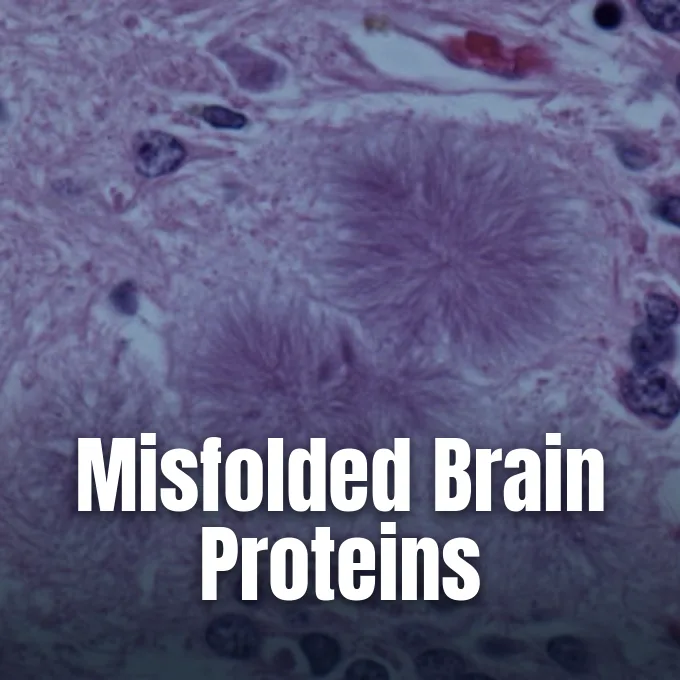
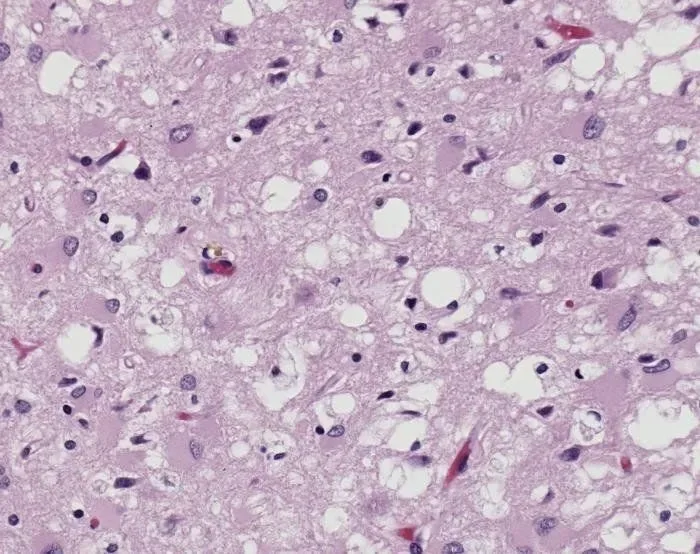
Biologists studying what are prions encounter an agent that completely lacks genetic material like DNA or RNA. These lethal agents are deformed shapes of a normal cell protein, known simply as PrP, found abundantly on the outer surface of nerve cells throughout mammalian brains. When a deformed particle touches a healthy copy, it forces the normal protein to fold into the same deadly shape. This self-driven chain reaction turns healthy brain units into clumps of amyloid fibers, leaving tiny sponge-like holes in brain tissue that lead inexorably to death [10].
- What Are Prions?
- Discovery of the Protein-Only Pathogen
- Structural Shift From Alpha Helices to Beta Sheets
- Mechanisms of Misfolding and Chain Replication
- Major Forms of Prion Disease in Humans
- Why Are Prions So Hard to Destroy?
- Similar Aggregation Patterns in Other Disorders
- Clinical Detection and Experimental Interventions
What Are Prions?
The word prion comes from proteinaceous infectious particle, a label that sets it apart from every other known pathogen in medicine. In healthy humans and animals, the normal cellular prion protein, called PrPC, sits on the outer membrane of neurons, where it binds copper ions and helps brain cells communicate with each other. The rogue form, named PrPSc after scrapie in sheep, shares the exact same string of amino acids as the healthy protein. The sole difference lies in how that string folds in three dimensions [13].
Because the pathogen is an altered shape of the host’s own cellular protein, the immune system mounts no defense against it. White blood cells make no antibodies. No fever occurs. Standard cooking heat cannot destroy it [7].
Human cells express this protein heavily within the central nervous system, which explains why clinical symptoms emerge as sudden memory loss, unsteady walking, and bodily tremors. The normal protein is encoded by the PRNP gene on human chromosome 20. When that gene carries an inherited fault, or when a person eats meat tainted with rogue animal tissue, the lethal chain begins. Sticky amyloid plaques pile up and kill neighboring neurons [12].

Discovery of the Protein-Only Pathogen
The medical riddle began in eighteenth-century Europe with scrapie, a fatal disease that caused sheep to rub their fleece raw against fences and stagger across pastures. For two centuries, researchers failed to find any culprit virus or bacterium in sheep brain extracts. In the 1950s, doctors observed an identical brain illness called kuru among the Fore people of Papua New Guinea [10].
Working at the National Institutes of Health, American physician Carleton Gajdusek showed in the 1960s that kuru and scrapie could jump to chimpanzees after injections of brain tissue from dead patients. This work proved the disease was transmissible and brought Gajdusek the 1976 Nobel Prize in Physiology or Medicine. However, the mystery deepened when lab tests showed the pathogen survived heavy doses of ultraviolet rays and ionizing radiation that destroy all DNA and RNA. In 1967, British mathematician J. S. Griffith proposed that an agent made purely of protein could copy itself without genes, but the medical establishment viewed the idea as heresy [14].
Biochemist Stanley B. Prusiner isolated the scrapie agent at the University of California, San Francisco, in 1982. He proved the isolate contained only protein and named it a prion. Prusiner won the 1997 Nobel Prize in Physiology or Medicine after experiments showed mice without the PrP gene stayed healthy [11].
Structural Shift From Alpha Helices to Beta Sheets
The shift from a harmless brain protein into an indestructible killer comes down to secondary folding. In 1993, K. M. Pan and colleagues at the University of California, San Francisco, used infrared spectroscopy to measure both protein shapes. Normal PrPC contained 42% alpha-helix coils and only 3% beta-sheet regions. The scrapie form PrPSc showed 43% beta-sheet sheets and 30% alpha-helices [2].
The human PrP precursor protein is made of 253 amino acids before cellular enzymes trim both ends to leave a mature chain of 208 amino acids tied to cell walls by a glycolipid anchor. The molecule carries five octapeptide repeats with the sequence PHGGGWGQ that trap copper ions, a hydrophobic stretch between residues 106 and 126, and a strong disulfide bridge linking cysteines 179 and 214. In healthy neurons, these flexible loops keep the unit water-soluble and easy for enzymes like proteinase K to break down. But when the loops untangle and flatten into parallel beta-sheets, the protein turns into an insoluble brick that stacks into dense fibrils and resists all natural digestive enzymes [13].
Healthy PrPC drops off cell walls when cut by the enzyme phosphoinositide phospholipase C, but rogue forms resist that release. Instead, the stacked sheets aggregate into fibers whose oily cores hide from cellular cleanup tools. The brain cannot break them [12].
Mechanisms of Misfolding and Chain Replication
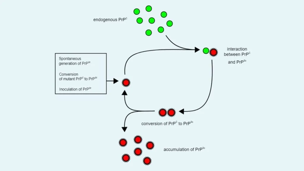
Prions do not copy themselves by building new chains from genetic code; they force existing healthy proteins to flip into their own deformed state. Early theorists suggested a heterodimer model, where one rogue PrPSc unit binds to a single healthy PrPC unit, alters its fold, and floats away to convert others. However, German biophysicist Manfred Eigen showed this simple model was mathematically impossible because it required a speed increase of 10^15 to work. Since free solo prions are never found in sick tissue, scientists looked at clustered fibers instead [14].
The fibril model solved the puzzle. In this framework, rogue proteins assemble into long linear amyloid fibers. The tips of these fibers act as templates, binding healthy proteins and locking them into the same beta-sheet pattern. As new units attach, the fiber grows like a stack of plates [13].
Linear growth alone would only cause a slow, steady rise in protein amounts, but animal brains show exponential surges in disease agents. Fibril breakage explains this sudden speed. As long amyloid filaments snap into shorter fragments, each break creates two fresh ends ready to recruit more healthy proteins. Host helper lipids, like phosphatidylethanolamine, along with cell RNA strands, bind to the fibers to speed up the folding switch [13].


Major Forms of Prion Disease in Humans
Human prion illnesses arise through three distinct paths: sporadic, genetic, and acquired. The sporadic kind, known as classic Creutzfeldt-Jakob disease, accounts for 85% of human cases and strikes 1 to 2 people per million each year worldwide. Sporadic CJD usually appears out of nowhere in adults aged 55 to 75 who have no family history or known exposure to tainted food. In these patients, a healthy PrP protein suddenly folds wrong by sheer chance, starting the deadly cascade in brain tissue [12].
Inherited forms stem from DNA faults in the PRNP gene and pass down in an autosomal dominant pattern. Over 50 different mutations are known. Fatal familial insomnia, caused by a mutation swapping aspartic acid for asparagine at codon 178, attacks the sleep-controlling thalamus and causes total sleeplessness, hallucinations, and death within two years. Case Western Reserve University researchers Dr Pierluigi Gambetti and Dr Piero Parchi also identified sporadic fatal insomnia in patients lacking the family gene fault. Meanwhile, a common switch at codon 129 between methionine and valine strongly shapes an individual’s personal risk of contracting disease [10].
Acquired prion disease happens when people eat or touch contaminated animal tissue. In the United Kingdom during the late 1980s and 1990s, an epidemic of Bovine Spongiform Encephalopathy (BSE) in cattle spread to people who ate beef, causing variant CJD in over 200 patients. By 2005, 24 countries had reported BSE among cattle. In North America, Canada identified BSE in 2003, and later that year a cow in Washington state tested positive. Chronic Wasting Disease (CWD) infects deer and elk across 32 U.S. states, Canada, South Korea, Norway, Finland, and Sweden. In medicine, tainted cadaveric growth hormone given to children before 1987 caused over 200 iatrogenic CJD cases worldwide [6].
Why Are Prions So Hard to Destroy?
Standard hospital cleaning rules that wipe out bacteria and viruses fail against prions. Because these rogue proteins carry no genetic code, ultraviolet light, gamma rays, and ionizing radiation leave them completely unharmed. Bathing contaminated tools in 70% alcohol, benzene, or formaldehyde does not kill the agent. Instead, formaldehyde locks the protein shape firmly in place. In laboratory trials, sheep brain slices preserved in formaldehyde and paraffin wax for years still infected mice when injected [9].
Prions survive dry oven heat up to 200 degrees Celsius for more than an hour. They bind tightly to stainless steel surgical tools within minutes of contact. Standard steam autoclaves fail to dislodge them, which has led to accidental disease spread during delicate brain operations [9].
Eliminating prions from steel and glass requires harsh chemicals that tear apart protein bonds. Effective hospital decontamination relies on soaking tools in strong sodium hypochlorite bleach or 1 molar sodium hydroxide, paired with long runs in high-pressure autoclaves. The World Health Organization recommends throwing away surgical tools used on high-risk brain patients rather than risking reuse. At Wageningen University in the Netherlands, J. P. M. Langeveld and co-workers showed in The Journal of Infectious Diseases that a bacterial keratinase enzyme from Bacillus licheniformis strain PWD-1 could digest scrapie and BSE prions in brain stems, but only when mixed with detergents and heated above 100 degrees Celsius [5].
Similar Aggregation Patterns in Other Disorders
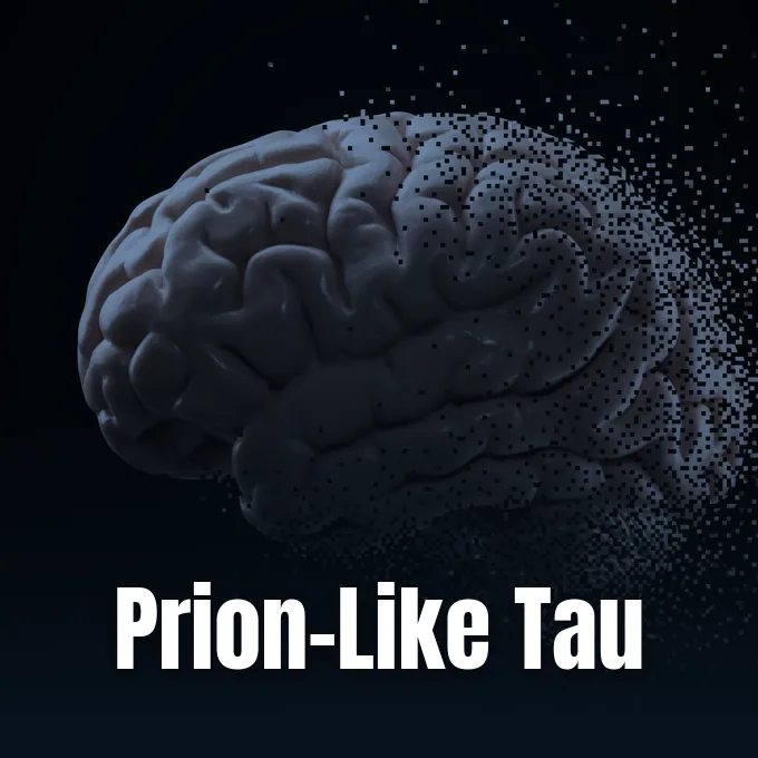
The concept of rogue proteins that spread by forcing normal copies to misfold has reshaped modern neuroscience. In a 2015 review for the Annual Review of Neuroscience, Lary Walker at Emory University and M. Jucker suggested calling prions “proteinaceous nucleating particles” to describe their basic physical action without causing panic about casual contagion. Today, researchers know that several common brain illnesses share this seeded clumping mechanism [3].
In Alzheimer’s disease, amyloid-beta proteins clump into outer plaques while tau proteins twist into inner tangles. Laboratory research on human tau strains demonstrates how specific protein shapes can induce spreading pathology along brain pathways. A similar templated conversion happens in Parkinson’s disease with alpha-synuclein proteins, which gather into toxic Lewy bodies. In 2015, Kurt Giles and Amanda Woerman in Stanley Prusiner’s laboratory at UCSF showed that alpha-synuclein prions from patients with Multiple System Atrophy (MSA) transmitted fatal brain degeneration to genetically altered mice within four months [8].
Because these illnesses share cascade patterns with general cognitive decline, studies into the biological mechanisms of dementia increasingly track how protein tangles poison synapses. Unlike kuru or BSE, Alzheimer’s and Parkinson’s do not spread through food or contact. But the core biophysics of templated clumping unites them all [3].
Clinical Detection and Experimental Interventions
Diagnosing prion disease in living patients has always been hard because early symptoms look like common dementias or stroke. For decades, doctors could only confirm a case after death by cutting brain tissue and viewing the spongy holes under a microscope. Today, clinical teams at the Cleveland Clinic and the National Prion Disease Pathology Surveillance Center use magnetic resonance imaging to spot bright signals in the cortex and run brain-wave tests to detect periodic sharp complexes [7].
A newer spinal fluid test called real-time quaking-induced conversion, or RT-QuIC, has changed medicine. This lab test takes a tiny drop of spinal fluid and mixes it with healthy recombinant PrPC. If even a trace of rogue prion is present, it forces the test protein to clump into amyloid strands, which glow under a fluorescent dye. RT-QuIC gives over 90% accuracy and lets doctors identify prion disease while patients are still alive [10].
Despite better tests, all prion diseases remain 100% fatal, usually taking life within months of diagnosis. Every human drug trial to date has failed to slow down brain damage, leaving medical teams to focus on gentle palliative care to keep patients comfortable. At the Paris Brain Institute in France, teams led by Marie-Claude Potier and Stéphane Haïk study how these rogue proteins spread between cells, seeking ways to block the fatal chain. New experimental paths include lab-made antibodies that grab onto amyloid fibers, small drugs that lock normal PrPC in its safe shape, and gene-silencing therapies designed to lower PrP production in brain cells [12].
- ACADEMIC JOURNAL Prusiner, S. B. (2013). Biology and genetics of prions causing neurodegeneration. Annual Review of Genetics, 47(1), 601-623. [Article Link]
- ACADEMIC JOURNAL Pan, K. M., Baldwin, M., Nguyen, J., Gasset, M., Serban, A., Groth, D., Mehlhorn, I., Huang, Z., Fletterick, R. J., & Cohen, F. E. (1993). Conversion of alpha-helices into beta-sheets features in the formation of the scrapie prion proteins. Proceedings of the National Academy of Sciences, 90(23), 10962-10966. [Article Link]
- ACADEMIC JOURNAL Walker, L. C., & Jucker, M. (2015). Neurodegenerative diseases: Expanding the prion concept. Annual Review of Neuroscience, 38(1), 87-103. [Article Link]
- ACADEMIC JOURNAL Race, B., Williams, K., Hughson, A. G., Jansen, C., Parchi, P., Rozemuller, A. J. M., & Chesebro, B. (2018). Familial human prion diseases associated with prion protein mutations Y226X and G131V are transmissible to transgenic mice expressing human prion protein. Acta Neuropathologica Communications, 6(1), 13. [Article Link]
- ACADEMIC JOURNAL Langeveld, J. P. M., Wang, J., Van de Wiel, D. F. M., Shih, G. C., Garssen, G. J., Bossers, A., & Shih, J. C. H. (2003). Enzymatic degradation of prion protein in brain stem from infected cattle and sheep. The Journal of Infectious Diseases, 188(11), 1782-1789. [Article Link]
- WEBSITE Centers for Disease Control and Prevention. (2026). About prion diseases. [Article Link]
- WEBSITE Cleveland Clinic. (2024). Prion disease. [Article Link]
- ONLINE NEWS Makin, S. (2015). A red flag for a neurodegenerative disease that may be transmissible. Scientific American. [Article Link]
- ONLINE NEWS Frazer, J. (2018). Prions are forever. Scientific American. [Article Link]
- WEBSITE Helmenstine, A. (2026). Prions: Definition, structure, diseases, and facts. Science Notes and Projects. [Article Link]
- WEBSITE Robertson, S. (2009). What is a prion? News-Medical. [Article Link]
- WEBSITE Dha, N. (2021). What are prions? News-Medical. [Article Link]
- WEBSITE Pan, S. (2024). Prions: Definition, structure, replication, function, disease. Biology Notes Online. [Article Link]
- WEBSITE Wikipedia contributors. (n.d.). Prion. Wikipedia, The Free Encyclopedia. [Article Link]


