




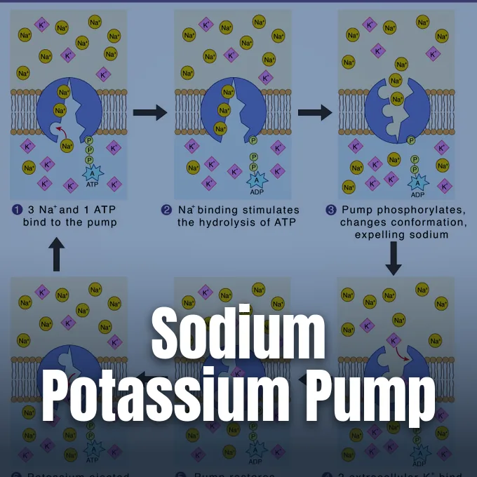
Every heartbeat and muscle twitch depends on a tiny protein engine embedded in cell membranes. The sodium potassium pump, known formally as Na+/K+-ATPase, moves three sodium ions out of animal cells while pulling two potassium ions inside against steep gradients. By splitting one molecule of ATP during each complete turn, it creates the resting voltage and ion balance that keep animal cells alive. Millions of these protein complexes operate in every tissue, consuming substantial metabolic fuel to maintain electrochemical stability. Without it, cells burst [10].
- Early Discovery by Jens Christian Skou
- Architecture of Catalytic Subunits
- Stages of the Post-Albers Cycle
- Why Does the Sodium Potassium Pump Require ATP?
- Membrane Potential in Excitable Tissues
- How the Sodium Potassium Pump Regulates Cell Volume
- Cardiac Glycosides and Cell Signals
- Human Diseases Linked to Na+/K+-ATPase Mutations
Early Discovery by Jens Christian Skou
In 1957, Danish researcher Jens Christian Skou isolated an enzyme from the leg nerves of shore crabs at Aarhus University, demonstrating that ATP breakdown occurred only when both sodium and potassium ions were present in the experimental bath. Before his laboratory trials, biologists knew that animal cells maintained uneven cation levels across their borders, but they lacked tangible evidence of a dedicated membrane carrier. Skou showed that the rate of chemical breakdown matched cation concentrations, identifying the first ion-transporting enzyme discovered in biology. The breakthrough transformed physiology. For this landmark achievement, Skou received the Nobel Prize in Chemistry in 1997, four decades after his first paper appeared in print [7].
Biochemists placed this molecule in the P-type ATPase family because it forms a transient aspartyl phosphate intermediate during ion transfer across the lipid bilayer. The reaction follows the Post-Albers cycle, named after researchers Albers and Post who traced its shifting shapes. In global biochemical archives, the KEGG database lists the enzyme as EC 7.2.2.13, grouping it with cation translocases that couple ATP hydrolysis to the active movement of sodium and potassium [13].
All animal cells rely on this pump. In 1966, researcher Schrier showed that red blood cell pumping couples directly to glycolysis, proving that cellular energy production tracks ion exchange. Millions of these pumps run without pause in human erythrocytes, cardiac muscle fibers, and cerebellar Purkinje neurons, ensuring that excitable tissues maintain their readiness to fire impulses [10].
Architecture of Catalytic Subunits
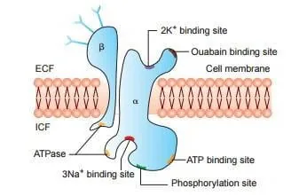
The complete enzyme is a heterodimer composed of a catalytic alpha subunit and a helper beta glycoprotein, accompanied in some tissues by a small FXYD regulator. The alpha subunit carries the catalytic core. It weighs roughly 100,000 Daltons and winds through the lipid bilayer ten times across transmembrane segments labeled M1 through M10. Both of its peptide ends sit in the cytoplasm, forming nucleotide-binding, phosphorylation, and actuator domains that bind ATP, coordinate three sodium ions, and capture two potassium ions [8].
The beta subunit is smaller, weighing about 55,000 Daltons with a single transmembrane span. It has no enzymatic activity of its own. Even so, this sugar-coated chain is indispensable because it helps the alpha subunit fold properly in the endoplasmic reticulum before guiding it to the outer surface of the cell. If the beta subunit is missing, alpha proteins misfold, aggregate in intracellular vesicles, and break down inside the cytoplasm before ever reaching the plasma membrane [3].
Mammals possess four alpha genes, designated ATP1A1 through ATP1A4, that show distinct tissue patterns mapped by Clausen and his coauthors. The ubiquitous alpha-1 variant maintains baseline ion levels in kidney, lung, and liver cells. In contrast, the alpha-2 isoform is concentrated in skeletal muscle, heart tissue, and vascular smooth muscle, while alpha-3 operates in brain neurons. The alpha-4 protein is unique to testis tissue, where it powers the swimming movement of sperm cells [3].
Stages of the Post-Albers Cycle
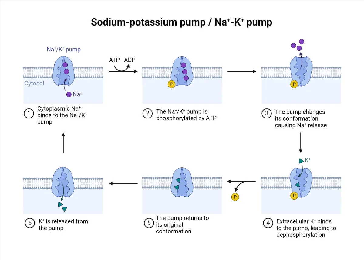
Modern structural studies reveal the pump cycle in atomic detail. In 2022, structural biologist Guo and his team used cryo-electron microscopy to capture recombinant human pump complexes in three conformations at resolutions between 2.7 and 3.2 Angstroms. Their reconstructions showed how the cytosolic gate opens in the E1 state to accept three sodium ions from the interior. Once sodium ions nestle into their binding sites, ATP docks at the cytoplasmic nucleotide domain, priming the enzyme for chemical catalysis [1].
Binding of three sodium ions triggers the transfer of a phosphate group from ATP to a conserved aspartate residue, generating a high-energy phosphorylated intermediate while releasing ADP into the cytosol. This phosphorylation forces a large structural tilt that closes the inner gate and opens an exit path toward the outside, shifting the enzyme into its E2 conformation. Because the outward-facing E2 state has a much lower affinity for sodium, all three sodium ions leave into the extracellular fluid. Two potassium ions from the outside then enter the exposed binding pocket, which induces rapid dephosphorylation of the aspartate residue. As the phosphate group departs, the pump returns to its inward-facing shape, carrying the two potassium ions across the membrane before releasing them into the cytoplasm [7].
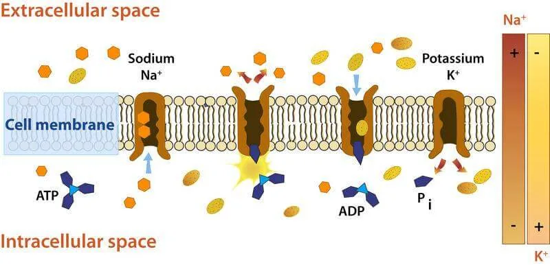
Potassium uptake takes another route. In 2015, biophysicist Castillo and his group proved that external potassium ions pass through a wide, water-filled channel, sensing only a small fraction of the electric field before occlusion. Computer models show that gating for potassium differs markedly from the steps that push sodium out. This unique inward pathway allows potassium ions to bind rapidly even when membrane voltages fluctuate wildly [2].
Why Does the Sodium Potassium Pump Require ATP?
The sodium potassium pump requires ATP because moving ions against their natural concentration gradients demands chemical energy. Cells must work against diffusion. In resting animal tissue, potassium is held at about 140 mM inside compared to just 4 mM outside, while sodium is kept near 12 mM inside versus 140 mM in extracellular fluid. Pushing three sodium ions out into a high-sodium environment while pulling two potassium ions into a crowded cytoplasm demands continuous thermodynamic work that only the cleavage of high-energy ATP bonds can supply [10].
This uphill transport consumes a huge share of cellular resources. Typical resting cells devote 30% of their total ATP production solely to keep these pumps running. The brain pays a heavy price. Cortical neurons spend up to 70% of their metabolic energy on sodium and potassium pumping to reset electrical gradients after firing impulses. Measurements indicate that the enzyme follows Michaelis-Menten kinetics, adjusting its turnover velocity to match fluctuations in cytoplasmic sodium levels [13].
Pump activity couples tightly to carbohydrate breakdown. In working skeletal muscles, blocking glycogenolysis reduces pump performance and leads to muscle fatigue. Red blood cells and vascular smooth muscle also feed their pumps from local glycolytic pools rather than waiting on distant mitochondrial diffusion. When energy pools run dry, ion exchange halts [10].

Membrane Potential in Excitable Tissues
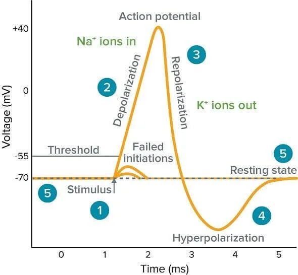
Because each cycle expels three sodium ions while bringing in only two potassium ions, the pump removes one positive charge with every turn. Physiologists classify this process as electrogenic transport. The net loss of positive charge, paired with passive potassium leakage through resting channels, creates a negative membrane voltage between -60 mV and -70 mV. This resting potential forms the electrical battery that powers nerves and muscles [7].
During an action potential, voltage-gated sodium channels open and sodium floods inward, driving the membrane voltage past +40 mV before potassium channels open to let potassium rush out for repolarization. A brief hyperpolarization follows. This dip below the resting baseline is vital for refractory timing, preventing backward firing and ensuring that electrical impulses travel strictly in one direction along nerve axons [7].
Action potentials rely on stored ion gradients rather than instantaneous pump output. Even so, repeated bursts of electrical signaling would quickly dissipate those gradients without ongoing maintenance. The sodium potassium pump works steadily behind the scenes, clearing excess sodium and replenishing potassium between nerve spikes. It keeps excitable membranes ready for the next incoming signal in central and peripheral neural pathways. These pumps never sleep [8].
How the Sodium Potassium Pump Regulates Cell Volume
Beyond membrane voltage, the sodium gradient drives secondary active transport throughout the body. Specialized carrier proteins like the SGLT sodium-glucose symporter harness the inward rush of sodium to pull glucose, amino acids, and vitamins into cells against concentration barriers. In renal nephrons, epithelial cells of the distal convoluted tubule run up to 50 million pumps per cell to reabsorb nutrients, balance blood pH, and keep fluid levels steady across daily filtration cycles [7].
The sodium potassium pump also protects animal cells from osmotic swelling. Cells contain proteins and organic molecules that can’t pass through the lipid membrane, creating an osmotic pull that draws water inward. By exporting three sodium ions for every two potassium ions brought inside, the pump decreases the total count of intracellular solutes. Water follows ions. Without this pump, animal cells swell and lyse [8].
In the digestive tract, epithelial polarity depends on this arrangement. Apical transporters import glucose from food using the sodium gradient, while pumps on the basolateral surface export sodium into the bloodstream. This setup keeps cytosolic sodium low and drives constant nutrient absorption into circulation, sustaining systemic metabolic balance. Epithelial cells depend entirely on this divided architecture [10].
Cardiac Glycosides and Cell Signals
Herbal extracts from foxglove plants have treated heart ailments for centuries. In 1785, English physician Whitering described the medical benefits of digitalis, and French pharmacist Nativelle isolated digitalin a century later. Today, Digoxin remains a standard cardiac glycoside used in cardiology. Pharmacorama explains that cardiac glycosides like Digoxin bind “to the extracellular part of enzyme i.e. that binds potassium, when it is in a phosphorylated state” to stop the pump cycle [9].
In heart muscle, slowing the pump increases contractile strength through linked calcium signaling. Under the Blaustein hypothesis, a small rise in cytosolic sodium slows the sodium-calcium exchanger NCX, leaving more calcium in cardiac myocytes to load the sarcoplasmic reticulum. Each subsequent contraction releases more calcium, boosting the mechanical force of ventricular heart muscle during systole [4]. Furthermore, researchers noted that clinical doses of Digoxin inhibit less than 5% of cardiac alpha-2 pumps, indicating that signaling events amplify the response. In membrane caveolae, a specialized subpopulation of pumps acts as a receptor that binds Ouabain, transactivates epidermal growth factor receptors, and stimulates downstream MAPK cascades that influence cell survival [10].

The pump also partners directly with Src kinase to regulate cell growth. When cardiotonic steroids bind to the alpha subunit, they release Src from inhibition, prompting reactive oxygen species generation and downstream signaling. Scientists developed NaKtide, a peptide derived from the alpha chain, to inhibit this pathway in laboratory models. Studies in the land snail Otala lactea demonstrate that reversible phosphorylation controls pump activity during metabolic dormancy [10].
Human Diseases Linked to Na+/K+-ATPase Mutations
Inherited defects in the sodium potassium pump cause serious human neurological conditions. Mutations in ATP1A3 cause rapid-onset dystonia-parkinsonism and alternating hemiplegia of childhood, producing abrupt movement failure under physical or emotional stress. Mutations in ATP1A2 impair potassium and sodium clearance in brain astrocytes, triggering familial hemiplegic migraine type 2. Patients carrying these alleles experience episodes of hemiparesis and prolonged cognitive aura [3].
Somatic mutations in ATP1A1 alter adrenal cortical cells, causing aldosterone-producing adenomas and secondary hypertension. In pigment cells, the transcription factor MITF regulates alpha-1 expression, linking ion transport to developmental signaling [11]. In cardiac tissue, research by Almilaji and his team revealed that human Parvovirus B19 capsid protein VP1 suppresses pump activity, contributing to endothelial dysfunction in viral cardiomyopathy. Chronic downregulation of the enzyme accelerates myocardial remodeling and heart failure [12].
Altered pump expression is also a hallmark of various human cancers. Loss of the beta-1 subunit weakens cell junctions and fosters tumor metastasis, whereas abnormal alpha subunit levels correlate with resistance to chemotherapy [5]. Because cardiac glycosides selectively drive apoptosis in cancer cells with altered pump profiles, researchers are testing targeted glycoside derivatives in oncology [6]. Skou’s crab nerve experiment in 1957 opened an enduring era in modern cellular physiology [7].
- ACADEMIC JOURNAL Guo, Y., Zhang, Y., Yan, R., Huang, B., Ye, F., Wu, L., Chi, X., Shi, Y., & Zhou, Q. (2022). Cryo-EM structures of recombinant human sodium-potassium pump determined in three different states. Nature Communications, 13(1). [Article Link]
- ACADEMIC JOURNAL Castillo, J. P., Rui, H., Basilio, D., Das, A., Roux, B., Latorre, R., Bezanilla, F., & Holmgren, M. (2015). Mechanism of potassium ion uptake by the Na+/K+-ATPase. Nature Communications, 6(1). [Article Link]
- ACADEMIC JOURNAL Clausen, M. V., Hilbers, F., & Poulsen, H. (2017). The Structure and Function of the Na,K-ATPase Isoforms in Health and Disease. Frontiers in Physiology, 8, 371. [Article Link]
- ACADEMIC JOURNAL Shattock, M. J., Ottolia, M., Bers, D. M., Blaustein, M. P., Boguslavskyi, A., Bossuyt, J., Bridge, J. H., Chen-Izu, Y., Clancy, C. E., Edwards, A. G., Goldhaber, J. I., Kaplan, J. H., Lingrel, J. B., Pavlović, D., Philipson, K. D., Sipido, K. R., & Xie, Z. (2015). Na+/Ca2+ exchange and Na+/K+-ATPase in the heart. The Journal of Physiology, 593(6), 1361-1382. [Article Link]
- ACADEMIC JOURNAL Mijatovic, T., Dufrasne, F., & Kiss, R. (2012). Na+/K+-ATPase and Cancer. Pharmaceutical Patent Analyst, 1(1), 91-106. [Article Link]
- ACADEMIC JOURNAL Li, Z., & Langhans, S. A. (2015). Transcriptional regulators of Na,K-ATPase subunits. Frontiers in Cell and Developmental Biology, 3, 66. [Article Link]
- WEBSITE Aryal, S., & Shakya, R. (2025). Sodium-Potassium (Na+/K+) Pump: Mechanism, Functions. Microbe Notes. [Article Link]
- WEBSITE Dhali, D. (2021). Sodium-Potassium Pump – Definition & Functions, with Diagram. Science Facts. [Article Link]
- WEBSITE Pharmacorama. (2026). NA+/K+-ATPase and inhibitors (Digoxin). [Article Link]
- WEBSITE Wikipedia contributors. (n.d.). Sodium–potassium pump. Wikipedia, The Free Encyclopedia. [Article Link]
- WEBSITE Wikipedia contributors. (n.d.). ATPase, Na+/K+ transporting, alpha 1. Wikipedia, The Free Encyclopedia. [Article Link]
- ACADEMIC JOURNAL Almilaji, A., Szteyn, K., Fein, E., Pakladok, T., Muñoz, C., Elvira, B., Towhid, S. T., Alesutan, I., Shumilina, E. V., Bock, C., Kandolf, R., & Lang, F. J. (2013). Down-Regulation of Na+/K+ATPase Activity by Human Parvovirus B19 Capsid Protein VP1. Cellular Physiology and Biochemistry, 31(4-5), 638-648. [Article Link]
- WEBSITE Kanehisa Laboratories. (2013). KEGG ENZYME: 7.2.2.13. Kyoto Encyclopedia of Genes and Genomes. [Article Link]


