


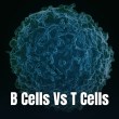

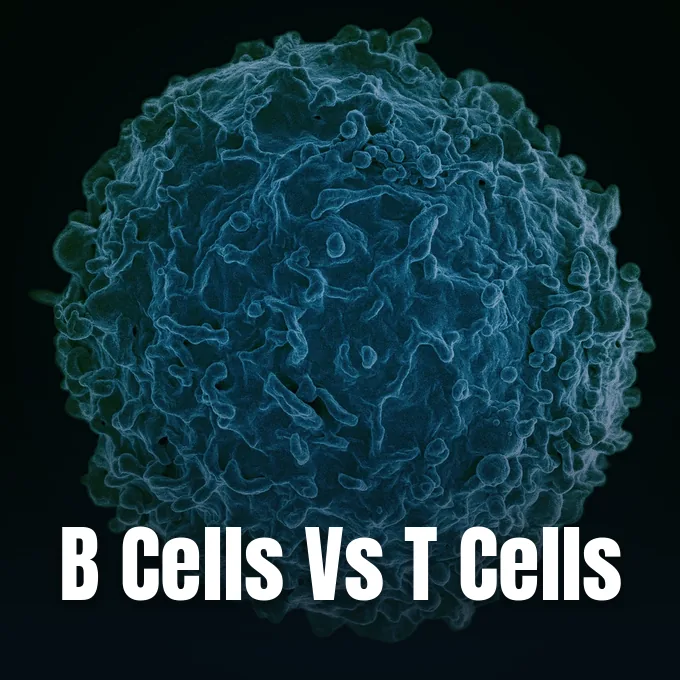
Adaptive immunity depends on a clear division of work between white blood cells that defend the body against distinct threats. The core distinction between b cells vs t cells centers on whether defensive action relies on free antibodies or direct cell combat. B cells govern humoral immunity by releasing targeted proteins into fluids to bind foreign germs, while T cells direct responses to kill infected host cells and manage signaling networks [9].
- How B Cells vs T Cells Divide Immune Labor
- Maturation Pathways in Bone Marrow and Thymus
- Antibody Secretion by Specialized B Lymphocytes
- Cell-Mediated Destruction by Cytotoxic T Cells
- Comparing B Cells vs T Cells in Clinical Blood Tests
- Why Lymphocyte Counts Fluctuate During Illness
- How B Cells vs T Cells Form Memory
- Diagnostic Ratios and Biomarkers in Systemic Disease
How B Cells vs T Cells Divide Immune Labor
The human immune system relies on white blood cells to find and clear foreign invaders with high biochemical precision. Innate cells offer fast, broad protection. In contrast, adaptive defenders build custom tools to eliminate specific bugs. Looking at b cells vs t cells shows this basic split, where antibody release guards open body fluids while cellular combat clears infected tissue sites throughout the body [9].
B cells act like tiny factories that spot free germs through receptors resting on their outer walls. When a naive B cell meets its matching invader, it clones itself and changes into an effector unit that makes thousands of antibodies each second. These Y-shaped proteins bind to toxins, coat viral coats, and mark invaders so larger scavenger cells can swallow them up without delay. This fluid-based defense shields the open spaces of the body, stopping circulating germs before they stick to healthy tissue and grow. Both lines arise from haematopoietic stem cells in marrow cavities, but their daily duties diverge across every stage of life [9].
T cells handle cellular defense by checking inside host cells rather than scanning free fluids. Germs that hide inside host cells escape free antibodies. Cytotoxic T cells spot viral fragments on host surfaces, releasing destructive chemicals that force sick cells to self-destruct before viruses spread, while helper T cells release cytokine signals that call in macrophages and help B cells make better antibodies [9].
Maturation Pathways in Bone Marrow and Thymus
Every lymphocyte begins its life as a common progenitor in spongy bone cavities. This growth pathway, termed lymphopoiesis, guides young cells along strict tracks toward mature roles. Mammalian B cells finish their full early training inside the core of the bone marrow, testing their surface sensors so self-attacking clones die off before any release into the blood stream occurs [9].
T cells take a much longer journey after leaving marrow spaces as fragile precursor cells. They enter blood vessels and migrate straight to the thymus, a chest organ where they build receptors and face harsh survival tests. Inside this thymic nursery, cells must recognize foreign peptides on histocompatibility proteins while sparing healthy self tissue. In a research review published in the journal Antioxidants, Gorkom and Bos at Maastricht showed that ascorbic acid, known as vitamin C, is needed for normal T cell development in cell culture and helps both T cells and natural killer cells divide during immune system recovery [2].
Historically, the anatomical origin of B lymphocytes was discovered in birds rather than humans. Researchers Chang and Glick showed that young avian B cells mature in the bursa of Fabricius, a gut-linked lymphoid sac that gave these defenders their B name. Mammals lack an avian bursa. Instead, their B cells mature directly within marrow spaces before moving out to colonize the spleen and lymph nodes [9].
Antibody Secretion by Specialized B Lymphocytes
Binding a foreign antigen wakes up a mature B cell and sets off rapid cell division. The activated cell soon turns into an antibody-making plasma cell packed with protein-building polyribosomes and rough endoplasmic reticulum. These cells pump out tailored antibodies that fit the exact shape of the invading pathogen. In birds and mammals alike, this outpouring of protein allows free antibodies to sweep mucosal barriers, catching bacteria and viruses before they slip past surface linings into inner organs [9].
Under a laboratory microscope, plasma cells can take on unusual forms in bone marrow smears. Pathologists describe variant shapes like flame cells, which show a pink fringe of outer cytoplasm, and Mott cells filled with round protein droplets. Flame cells appear often in feline marrow samples and in human plasma cell disorders, marking altered antibody storage inside expanding cell pockets during prolonged immune stimulation [10].
Uncontrolled expansion of identical B cell clones can lead to serious blood disorders. Medical specialists at the Cleveland Clinic track a condition called monoclonal B-cell lymphocytosis, where identical B cell clones multiply in blood samples. While people with this condition often feel healthy for years, carrying high numbers of identical B cells raises their risk of developing chronic lymphocytic leukemia, particularly among patients with a family history of blood cancer [7].
Cell-Mediated Destruction by Cytotoxic T Cells
Cellular immunity relies on direct physical touch rather than free antibodies. Cytotoxic T cells carry CD8 surface markers and patrol tissues looking for sick host cells displaying viral peptides. When a cytotoxic cell locks onto its target, it dumps toxic granules containing perforin and granzymes directly onto the target wall, forcing the sick cell to commit suicide without harming healthy tissue nearby [9].
Helper T cells carry CD4 receptors and steer the wider immune response by reading antigen cues and sending chemical orders to other cells. They wake up macrophages, support cytotoxic T cells, and push B cells to produce stronger antibodies. Runaway chemical signals can damage healthy organs, a hazard detailed in reports on how coronavirus causes cytokine storm at the molecular level during severe viral infection. When HIV infects and wipes out CD4+ helper T cells, the entire adaptive network breaks down, leaving patients open to fatal opportunistic bugs [9].
Solid tumors present a harsh environment that blocks infiltrating T cells. In a review in Frontiers in Oncology, Bellone and Calcinotto showed how leaky tumor vessels, poor blood flow, and endothelial cell shutdown prevent cytotoxic T cells from entering tumor tissue. Reversing this barrier helps restore T cell fighting fitness inside tumors [6].



Comparing B Cells vs T Cells in Clinical Blood Tests
Routine laboratory tests count white blood cells to check overall immune health during illness. Under a light microscope using Wright’s stain, normal lymphocytes look like small round cells with dark nuclei about 7 μm in diameter. Because mature B cells and T cells look alike under a standard lens, lab technicians cannot tell them apart by eye alone [9].
To separate and count b cells vs t cells accurately, diagnostic labs use flow cytometry machines. This method tags cell surface markers with glowing antibodies, letting lasers scan thousands of single cells each second as they flow past a sensor. Clinicians use these readouts to measure helper T cells, killer T cells, and antibody-producing B cells when evaluating immune disorders or tracking leukemia treatments. Across healthy adult blood counts, lymphocytes make up between 18% and 42% of all circulating white blood cells [9].
Reference counts for white blood cells shift with age. Guidance from the Cleveland Clinic states that healthy adults normally carry from 1,000 up to 4,800 lymphocytes in every single microliter of blood. Pediatric counts run much higher, with normal ranges for children sitting between 3,000 and 9,500 cells per microliter as growing bodies build immune defenses [7].
Why Lymphocyte Counts Fluctuate During Illness
Lymphocyte counts rise and fall in the blood because bone marrow output, tissue migration, and cell destruction shift quickly during sickness. A sharp jump in lymphocyte numbers, called lymphocytosis, usually means the immune system is actively fighting off an infection. Viral illnesses like mononucleosis cause most count spikes, but some bacteria also push counts upward [7]. The whooping cough bacterium Bordetella pertussis makes a toxin that stops lymphocytes from entering lymph nodes, driving adult counts above 4,000 cells per microliter and pushing child counts past 8,000 cells [9].
Cell shapes change during heavy viral battles when CD8+ cytotoxic T cells swell up into larger forms. Described by W. Türk in 1907 and classified by Hal Downey and C.A. McKinlay in 1923, these reactive lymphocytes grow wider than 30 microns with pale blue cytoplasm. Downey Type II cells appear most often during Epstein-Barr virus, cytomegalovirus, and hepatitis infections, taking on odd shapes as they press against neighboring red blood cells [10].
Low lymphocyte levels, known as lymphocytopenia, follow bodily trauma, surgery, or bone marrow suppression. Low numbers leave the body weak against new bugs, increasing infection rates after hospital operations. Research into why some people never get Covid-19 pointed toward early innate and cellular barriers that clear viral particles before symptoms start. When cell counts fall below normal lines, doctors check whether nutrition, drug treatments, or viral damage caused the drop [9].
How B Cells vs T Cells Form Memory
The main strength of adaptive immunity is building long-term memory after an infection clears. Most short-lived fighting cells die by apoptosis once germs disappear, but a small pool survives as resting memory cells. Looking at b cells vs t cells during this quiet phase shows two distinct survival paths, where memory B cells hold antibody blueprints while memory T cells patrol deep tissues for years [9].
Memory B cells wait inside the spleen and lymph nodes for their target germ to return. When re-exposed to the same pathogen, these cells wake up quickly, dividing and changing into plasma cells that pour fresh antibodies into the blood within hours. Memory T cells stay scattered across mucosal linings and skin layers. They kill reinfected cells on the spot before pathogens gain ground, lasting for years in healthy tissues [9].
Nerve signals also guide lymphocyte survival and daily work. In a review in the Journal of Neuroimmunology, Kerage, Sloan, Mattarollo, and McCombe showed that lymphocytes express surface receptors for neurotransmitters, allowing brain signals to tune immune cell movement. Work on reduced serotonin levels and long Covid symptoms showed how chemical shifts accompany altered recovery after viral sickness [4].
Diagnostic Ratios and Biomarkers in Systemic Disease
Because lymphocyte counts drop during physical stress, doctors calculate blood ratios to gauge body-wide inflammation. The neutrophil-to-lymphocyte ratio divides absolute neutrophils by absolute lymphocytes from a routine blood draw. In healthy adults, baseline ratios range from 0.78 to 3.53 according to reference studies. Zahorec proposed using this ratio as a stress score in intensive care units in 2001 at the St. Elizabeth Cancer Institute in Bratislava, while Hiroshi Sato, Yasuhiro Tsubosa, and Tatsuyuki Kawano showed in the World Journal of Surgery in 2012 that the ratio predicts chemotherapy response in esophageal cancer [11].
During the coronavirus pandemic, blood ratios offered cheap prognostic clues for hospital staff. In the Caspian Journal of Internal Medicine, Eslamijouybari, Heydari, and Maleki studied 527 patients at Imam Hospital in Sari, reporting that neutrophil-to-lymphocyte ratios were two times higher in patients who died of COVID-19 compared to those who recovered [8]. In the Journal of Clinical Medicine Research, Chan and Rout ran a meta-analysis of 20 studies with 3,508 patients, confirming that higher ratios mark severe disease [1].
Blood ratios also help track stroke recovery and mood disorders. In the Journal of Neuroinflammation, Gong, Liu, and Chen studied 1,060 stroke patients, linking high ratios to early brain deterioration after clot-busting therapy [5]. In Brain Sciences, Fusar-Poli and Natale studied 294 patients with bipolar disorder, finding higher ratios during manic phases than depressive phases. These simple cell ratios help clinicians track systemic inflammation across diverse medical conditions [3].
- ACADEMIC JOURNAL Chan, A. S., & Rout, A. (2020). Use of Neutrophil-to-Lymphocyte and Platelet-to-Lymphocyte Ratios in COVID-19. Journal of Clinical Medicine Research, 12(7), 448-453. [Article Link]
- ACADEMIC JOURNAL Gorkom, G. N. Y. v., Wolterink, R. G. J. K., Elssen, C. H. M. J. v., Wieten, L., Germeraad, W. T., & Bos, G. M. (2018). Influence of Vitamin C on Lymphocytes: An Overview. Antioxidants, 7(3), 41. [Article Link]
- ACADEMIC JOURNAL Fusar‐Poli, L., Natale, A., Amerio, A., Cimpoesu, P., Filioli, P. G., Aguglia, E., Amore, M., Serafini, G., & Aguglia, A. (2021). Neutrophil-to-Lymphocyte, Platelet-to-Lymphocyte and Monocyte-to-Lymphocyte Ratio in Bipolar Disorder. Brain Sciences, 11(1), 58. [Article Link]
- ACADEMIC JOURNAL Kerage, D., Sloan, E. K., Mattarollo, S. R., & McCombe, P. (2019). Interaction of neurotransmitters and neurochemicals with lymphocytes. Journal of Neuroimmunology, 332, 99-111. [Article Link]
- ACADEMIC JOURNAL Gong, P., Liu, Y., Gong, Y., Chen, G., Zhang, X., Wang, S., Zhou, F., Duan, R., Chen, W., Huang, T., Wang, M., Deng, Q., Shi, H., Zhou, J., Jiang, T., & Zhang, Y. (2021). The association of neutrophil to lymphocyte ratio, platelet to lymphocyte ratio, and lymphocyte to monocyte ratio with post-thrombolysis early neurological outcomes in patients with acute ischemic stroke. Journal of Neuroinflammation, 18(1), 51. [Article Link]
- ACADEMIC JOURNAL Bellone, M., & Calcinotto, A. (2013). Ways to Enhance Lymphocyte Trafficking into Tumors and Fitness of Tumor Infiltrating Lymphocytes. Frontiers in Oncology, 3, 231. [Article Link]
- WEBSITE Cleveland Clinic. (2023). Why Do I Have a High Lymphocyte Count? Cleveland Clinic. [Article Link]
- ACADEMIC JOURNAL Eslamijouybari, M., Heydari, K., Maleki, I., Moosazadeh, M., Hedayatizadeh-Omran, A., Vahedi, L., Ghasemian, R., Sharifpour, & Alizadeh-Navaei, R. (2020). Neutrophil-to-Lymphocyte and Platelet-to-Lymphocyte Ratios in COVID-19 Patients and Control Group and Relationship with Disease Prognosis. Caspian Journal of Internal Medicine, 11(Suppl 1), 531-535. [Article Link]
- WEBSITE Wikipedia contributors. (2023). Lymphocyte. In Wikipedia, The Free Encyclopedia. [Article Link]
- WEBSITE Wikipedia contributors. (2023). Reactive lymphocyte. In Wikipedia, The Free Encyclopedia. [Article Link]
- WEBSITE Wikipedia contributors. (2023). Neutrophil to lymphocyte ratio. In Wikipedia, The Free Encyclopedia. [Article Link]
Cover image: NIAID / Wikimedia Commons, CC BY 2.0


