





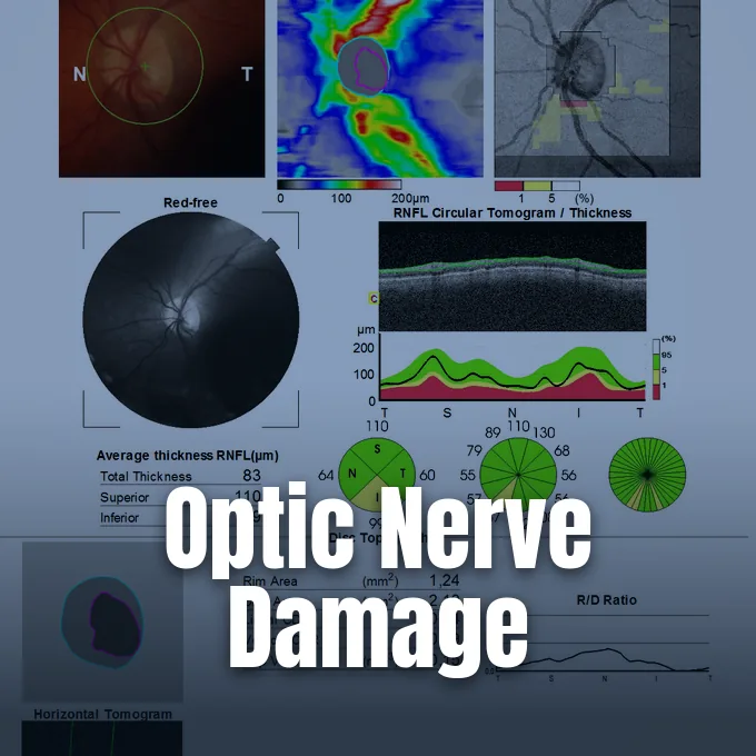
Glaucoma is a group of chronic eye conditions where progressive damage to the optic nerve compromises visual perception. Inside the eye, clear fluid continually circulates to nourish transparent tissues, but drainage resistance can cause internal pressure to rise. When this intraocular pressure pushes against delicate neural cables at the rear of the eye, nerve fibers slowly deteriorate and cause permanent vision loss. Because the disease often advances without early pain or blurred central vision, people frequently lose peripheral sight before noticing any problem [1].
- What Is Glaucoma?
- How Aqueous Humor Circulates and Builds Pressure
- The Biological Cascade Damaging the Optic Nerve
- Open-Angle and Angle-Closure Classifications
- Congenital and Secondary Glaucoma Forms
- Mapping Visual Field Defects and Progression Rates
- Why Glaucoma Cannot Be Cured but Can Be Managed
- Surgical Drainage Pathways and Clinical Outcomes
What Is Glaucoma?
The word comes from the Ancient Greek term glaukós, meaning gleaming or gray-blue. In modern ophthalmology, the term describes progressive optic neuropathies defined by retinal ganglion cell degeneration and optic disc cupping. Central acuity often stays normal while outer boundaries vanish [13].
The condition represents a major worldwide public health concern that affects millions of households across every continent. More than 2.7 million people in the United States and approximately 64 million people worldwide live with optic nerve damage from this disease. By 2040, global prevalence is projected to climb past 111 million individuals as populations live longer. Cataracts remain more common across the globe, but they can be treated surgically, whereas blindness resulting from optic neuropathy cannot be reversed once neural tissue has been lost. In the United States, it is the leading cause of irreversible blindness among African American and Hispanic populations [12].
Early detection remains difficult because the most common form develops silently without discomfort. Most people feel no eye pain and experience no changes in their reading clarity during the early stages of disease. Subtle blind spots develop in side vision, but the brain compensates by blending images from both eyes. Without regular comprehensive eye exams that measure internal pressure and examine the optic disc, structural damage continues unchecked [2].

How Aqueous Humor Circulates and Builds Pressure
To understand how internal pressure rises, clinicians examine how clear fluid continuously circulates through the front segment of the human eye to support avascular structures. The ciliary body, located directly behind the colored iris, continuously secretes a nourishing fluid called aqueous humor into the posterior chamber. This fluid moves forward through the pupil into the anterior chamber, delivering oxygen, glucose, and amino acids to the cornea and lens. These structures lack blood vessels so they can stay clear and refract incoming light cleanly. After circulating across the front of the eye, aqueous humor flows into the drainage angle formed where the iris meets the outer cornea [13].
The primary exit route runs through a porous tissue band known as the trabecular meshwork. Fluid filters through these tiny connective channels into Schlemm’s canal, which connects to episcleral veins that carry fluid back into the bloodstream. In healthy eyes, fluid outflow precisely matches fluid creation, keeping intraocular pressure (IOP) in equilibrium. If microscopic outflow channels become clogged with extracellular matrix debris or collapse under cellular stress, fluid accumulates in the anterior chamber and drives intraocular pressure above physiological limits [12].
Clinical standards traditionally treat an intraocular reading above 21 mmHg as ocular hypertension. Yet eye pressure varies between individuals and changes during the day. Some people tolerate readings above 24 mmHg without losing nerve fibers, while others suffer optic neuropathy at 15 mmHg. Pressure level is a primary risk factor, but it does not tell the whole story [13].
The Biological Cascade Damaging the Optic Nerve
Visual information captured by photoreceptor cells travels through roughly one million nerve fibers formed by retinal ganglion cells. These axons stretch across the inner surface of the retina and gather at the optic disc to form the optic nerve. To leave the eyeball and enter the brain, these fibers must pass through the lamina cribrosa, a mesh-like plate of collagenous connective tissue. When pressure inside the eye climbs, physical tension pushes against the lamina cribrosa, compressing the narrow pores through which axon bundles travel. This mechanical deformation pinches axon bundles as they pass through the connective sieve, halting axoplasmic transport of essential neurotrophic signals between the brain and retinal ganglion cells [12].
Mechanical strain explains only part of the damage. Vascular factors also dictate how well retinal neurons survive. In the Los Angeles Latino Eye Study (LALES), which evaluated 6,357 adult participants, researchers discovered that ocular perfusion pressure plays an influential role in the development and progression of optic neuropathy. Perfusion pressure equals systemic blood pressure minus intraocular pressure. When diastolic blood pressure drops too low, or when eye pressure rises, blood flow to the optic disc slows down. “Check the BP,” said Dr. Rohit Varma, noting that keeping a healthy balance between blood pressure and eye pressure protects delicate optic tissue [8].

Why do some people lose sight when their eye pressure stays normal? Cellular studies show that oxidative stress, local inflammation, and toxic glutamate buildup can trigger cell suicide in retinal ganglion cells. These biochemical processes resemble mechanisms seen in Alzheimer’s and other brain disorders. Nerve cells die even when physical fluid pressure appears completely normal [8].
Open-Angle and Angle-Closure Classifications
Eye care professionals divide primary cases into two major anatomical forms: open-angle and angle-closure disease. Primary open-angle glaucoma is the most frequent variety, accounting for roughly three-quarters of all diagnoses in North America. In open-angle disease, the physical space between the iris and cornea looks completely wide and unobstructed during an examination. However, microscopic resistance builds deep within the trabecular meshwork itself. Eye pressure rises slowly over many years without giving any outward sign of trouble. Because the cornea stays clear and no pain occurs, individuals usually do not realize that their peripheral visual field is narrowing until substantial optic nerve fibers have perished [1].
In angle-closure disease, the drainage route becomes physically blocked because the iris bunches forward against the cornea. When this blockage happens suddenly, fluid cannot escape and eye pressure spikes to extreme levels within minutes. This acute closed-angle event causes intense eye pain, nausea, vomiting, cloudy vision, and bright rainbow halos around lamps. Acute angle-closure is an urgent medical emergency. Without prompt medical treatment to reopen fluid drainage pathways and relieve internal pressure, permanent blindness can take hold in the affected eye in only a few days [2].
Anatomical features and population ancestry strongly influence which form develops. Angle-closure is four times more common in Asian countries than in North America, and women experience it more often than men due to narrower anterior chambers. Meanwhile, open-angle disease occurs roughly five times more often among African American and Hispanic adults than among Caucasian adults. Data from the African Descent and Glaucoma Evaluation Study (ADAGES) revealed that Black patients often possess thinner corneas and larger optic cups, which may increase structural susceptibility to pressure [8].



Congenital and Secondary Glaucoma Forms
Primary congenital glaucoma is a rare inherited condition that appears at birth or develops during early infancy, occurring in roughly 1 in 10,000 births in the United States. Across the world, incidence rates fluctuate widely from 1 in 22,000 live births in Northern Ireland to 1 in 2,500 in Saudi Arabia and 1 in 1,250 in Romania. Infants present with a classic triad of symptoms: epiphora (excessive tearing), photophobia (light sensitivity), and blepharospasm, where the eyelids squeeze shut involuntarily. Because infant eye tissues remain pliable, elevated intraocular pressure stretches the cornea and eyeball into an enlarged globe, a visible condition known as buphthalmos [14].
Genetic breakthroughs have uncovered key biological mechanisms that cause congenital outflow failure. A research team led by Dr. Janey L. Wiggs at Harvard Medical School and Mass Eye and Ear examined families with childhood optic neuropathy and discovered mutations in the THBS1 gene. Working with Dr. Robert J. D’Amato at Boston Children’s Hospital and Dr. Owen M. Siggs at Flinders University in Australia, the team confirmed their findings across three unrelated families of European, Indian, and Sudanese descent. Rather than altering blood vessel growth, the mutated thrombospondin-1 protein misfolds and accumulates in the trabecular meshwork. This abnormal protein buildup clogs outflow channels and drives eye pressure upward during early development [5].
Secondary glaucoma arises from recognizable external insults, medications, or concurrent illnesses. Extended use of steroid eye drops can reduce trabecular permeability and trigger pressure spikes. Eye injuries that tear internal angle structures can cause angle-recession glaucoma years after trauma occurred. Diabetic retinopathy and central retinal vein occlusions can stimulate abnormal blood vessel growth over the drainage angle, causing severe neovascular disease [13].

Mapping Visual Field Defects and Progression Rates
In 1928, Harry Moss Traquair described the normal human visual field as an “island of vision in a sea of darkness”, peaking sharply at the fovea where vision is sharpest and sloping downward toward the periphery. Under normal conditions, vision extends 60° upward, 75° downward, 110° outward toward the temple, and 60° inward toward the nose. In 1972, Franz Fankhauser developed computerized automated perimetry at the University of Berne, allowing clinicians to test retinal sensitivity at dozens of fixed points. In glaucomatous neuropathy, nerve damage typically starts in the arcuate nerve bundles, producing subtle paracentral depressions and nasal steps in side vision before patients ever notice visual changes [9].
Prospective natural history data from the Early Manifest Glaucoma Trial (EMGT) documented untreated rates of disease progression across 118 control patients who were monitored regularly with automated perimetric testing. Researchers evaluated 46 patients with high-tension glaucoma, 57 with normal-tension glaucoma, and 15 with pseudoexfoliation glaucoma. Across all untreated eyes, the median rate of visual loss was -0.40 dB/year. Median rates varied significantly among subtypes, measuring -0.46 dB/year in high-tension patients, -0.22 dB/year in normal-tension patients, and -1.13 dB/year in pseudoexfoliation cases [10].
Over the six-year monitoring period, 68% of untreated patients showed measurable progression. Rates reached 74% in high-tension cases, 56% in normal-tension cases, and 93% in pseudoexfoliation cases. Time to confirmed deterioration was 19.5 months for pseudoexfoliation, 44.8 months for high-tension, and 61.1 months for normal-tension glaucoma. Older age consistently accelerated the speed of visual decline [10].
Why Glaucoma Cannot Be Cured but Can Be Managed
Because mature retinal ganglion cells lack the biological capacity to regenerate within the human central nervous system, vision lost to progressive axonal decay cannot be recovered by any existing medical intervention. Medical science has no current therapy that can bring back lost visual field or restore a cupped optic nerve. For that reason, clinicians cannot speak of a cure. Instead, the central goal of all management is to lower intraocular pressure to a safe target level that halts or slows further nerve fiber loss. Lowering eye pressure relieves physical strain at the optic nerve head and helps preserve surviving sight [2].
Medicated eye drops remain the initial medical treatment, working through five main drug classes to either decrease fluid generation or speed drainage. Prostaglandin analogues improve fluid outflow, while beta-blockers and alpha-agonists reduce aqueous production by the ciliary body. Yet keeping up with daily drops presents real challenges. Studies show that approximately one-third of patients stop collecting their prescribed eye drop refills within their first year of treatment, leaving eyes vulnerable to ongoing damage [3].

To overcome the hurdle of medication adherence, the randomized LiGHT trial compared selective laser trabeculoplasty (SLT) against eye drops across 718 patients at six hospitals in the United Kingdom. The study found that 74.2% of patients in the laser group maintained their target eye pressure without needing any eye drops for three years after treatment. Health-related quality of life scores were nearly identical, registering 0.89 in the laser cohort and 0.90 in the eye drops cohort. Furthermore, using laser trabeculoplasty as a first-line intervention saved the National Health Service an average of £451 per patient in eye specialist appointments. This evidence demonstrates that early laser intervention provides safe, effective, and cost-efficient pressure reduction without placing the daily burden of drop compliance onto patients [3].
Surgical Drainage Pathways and Clinical Outcomes
When medications and laser trabeculoplasty fail to keep intraocular pressure at safe levels, surgeons turn to operating procedures that build new drainage channels. Trabeculectomy has served as the conventional standard filtering surgery for decades. In this procedure, the surgeon cuts a partial-thickness flap into the white sclera and removes a tiny piece of trabecular meshwork to create an opening into the anterior chamber. Fluid flows out through this opening into a blister-like pocket called a bleb under the conjunctiva, where surrounding capillaries slowly absorb it. While trabeculectomy lowers pressure reliably, entering the eye carries hazards of hypotony, leaks, and severe intraocular infections [15].
To avoid penetrating the front chamber, surgeons developed nonpenetrating techniques such as viscocanalostomy and deep sclerectomy. In these procedures, surgeons unroof Schlemm’s canal and thin the outer wall while leaving an intact membrane between the cornea and trabecular space. As Dr. Iqbal K. Ahmed explained, leaving this delicate barrier preserves natural outflow resistance, preventing sudden pressure drops immediately after surgery. Surgeons often insert a tiny collagen spacer, such as the AquaFlow device, into the scleral space to hold the drainage reservoir open and prevent scar formation [11].
If eye pressure remains too high later, clinicians can perform Nd:YAG laser goniopuncture to open the thin membrane into a full filtering route. Dr. Ahmed noted that this approach functions as a staged procedure that offers lower complication rates during initial healing. A survey of American Glaucoma Society specialists found that 16% of respondents perform nonpenetrating procedures in their practices. Selecting between medical, laser, and surgical options enables clinicians to tailor pressure reduction to each individual’s anatomical needs [11].
- WEBSITE National Eye Institute. (2014, January 21). Glaucoma: The ‘silent thief’ begins to tell its secrets. [Article Link]
- WEBSITE MedlinePlus. (2024, September 7). Glaucoma: MedlinePlus medical encyclopedia. [Article Link]
- ONLINE NEWS NIHR Evidence. (2019, June 3). A laser eye procedure can be effective and safe if used early as treatment for glaucoma. [Article Link]
- WEBSITE Cleveland Clinic. (2024, October 8). Recognizing congenital glaucoma in infants. [Article Link]
- PRESS RELEASE Jaslow, R. (2022, November 30). New genetic mutation behind childhood glaucoma identified. Harvard Medical School. [Article Link]
- WEBSITE Guy’s and St Thomas’ NHS Foundation Trust. (2026, January 1). Viscocanalostomy glaucoma surgery – Overview. [Article Link]
- ONLINE NEWS ScienceDaily. (2022, December 2). New genetic mutation behind childhood glaucoma identified. [Article Link]
- ONLINE NEWS Karmel, M. (2010, January 6). Glaucoma in the African-American and Latino communities: Studies release more data. EyeNet Magazine, American Academy of Ophthalmology. [Article Link]
- BOOK CHAPTER Piltz-Seymour, J. R., Tai, T. Y. T., Smith, S., & Drance, S. M. (2016, July 11). Visual fields in glaucoma. Ento Key. [Article Link]
- ACADEMIC JOURNAL Heijl, A., Bengtsson, B., Hyman, L., & Leske, M. C. (2009). Natural history of open-angle glaucoma. Ophthalmology, 116(12), 2271–2276. [Article Link]
- ONLINE NEWS Ahmed, I. K. (2005, September 15). Making the case for nonpenetrating surgery. Review of Ophthalmology. [Article Link]
- WEBSITE Medscape. (2024, October 29). Glaucoma, suspect, adult: Background, pathophysiology, epidemiology. [Article Link]
- WEBSITE Wikipedia contributors. Glaucoma. Wikipedia, The Free Encyclopedia. [Article Link]
- WEBSITE Wikipedia contributors. Primary congenital glaucoma. Wikipedia, The Free Encyclopedia. [Article Link]
- WEBSITE Wikipedia contributors. Glaucoma surgery. Wikipedia, The Free Encyclopedia. [Article Link]
Cover image: Jmarchn / Wikimedia Commons, CC BY-SA 3.0


