





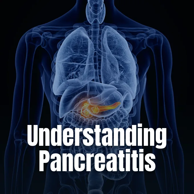
When patients explore what is pancreatitis, clinical guides describe a damaging process where digestive juices attack the very organ that produced them. Located behind the stomach, the pancreas produces enzymes to digest fats, proteins, and sugars, while endocrine cells make insulin and glucagon to control blood sugar [10, 14]. In healthy bodies, these digestive fluids stay inactive until they flow through the main duct into the small intestine. In pancreatitis, normal cell defenses fail, causing early autodigestion within the gland itself. Across the United States, acute attacks drive about 350,000 emergency department visits each year, producing hospital stays that range from mild swelling to life-threatening illnesses with multiorgan failure [5, 14].
- What Is Pancreatitis?
- How Trapped Enzymes Cause Auto-Digestion
- Common Triggers Leading to Glandular Injury
- Differences Between Acute and Chronic Pancreatitis
- Recognizing Primary Warning Signs
- Diagnostic Criteria and Imaging Protocols
- Medical Treatments for Pancreatitis
- Potential Complications Affecting Long-Term Health
What Is Pancreatitis?
The pancreas is a vital gland situated behind the stomach [10]. Its tissue consists largely of exocrine acinar cells, which manufacture digestive enzymes in the form of inactive zymogens before packaging them into protective granules [8, 14]. The gland moves these proenzymes through the duct into the duodenum to support normal digestion [14].
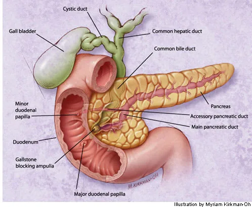
Why do these enzymes activate early inside the gland instead of the bowel? In a healthy intestine, a brush-border enzyme called enteropeptidase cleaves inactive trypsinogen into active trypsin, which then starts the orderly breakdown of dietary proteins. Cellular injury disrupts this containment. Digestive zymogen granules fuse with lysosomes inside acinar cells, allowing lysosomal enzymes to turn inactive precursors into active trypsin early, which unleashes a destructive enzymatic cascade that damages cell walls, causes severe tissue breakdown, and digests the gland from within. Once active trypsin escapes into surrounding tissue, it triggers other proenzymes to amplify the injury [8, 14].
Early enzyme release causes cell death, fluid leakage, and tissue swelling in the pancreas. Medical records show that autopsy reports first identified acute pancreatitis in 1882, while chronic pancreatitis gained separate clinical standing in 1946. Both forms share the same core danger, occurring when the gland is overwhelmed and injured by its own digestive juices [10, 14].

How Trapped Enzymes Cause Auto-Digestion
The cellular start of pancreatitis depends on disrupted calcium signals inside acinar cells. Pathological triggers cause sustained calcium release from internal storage pools, producing an abnormal calcium overload that halts normal enzyme secretion into ductal branches [7]. Trapped zymogens gather in large vacuoles with acidic lysosomal enzymes inside acinar cells, creating ideal biochemical conditions for early trypsin release [8, 14].
Once cell walls break, injured tissue releases active proteases that stimulate nearby immune cells. Macrophages and blood vessels release early messengers, including tumor necrosis factor-alpha and interleukin-6, which draw white blood cells into the injured gland. Activated neutrophils invade the gland tissue and release superoxide radicals with elastase enzymes, breaking down connective fibers and eroding small blood vessel walls. This cellular influx raises capillary leakiness, causing swelling, bleeding, and poor blood flow [8, 14]. If these chemical messengers flood the bloodstream in large amounts, they trigger systemic inflammatory response syndrome (SIRS), spreading tissue injury to distant organs and threatening vital organ systems [2, 14].
Enzymatic damage also spreads into nearby fat deposits, causing localized fat necrosis. Leaked lipase splits neutral fats into free fatty acids, which bind with calcium ions to form chalky deposits. This reaction, known as saponification, consumes calcium from the blood and often causes low calcium levels in sick patients [14]. In mild attacks, local defenses calm the swelling in a few days [10].
Common Triggers Leading to Glandular Injury
Biliary stones cause roughly 40 percent of acute cases in hospital registries. When a gallstone moves from the gallbladder into the common bile duct, it lodges near the ampulla of Vater to block the shared drainage path for bile and digestive juices, creating acute duct pressure and fluid backup [1, 13, 14]. Trapped juices back up into the gland, injuring cells and triggering early enzyme release [13, 14]. Records from the National Library of Medicine show that women develop gallstone pancreatitis more often than men, reflecting higher rates of gallstone disease in women [10, 13]. These stones trigger acute swelling by halting the natural flow of secretions [1, 10].
Heavy alcohol consumption ranks as the second leading etiology. In the United States, prolonged alcohol use is responsible for 17% to 25% of acute cases and between 42% and 77% of chronic pancreatitis cases, typically requiring four to five drinks daily for five or more years to induce lasting damage [10, 13, 14]. Ethanol directly destabilizes acinar cell membranes through oxidative stress [14].
Metabolic disorders, clinical procedures, and inherited genetic mutations contribute additional recognized triggers for acute and chronic disease. Serum triglyceride levels exceeding 1,000 mg/dL account for 2 to 7 percent of attacks, while familial chylomicronemia syndrome elevates lifetime risk up to 76 percent. Endoscopic retrograde cholangiopancreatography (ERCP) triggers post-procedure inflammation in 3 to 10 percent of examinations. Genetic disorders such as cystic fibrosis, autoimmune pancreatitis, and gain-of-function mutations in the cationic trypsinogen gene PRSS1, the SPINK1 gene, or the CFTR gene lower activation thresholds to induce hereditary pancreatitis [5, 14].
Differences Between Acute and Chronic Pancreatitis
Doctors divide pancreatitis into acute and chronic forms based on clinical course, tissue healing, and lasting damage. The revised Atlanta classification groups acute pancreatitis into mild, moderately severe, and severe forms based on whether organ failure is fleeting or persistent [1, 14]. Mild acute cases heal within a week without lasting injury, while severe cases cause tissue death [1, 10].
Chronic pancreatitis is a progressive disorder marked by irreversible fibrotic scarring and permanent loss of exocrine function [6, 11]. Instead of healing cleanly after an attack, the gland undergoes ongoing fibrotic replacement of healthy tissue driven by persistently active pancreatic stellate cells that generate excess collagen [8, 14]. Repeated acute flares often trigger this lasting structural decline [6, 11]. Cigarette smoking acts synergistically with alcohol consumption, accelerating parenchymal calcification, ductal stone formation, and irreversible glandular atrophy until the organ loses both digestive enzyme synthesis and insulin production [6, 14]. Over years of injury, healthy lobular architecture is replaced by dense, non-functional fibrous bands [6, 8].
Lasting gland damage creates deep digestive problems over time [6, 11]. When enzyme output drops too low, patients develop malabsorption and steatorrhea, passing bulky, pale, greasy stools [11, 12]. Furthermore, loss of insulin-producing cells leads to type 3c diabetes in 38 to 40 percent of chronic patients [6, 12]. Clinical specialists at the University of Chicago Medicine note that managing chronic disease needs lifelong care, enzyme pills, and diet changes [11, 12].

Recognizing Primary Warning Signs
Abdominal pain is the cardinal symptom of pancreatitis [9, 14]. In acute attacks, severe pain begins suddenly in the epigastric region or left upper quadrant, characteristically radiating straight through to the mid-back within minutes after eating high-fat meals that trigger digestive enzyme release [10, 13, 14]. Patients typically describe the ache as a severe, relentless pressure that grows increasingly intense when lying flat. To find relief from this boring abdominal pressure, patients often sit upright and lean forward, because lying supine compresses sensitive retroperitoneal nerve pathways against the spine [10, 13]. The discomfort can persist steadily for days without relief [9, 10].
Acute flares also cause full-body symptoms linked to gut inflammation. Nausea and vomiting happen in 75 to 90 percent of acute patients, accompanied by mild fever, fast heart rate, and dehydration [10, 13]. In rare severe cases with dying tissue, bruising appears on the flanks (Grey Turner sign) or around the navel (Cullen sign) [14].
Diagnostic Criteria and Imaging Protocols
Doctors diagnose acute pancreatitis using international consensus criteria [1, 7]. Under the revised Atlanta classification, confirming a diagnosis requires verifying at least two of three criteria: characteristic upper abdominal pain, serum amylase or lipase elevated three times normal thresholds, or characteristic cross-sectional imaging findings [1, 14]. Requiring two matching signs prevents diagnostic confusion with peptic ulcers or acute cholecystitis [14].
Blood tests provide a fast and reliable way to check for pancreatic injury. Serum lipase is the preferred enzyme marker because it provides higher accuracy and sensitivity than serum amylase [10, 13]. Lipase levels climb within four to eight hours of symptom onset, peak near 24 hours, and stay high for 8 to 14 days, whereas amylase returns to normal within three to five days [13]. Doctors also check liver enzymes, blood calcium, lipids, and kidney markers to spot causes and organ stress [7, 14]. In chronic pancreatitis, routine amylase and lipase levels often look normal because scarred tissue can no longer produce large amounts of enzymes [11, 14]. Clinicians must rely on imaging and functional tests in these advanced cases [6, 11].
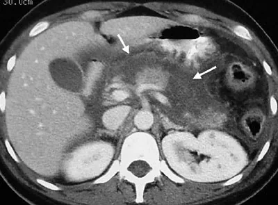
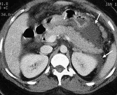
Diagnostic imaging establishes underlying etiology and detects local complications [1, 14]. Transabdominal ultrasound is the initial study to detect gallstones, though bowel gas obscures the pancreas in 25 to 35 percent of examinations [13, 14]. In rare international cases, ultrasound identifies migrating Ascaris lumbricoides worms blocking the biliary duct [14]. Contrast-enhanced computed tomography (CT) performed 48 to 72 hours after symptom onset provides definitive staging of parenchymal perfusion, peripancreatic fluid collections, and areas of tissue necrosis [1, 13].
Medical Treatments for Pancreatitis
Early medical care centers on prompt intravenous fluid hydration [1, 7]. Prompt fluid therapy counteracts fluid shifts, restores blood volume, and protects blood flow within the inflamed gland to prevent tissue death [1, 14]. Official clinical practice guidelines from the American College of Gastroenterology strongly recommend balanced crystalloid fluids, specifically lactated Ringer solution, given at controlled infusion rates during the first 24 hours [1, 3]. Doctors track blood pressure, pulse, and urine output closely to avoid fluid overload while keeping good blood flow to vital organs [1, 7]. Pain control with intravenous opioids gives fast comfort during acute attacks without hiding key exam findings [14]. This careful fluid and pain protocol stabilizes patients during the early hours of hospital care [1, 7].
In mild acute pancreatitis, patients resume oral feeding with low-fat foods as soon as abdominal pain and nausea begin to improve, moving away from historical routines of prolonged fasting. For patients with severe illness unable to tolerate oral meals, early enteral tube feeding into the stomach or small intestine maintains the mucosal barrier and reduces systemic infectious complications compared to total parenteral nutrition [1, 4, 5].

Definitive management treats underlying causes to prevent future attacks [1, 10]. Patients with gallstone pancreatitis should undergo cholecystectomy during the index admission once acute inflammation resolves, while those with concurrent cholangitis require urgent ERCP for stone extraction [1, 13]. Chronic pancreatitis requires complete alcohol and tobacco cessation. Patients take oral pancreatic enzyme capsules with meals to correct malabsorption, adhere to a low-fat diet, and manage secondary pancreatogenic diabetes with custom insulin regimens [11, 12].
Potential Complications Affecting Long-Term Health
Local complications develop as inflammatory fluid pools around the pancreas [2, 14]. During the first four weeks, these collections remain unencapsulated as acute peripancreatic fluid or acute necrotic collections. When fluid collections persist beyond four weeks without resolving, they develop a fibrous non-epithelial capsule, forming a mature pancreatic pseudocyst containing concentrated digestive enzymes that may require endoscopic drainage. Pseudocysts can cause persistent pain, rupture into the peritoneal cavity, or compress the stomach and duodenum [14].
In necrotizing disease, dead tissue matures after four weeks into walled-off pancreatic necrosis (WOPN). Evidence-based consensus statements from the World Society of Emergency Surgery emphasize that bacterial invasion of necrotic collections occurs in 20 to 40 percent of severe cases, sharply increasing patient mortality and necessitating urgent clinical intervention [2, 14]. Treating infected collections follows a step-up approach. Medical teams begin with targeted antibiotics and percutaneous catheter drainage, reserving minimally invasive endoscopic necrosectomy or open surgical debridement for patients who fail to improve. This conservative step-up strategy minimizes surgical trauma while clearing infected tissue [2, 3].
Systemic complications occur when circulating inflammatory mediators trigger multiorgan failure. Severe attacks can cause acute respiratory distress syndrome (ARDS), systemic shock, or acute kidney injury, accounting for most hospital fatalities in acute pancreatitis [2, 14]. Over decades, chronic pancreatitis causes permanent digestive failure, brittle diabetes, and a heightened risk of pancreatic ductal adenocarcinoma that requires structured long-term multidisciplinary monitoring [6, 11, 12].
- ACADEMIC JOURNAL Tenner, S., Vege, S. S., Sheth, S. G., Sauer, B., Yang, A., Conwell, D. L., Yadlapati, R. H., & Gardner, T. B. (2024). American College of Gastroenterology Guidelines: Management of Acute Pancreatitis. The American Journal of Gastroenterology, 119(3), 419-437. [Article Link]
- ACADEMIC JOURNAL Leppäniemi, A., Tolonen, M., Tarasconi, A., Segovia-Lohse, H., Gamberini, E., Kirkpatrick, A. W., Ball, C. G., Parry, N., Sartelli, M., Wolbrink, D., van Goor, H., Baiocchi, G., Ansaloni, L., Biffl, W., Coccolini, F., Di Saverio, S., Kluger, Y., Moore, E., & Catena, F. (2019). 2019 WSES guidelines for the management of severe acute pancreatitis. World Journal of Emergency Surgery, 14(1). [Article Link]
- ACADEMIC JOURNAL Arvanitakis, M., Dumonceau, J., Albert, J., Badaoui, A., Bali, M., Barthet, M., Besselink, M., Deviere, J., Oliveira Ferreira, A., Gyökeres, T., Hritz, I., Hucl, T., Milashka, M., Papanikolaou, I., Poley, J., Seewald, S., Vanbiervliet, G., van Lienden, K., van Santvoort, H., & van Hooft, J. (2018). Endoscopic management of acute necrotizing pancreatitis: European Society of Gastrointestinal Endoscopy (ESGE) evidence-based multidisciplinary guidelines. Endoscopy, 50(5), 524-546. [Article Link]
- ACADEMIC JOURNAL Forsmark, C. E., Vege, S. S., & Wilcox, C. M. (2016). Acute Pancreatitis. New England Journal of Medicine, 375(20), 1972-1981. [Article Link]
- ACADEMIC JOURNAL Mederos, M. A., Reber, H. A., & Girgis, M. D. (2021). Acute Pancreatitis: A Review. JAMA, 325(4), 382. [Article Link]
- ACADEMIC JOURNAL Singh, V. K., Yadav, D. K., & Garg, P. K. (2019). Diagnosis and Management of Chronic Pancreatitis: A Review. JAMA, 322(24), 2422. [Article Link]
- ACADEMIC JOURNAL Szatmary, P., Grammatikopoulos, T., Cai, W., Huang, W., Mukherjee, R., Halloran, C. M., Beyer, G., & Sutton, R. (2022). Acute Pancreatitis: Diagnosis and Treatment. Drugs, 82(12), 1251-1276. [Article Link]
- ACADEMIC JOURNAL Brock, C. (2013). Pathophysiology of chronic pancreatitis. World Journal of Gastroenterology, 19(42), 7231. [Article Link]
- WEBSITE National Institute of Diabetes and Digestive and Kidney Diseases. (2025). Symptoms & Causes of Pancreatitis. NIDDK, National Institutes of Health. [Article Link]
- WEBSITE National Library of Medicine. (2025). Acute Pancreatitis. MedlinePlus Medical Encyclopedia. [Article Link]
- WEBSITE National Library of Medicine. (2025). Chronic Pancreatitis. MedlinePlus Medical Encyclopedia. [Article Link]
- WEBSITE University of Chicago Medicine. (2023). Chronic Pancreatitis Treatment. [Article Link]
- ACADEMIC JOURNAL Munoz, A., & Katerndahl, D. A. (2000). Diagnosis and Management of Acute Pancreatitis. American Family Physician, 62(1), 164-174. [Article Link]
- WEBSITE Wikipedia contributors. Pancreatitis. Wikipedia, The Free Encyclopedia. [Article Link]


