





What is hemophilia? The medical diagnosis identifies a rare, lifelong bleeding disorder where the blood cannot form firm clots to stop bleeding after injury or surgery [1]. When an individual sustains a cut or internal bruise, normal platelets and plasma proteins work in close coordination to seal the damaged blood vessel wall. People born with hemophilia lack sufficient amounts of key clotting factors, allowing blood to ooze slowly into surrounding tissues over days or weeks. Modern therapies are transforming this chronic condition into a manageable illness.
- What Is Hemophilia?
- Why Hemophilia Is More Common in Males
- How Clotting Factor Deficiencies Cause Bleeding
- Recognizing Symptoms of Internal Bleeding
- Diagnostic Blood Tests and Factor Assays
- Why Hemophilia B Is Called Christmas Disease
- Factor Replacement and Non-Factor Treatments
- Clinical Advances in Gene Therapy
What Is Hemophilia?
Blood coagulation relies on a complex series of chemical interactions known as the coagulation cascade, involving roughly twenty distinct plasma proteins that circulate continuously through the vascular system [3]. When a blood vessel tears, these proteins activate sequentially in a rapid chain reaction, reinforcing the initial platelet plug with a resilient network of insoluble fibrin strands. In patients with hereditary hemophilia, a genetic alteration disrupts the synthesis of either factor VIII or factor IX, leaving the biochemical cascade incomplete and fragile. Without these essential proteins, the body cannot form durable seals over damaged blood vessels.
Factor VIII and factor IX function together at a pivotal junction within the intrinsic coagulation pathway, assembling into a catalytic complex on platelet surfaces to cleave factor X into its active form [13]. Activated factor X then converts prothrombin into thrombin, the central enzyme responsible for transforming soluble fibrinogen into a rigid mesh of polymerized fibrin strands. When either cofactor is missing or structurally defective, thrombin generation slows to a fraction of its normal rate. The resulting blood clot lacks mechanical stability, allowing hematomas to expand deep within muscle beds and soft tissues.
Normal human blood contains clotting factor concentrations that range between 50 and 150 percent of standard laboratory reference values [9]. When an inherited genetic change lowers these levels, clinical symptoms emerge in direct proportion to the remaining amount of functional protein circulating in the bloodstream. Medical registries across the world confirm that this deficit impairs natural healing after routine dental work, minor accidents, or invasive surgery.
Why Hemophilia Is More Common in Males
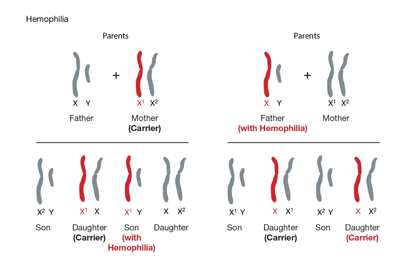
Gender genetics explain why inherited coagulation disorders appear almost exclusively among male infants [2]. The specific genes responsible for producing factor VIII and factor IX are located on the long arm of the X chromosome. Human biological sex is determined by the twenty-third pair of chromosomes: females have two X chromosomes, while males have one X and one Y chromosome. A male inherits his single X chromosome from his mother and a Y chromosome from his father. If that single maternal chromosome carries a mutated gene, he will develop hemophilia because the Y chromosome carries no matching gene to balance the deficiency.
Females inherit two X chromosomes, which usually protects them from severe bleeding complications [4]. A female who receives a mutated clotting gene on one X chromosome generally has a healthy copy on her second chromosome that produces enough clotting factor. These women are known as carriers. A carrier mother faces a 50 percent chance at each birth of passing the variant gene to her child. A son who inherits the gene will have hemophilia, whereas a daughter will become a carrier. A father with the condition passes the mutated X chromosome to all his daughters, but never to his sons.
Protection in female carriers is not absolute because cells undergo an embryonic process called lyonization, or random X-inactivation [3]. In early development, female cells turn off one X chromosome. When this process is skewed against the healthy gene, plasma factor levels can fall below 40 percent. Carrier women then face noticeable bleeding risks during childbirth, dental extractions, and major surgical procedures.
How Clotting Factor Deficiencies Cause Bleeding
When tissue injury tears a microscopic capillary, endothelial cells release tissue factor to initiate local blood clotting [13]. Circulating platelets adhere rapidly to the damaged collagen fibers, secreting chemical signals that recruit additional platelets to form a temporary mechanical barrier. In healthy individuals, the intrinsic pathway accelerates this process when factor IXa pairs with activated factor VIII on the platelet surface, creating the tenase complex. This molecular assembly activates factor X thousands of times faster than solitary enzymes can achieve alone. In individuals lacking factor VIII or IX, this amplification loop fails to engage, leaving the clotting response weak and incomplete.
Superficial scratches on the skin often stop bleeding quickly in people with hemophilia because platelets alone can seal minor breaks in tiny capillary walls [18]. The primary clinical hazard arises hours or days later when ordinary mechanical movement dislodges that initial, fragile platelet plug. Without dense fibrin strands to stabilize the clot, healing tissue begins to ooze continuously. A simple tooth extraction or minor dental scrape can bleed for multiple days if medical staff do not administer targeted factor concentrates.
Deep tissue bleeding can also occur spontaneously without any obvious blow or physical collision [1]. Blood can pool inside large muscle groups such as the calves, thighs, and the deep iliopsoas muscle located near the hip joint. These expanding intramuscular hematomas generate intense pressure on surrounding peripheral nerves, causing numbness, severe local pain, and temporary paralysis if left untreated. Prompt infusion of missing clotting factors relieves tissue pressure and prevents long-term nerve damage.
Recognizing Symptoms of Internal Bleeding
Internal bleeding into joint cavities, clinically termed hemarthrosis, represents the most frequent and disabling complication of severe hemophilia [16]. These bleeds occur most often in hinge joints that bear weight or absorb repetitive movement, especially the knees, ankles, and elbows. Young children often notice a tingling or warm sensation inside the joint before any swelling becomes visible to their parents. As blood fills the closed synovial capsule, the joint swells, stiffens, and becomes intensely painful to touch or flex. Toddlers often become unusually fussy and refuse to crawl, stand, or walk on the affected limb.
When an individual joint bleeds repeatedly, it becomes a target joint, triggering a debilitating chronic cycle of inflammation [17]. Free iron deposited from pooled blood induces synovial hypertrophy and chronic inflammation, termed haemophilic arthropathy. Over repeated episodes, destructive enzymes break down smooth articular cartilage and erode the underlying subchondral bone. This destructive progression eventually leads to severe joint deformities, chronic pain, and limited physical mobility. Orthopedic specialists often have to perform corrective joint replacement surgery in middle-aged adults who suffered recurrent childhood joint bleeds.
Intracranial bleeding inside the skull constitutes the most dangerous emergency associated with inherited coagulation defects [1]. Bleeding can occur inside the brain tissue or beneath the surrounding membranes following traumatic delivery or childhood head bumps. Warning signs include persistent headaches, lethargy, repeated vomiting, sudden behavioral changes, and seizures. Healthcare providers must administer high-dose clotting factor concentrates immediately before ordering brain imaging scans to reduce the risk of permanent neurological deficits or death.


Diagnostic Blood Tests and Factor Assays
Accurate diagnosis of a suspected bleeding disorder begins with baseline coagulation screening tests that evaluate how quickly blood forms a clot [16]. Clinicians order a complete blood count along with a prothrombin time (PT) test and an activated partial thromboplastin time (aPTT) test. In both hemophilia A and B, the prothrombin time and platelet counts fall within normal ranges because extrinsic clotting factors remain intact. In contrast, the partial thromboplastin time shows significant prolongation because factor VIII and factor IX are vital components of the intrinsic pathway.
When screening tests reveal an isolated prolongation of the intrinsic clotting time, hematologists perform specific factor assays to measure exact protein activity in blood plasma [9]. These assays determine whether the deficiency involves factor VIII or factor IX, allowing doctors to categorize clinical severity into three established grades. Patients with severe hemophilia possess less than 1 percent of normal factor activity, experiencing frequent spontaneous bleeds into joints and muscles. Moderate hemophilia involves factor activity between 1 and 5 percent, while mild hemophilia involves activity between 5 and 40 percent of normal levels.
In families with a confirmed genetic history, diagnostic testing can take place before birth or immediately following delivery [4]. Obstetricians collect umbilical cord blood at birth to test for factor VIII levels, which match adult reference ranges in healthy newborns. Testing for factor IX deficiency is often repeated when the infant reaches six months of age because newborn factor IX production depends on infant liver development and circulating vitamin K. Routine neonatal procedures like circumcision should be delayed whenever a family history suggests an undiagnosed bleeding disorder.

Why Hemophilia B Is Called Christmas Disease
Medical practitioners historically viewed all severe inherited bleeding disorders as a single condition until laboratory discoveries in 1952 revealed that different protein deficiencies produce identical clinical signs [13]. Early researchers discovered that mixing plasma samples from two different patients with severe bleeding could correct their respective clotting times in the test tube. This cross-correction proved that each patient suffered from a distinct molecular defect. The newly isolated condition was named Christmas disease after Stephen Christmas, a young boy who was the first individual thoroughly evaluated with this specific clotting deficiency.
Formal scientific descriptions of this newly discovered factor IX deficiency appeared in the December 1952 Christmas issue of the British Medical Journal [19]. The convergence of the patient’s family surname and the holiday publication date permanently fixed the nickname in medical history. Hemophilia B occurs in roughly 1 in 20,000 to 1 in 40,000 newborn males, representing approximately 15 percent of all hemophilia diagnoses worldwide. Despite having an identical clinical appearance to hemophilia A, it requires entirely separate replacement therapies because factor VIII products have no therapeutic effect on factor IX deficiency.
Hematologists also identify factor XI deficiency, a distinct condition recognized as hemophilia C [14]. Unlike types A and B, factor XI deficiency is an autosomal condition located on chromosome 4, meaning it affects men and women with equal frequency. The condition is most prevalent among Ashkenazi Jewish populations, where carrier frequencies range between 8 and 13.4 percent. Bleeding in factor XI deficiency is usually mild, primarily affecting mucosal tissues like the nose, mouth, and urinary tract after surgical procedures.
Factor Replacement and Non-Factor Treatments
For decades, the cornerstone of medical management has been prophylactic factor replacement therapy, which involves regular intravenous infusions of missing coagulation proteins [13]. Before the development of concentrated therapies, patients relied on whole blood or crude cryoprecipitate transfusions that carried serious risks of viral infection before viricidal testing standards took effect in 1985. Modern recombinant factor concentrates are produced using advanced cell cultures without human albumin, providing high purity and safety. Patients and families routinely learn to perform these intravenous infusions at home, administering factor several times a week to prevent spontaneous bleeding.
Around 15 to 30 percent of patients with severe hemophilia A develop neutralizing antibodies known as inhibitors, which destroy infused factor VIII proteins [7]. To manage bleeding in these patients, clinicians historically relied on bypassing agents like recombinant activated factor VII. In 2018, the U.S. Food and Drug Administration approved emicizumab (Hemlibra), a bispecific monoclonal antibody administered under the skin. In the pivotal HAVEN 3 clinical trial, weekly emicizumab prophylaxis achieved a 96 percent reduction in annualized bleeding rates among patients with severe hemophilia A without inhibitors.
Treatment options expanded on March 28, 2025, when the FDA approved Qfitlia (fitusiran), a first-in-class antithrombin-lowering therapy developed by Sanofi [15]. Administered by subcutaneous injection as infrequently as once every two months, the small interfering RNA medicine lowers antithrombin to promote thrombin generation in patients with hemophilia A or B. Tanya Wroblewski, M.D., deputy director in the FDA’s Center for Drug Evaluation and Research, explained: “Today’s approval of Qfitlia is significant for patients with hemophilia because it can be administered less frequently than other existing options.” In the ATLAS trials, fitusiran reduced annualized bleed rates by 71 to 73 percent.
Clinical Advances in Gene Therapy
Gene therapy offers the potential for long-term correction of inherited bleeding disorders by delivering functional clotting genes directly into patient liver cells [8]. Clinical trials led by Professor John Pasi at Barts Health NHS Trust and Queen Mary University of London evaluated adeno-associated virus vectors designed to introduce the factor VIII gene. Following a single infusion, eleven of thirteen trial participants achieved normal or near-normal factor VIII levels and stopped routine preventive injections. Professor Pasi stated: “We have seen mind-blowing results which have far exceeded our expectations.” Jake Omer, a 29-year-old participant who previously injected factor three times weekly, reported that the single treatment freed him from chronic ankle pain.
Remarkable results have also emerged for factor IX deficiency through liver-directed gene delivery platforms [20]. In a landmark study published in the New England Journal of Medicine, lead author Professor Pratima Chowdary of Royal Free Hospital and University College London tested the FLT180a vector in patients with severe hemophilia B. The trial demonstrated sustained factor IX expression in nine out of ten enrolled participants, eliminating their need for weekly intravenous infusions. Professor Chowdary noted: “Removing the need for haemophilia patients to regularly inject themselves with the missing protein is an important step in improving their quality of life.”
Elliott Mason, a trial participant who received the single factor IX gene infusion, described his recovery to BBC News: “My life is completely normal, there’s nothing that I have to stop and think “how might my haemophilia affect this?”.” [11]. The U.S. Food and Drug Administration granted commercial approval to etranacogene dezaparvovec (Hemgenix) in November 2022, marking the first approved gene therapy for hemophilia B. While hematologists continue monitoring patient registries to assess how long viral expression lasts, these clinical breakthroughs offer real hope for a permanent functional cure.
- WEBSITE Centers for Disease Control and Prevention. (2025). About Hemophilia. [Article Link]
- WEBSITE National Human Genome Research Institute. (2026). Hemophilia. [Article Link]
- WEBSITE MedlinePlus. (2022). Hemophilia: MedlinePlus Genetics. [Article Link]
- WEBSITE Centers for Disease Control and Prevention. (2024). Information on Hemophilia for Women. [Article Link]
- WEBSITE MedlinePlus Medical Encyclopedia. (2026). Hemophilia A. [Article Link]
- PRESS RELEASE U.S. Food and Drug Administration. (2025). FDA Approves Novel Treatment for Hemophilia A or B, with or without Factor Inhibitors. [Article Link]
- PRESS RELEASE U.S. Food and Drug Administration. (2018). FDA approves emicizumab-kxwh for hemophilia A with or without factor VIII inhibitors. [Article Link]
- PRESS RELEASE Barts Health NHS Trust. (2017). Groundbreaking gene therapy trial set to cure haemophilia A. [Article Link]
- WEBSITE Department of Health & Human Services, State Government of Victoria. (1999). Haemophilia. [Article Link]
- WEBSITE MedlinePlus Medical Encyclopedia. (2026). Hemophilia B. [Article Link]
- ONLINE NEWS Gallagher, J. (2022). Transformational therapy cures haemophilia B. BBC News. [Article Link]
- ONLINE NEWS Gallagher, J. (2017). Haemophilia A trial results ‘mind-blowing’. BBC News. [Article Link]
- WEBSITE Medscape. (2026). Hemophilia A (Factor VIII Deficiency): Background, Pathophysiology, Etiology. [Article Link]
- WEBSITE Medscape. (2025). Hemophilia C (Factor XI Deficiency): Background, Pathophysiology, Etiology. [Article Link]
- PRESS RELEASE Sanofi. (2025). Qfitlia approved as the first therapy in the US to treat hemophilia A or B with or without inhibitors. [Article Link]
- WEBSITE KidsHealth. (2026). Hemophilia. [Article Link]
- WEBSITE Wikipedia. (n.d.). Haemophilia. [Article Link]
- WEBSITE Wikipedia. (n.d.). Haemophilia A. [Article Link]
- WEBSITE Wikipedia. (n.d.). Haemophilia B. [Article Link]
- ACADEMIC JOURNAL Chowdary, P., Shapiro, S., Makris, M., Evans, G., Boyce, S., Talks, K., Dolan, G., Reiss, U., Phillips, M., Riddell, A., Peralta, M. R., Quaye, M., Patch, D. W., Tuddenham, E., Dane, A., Watissée, M., Long, A., & Nathwani, A. (2022). Phase 1–2 Trial of AAVS3 Gene Therapy in Patients with Hemophilia B. New England Journal of Medicine, 387(3), 237-247. [Article Link]


1 comment